Beyond AlphaFold: Benchmarking Evolutionary Algorithms Against Machine Learning for Novel Protein Folding and Design
This article provides a comprehensive benchmark of Evolutionary Algorithms (EAs) and Machine Learning (ML) models for protein structure prediction and design.
Beyond AlphaFold: Benchmarking Evolutionary Algorithms Against Machine Learning for Novel Protein Folding and Design
Abstract
This article provides a comprehensive benchmark of Evolutionary Algorithms (EAs) and Machine Learning (ML) models for protein structure prediction and design. Targeting researchers and drug development professionals, it explores the foundational principles of both approaches, detailing their methodological applications and inherent strengths. The analysis delves into critical troubleshooting and optimization strategies for deploying these computational tools effectively. Through a rigorous validation and comparative framework, assessing metrics like accuracy, novelty, and resource efficiency, the article synthesizes key takeaways. It concludes that a hybrid AI future, leveraging the complementary strengths of EAs and ML, holds the greatest promise for unlocking novel protein functions and accelerating biomedical discovery.
The Computational Protein Folding Landscape: From Physical Principles to AI-Driven Prediction
Biological Context: From Linear Chains to Functional Machines
The "protein folding problem" is one of biology's greatest unsolved mysteries. It refers to the challenge of predicting how a linear sequence of amino acids folds into a specific, three-dimensional structure that dictates its function [1]. Proteins are the primary architects of cellular activity, catalyzing reactions, providing structural support, and regulating biochemical processes. A protein's final, functional (native) tertiary structure is typically achieved through a stepwise establishment of regular secondary structures like α-helices and β-sheets, which then form the complete 3D architecture [2].
The precise final structure is not random; it is encoded in the amino acid sequence. This structure is crucial because it enables the protein to interact with other molecules and perform its role. Protein misfolding occurs when this process goes awry, and it is directly linked to severe diseases. Misfolded proteins can aggregate, leading to conditions such as Alzheimer's disease, Type II Diabetes, and cardiovascular diseases [3] [1]. For instance, in cardiovascular disease, misfolding of proteins like Apolipoprotein B (ApoB) can lead to atherosclerosis, where fatty acids accumulate in arteries, increasing the risk of heart attack and stroke [1].
The AI Revolution: Benchmarking Modern Protein Structure Prediction Tools
The field of protein structure prediction was revolutionized by artificial intelligence (AI), particularly with the introduction of AlphaFold2. Today, several AI models offer different trade-offs in accuracy, speed, and resource requirements, which are critical for researchers to consider.
The following table provides a quantitative comparison of three prominent ML-based protein folding methods, benchmarking their performance on key operational metrics.
Table 1: Performance Benchmarking of Machine Learning Protein Folding Tools
| Model | Developer | Key Strength | Running Time (for 400aa sequence) | PLDDT Accuracy (for 400aa sequence) | GPU Memory Usage |
|---|---|---|---|---|---|
| ESMFold | Meta AI | Exceptional speed | ~20 seconds | 0.93 [4] | 18 GB [4] |
| OmegaFold | HelixFold | Balance of speed and accuracy for shorter sequences | ~110 seconds | 0.76 [4] | 10 GB [4] |
| AlphaFold (via ColabFold) | Google DeepMind | High overall accuracy | ~210 seconds | 0.82 [4] | 10 GB [4] |
| OpenFold3 | Academic Consortium | Open-source, aims to match AlphaFold3 performance | Information Not Shown | Information Not Shown | Information Not Shown |
| SimpleFold | Apple | Uses general-purpose transformers, challenges need for complex custom architectures | Information Not Shown | Information Not Shown | Information Not Shown |
Analysis for Tool Selection
- For High-Throughput Screening: ESMFold's remarkable speed makes it ideal for tasks requiring rapid analysis of large numbers of sequences, such as initial characterization of genomic data [4].
- For Maximum Accuracy on Complex Targets: AlphaFold remains the gold standard for overall accuracy, often matching the precision of experimental methods. It is the best choice when the highest confidence prediction is required [1] [4].
- For Resource-Constrained Environments or Shorter Sequences: OmegaFold provides a compelling balance, offering good accuracy with lower computational cost, making it suitable for labs with limited GPU resources, especially for sequences under 400 amino acids [4].
- For Open-Source and Collaborative Science: The development of OpenFold3 is a significant move towards creating a powerful, open-source alternative to proprietary models, which can foster greater collaboration and transparency in research [5].
Experimental Paradigms: From Standardized Bench Experiments to Mega-Scale Assays
Understanding protein folding requires robust experimental data. The field has established standardized protocols for traditional kinetics studies and developed novel high-throughput methods to generate data on an unprecedented scale.
Consensus Experimental Conditions for Folding Kinetics
To enable meaningful comparison of folding data across different laboratories, the scientific community has proposed a set of consensus conditions for in vitro experiments [6].
Table 2: Standardized Experimental Conditions for Protein Folding Kinetics
| Experimental Parameter | Consensus Standard | Rationale |
|---|---|---|
| Temperature | 25°C | Easily maintained, maximizes backward compatibility with existing literature [6]. |
| Buffer | 50 mM Phosphate or HEPES (pH 7.0) | Buffers effectively at neutral pH; a common baseline for experimental comparison [6]. |
| Denaturant | Urea | Preferred over guanidinium salts due to fewer confounding ionic strength effects [6]. |
| Data Reporting | lnkf (sec⁻¹) and m-values in (kJ/mol)/M | Standardized units ensure consistency and prevent errors in comparative analysis [3] [6]. |
High-Throughput Workflow: cDNA Display Proteolysis
Recent advances have enabled massively parallel measurement of protein stability. The cDNA display proteolysis method is a powerful high-throughput assay that can measure thermodynamic folding stability for hundreds of thousands of protein domains in a single experiment [7].
The diagram below illustrates the integrated experimental and computational workflow of this method.
This workflow begins with a synthetic DNA library where each oligonucleotide encodes a test protein. The DNA is transcribed and translated in vitro using cell-free cDNA display, resulting in proteins covalently attached to their encoding cDNA. This pool of protein-cDNA complexes is then subjected to protease digestion. The key principle is that unfolded proteins are cleaved more rapidly than folded ones. The intact (protease-resistant) complexes are purified, and the surviving sequences are quantified using deep sequencing. Finally, a Bayesian kinetic model uses the sequencing counts to infer the thermodynamic folding stability (ΔG) for each of the hundreds of thousands of protein variants [7].
Researchers in protein folding and design rely on a suite of databases, software, and experimental resources.
Table 3: Essential Research Reagents and Resources for Protein Folding Research
| Resource Name | Type | Function and Application |
|---|---|---|
| ACPro Database [3] | Data Repository | A curated database of verified protein folding kinetics data, used for testing predictive models. |
| cDNA Display Proteolysis [7] | Experimental Assay | A high-throughput method for measuring thermodynamic folding stability for up to 900,000 protein variants. |
| Evolutionary Algorithms (DAO-MOGA) [8] | Computational Tool | A genetic algorithm for the inverse protein folding problem, optimizing for sequence diversity and structure. |
| Protein Data Bank (PDB) | Data Repository | The global repository for experimentally-determined 3D structures of proteins, used for training and validation. |
| 3D Profile (3D-1D Scoring) [8] | Computational Metric | A score evaluating the compatibility of an amino acid sequence with a target 3D structure for protein design. |
The integration of AI-based structure prediction with high-throughput experimental data is shaping the future of protein science. While AI tools like AlphaFold, ESMFold, and OmegaFold provide rapid structural models, large-scale experimental data remains crucial for understanding the hidden thermodynamics of folding—the energetics that drive the process and are invisible in static structures [7]. This synergy is particularly powerful for tackling the inverse folding problem, where evolutionary algorithms and other computational methods are used to design novel sequences that fold into a desired structure [8]. As both AI models and experimental techniques continue to evolve, they promise to unlock deeper insights into protein misfolding diseases and accelerate the rational design of proteins for therapeutic and biotechnology applications.
The protein folding problem represents one of the central challenges in structural biology, seeking to understand how a linear amino acid sequence spontaneously folds into a unique three-dimensional functional structure [9]. The energy landscape theory provides a powerful conceptual framework for understanding this process, proposing that natural proteins have evolved "minimally frustrated" folding landscapes that are funneled toward the native state [10]. This funneling allows proteins to avoid the kinetic traps that would be inevitable in a random heteropolymer and to fold efficiently on biological timescales.
In this framework, the molten globule represents a crucial intermediate state—a compact, partially organized ensemble of structures that retains significant secondary structure but lacks fixed tertiary side-chain packing [10]. The characterization of these landscapes involves both physical energy landscapes (derived from atomic interactions and physics-based models) and evolutionary energy landscapes (inferred from statistical analysis of homologous protein sequences) [10]. This article examines how modern machine learning methods for protein structure prediction navigate these landscapes, benchmarking their performance against physical principles and each other.
Theoretical Framework: Physical and Evolutionary Energies
The Principle of Minimal Frustration
The principle of minimal frustration posits that natural protein sequences have been evolutionarily selected to encode energy landscapes where interactions stabilizing the native state are mutually reinforcing rather than competing [10]. This stands in contrast to random amino acid sequences, which typically exhibit rugged landscapes with numerous deep kinetic traps. In minimally frustrated systems, the energetic bias toward the native state is sufficiently strong that the protein can rapidly fold without becoming trapped in non-native configurations.
Quantitatively, this relationship can be expressed through the equation:
[ 2\left(\frac{Tf}{T{sel}}\right) = \left(\frac{1}{Tg^2} + \frac{1}{Tf^2}\right) ]
Where (Tf) represents the protein's folding temperature, (Tg) indicates the glass transition temperature below which the protein would become trapped in non-native states, and (T{sel}) represents the evolutionary selection temperature [10]. For natural proteins, (Tf/T_g > 1), ensuring that folding occurs before the system becomes trapped in misfolded states.
Evolutionary Energy Landscapes from Sequence Coevolution
Direct coupling analysis (DCA) and other coevolution-based methods leverage the evolutionary record encoded in multiple sequence alignments to infer structural constraints [10]. The underlying assumption is that pairs of residues that interact in the tertiary structure will show correlated evolutionary patterns to maintain functional folds. These methods parameterize a Potts model Hamiltonian that assigns an evolutionary energy to any given sequence, effectively defining the evolutionary landscape [10].
The relationship between physical and evolutionary energies can be described by:
[ P(S) = \frac{e^{-\beta E(S)}}{Z} ]
Where (P(S)) represents the probability that sequence (S) adopts the folded structure, (E(S)) is the energy of the folded structure, (\beta = (kB T{sel})^{-1}), and (Z) is the partition function [10]. This formalism demonstrates how evolutionary constraints shape foldable sequences.
Pseudogenes as Natural Experiments in Landscape Devolution
Pseudogenes—formerly protein-coding sequences that have accumulated degenerative mutations—provide natural experiments for testing energy landscape theory [10]. When selective pressure to maintain a functional fold is removed, pseudogene sequences typically accumulate mutations that disrupt the native global network of stabilizing residue interactions, increasing frustration and decreasing foldability [10].
Interestingly, in some cases, pseudogene mutations actually decrease energetic frustration while simultaneously altering biological function, particularly in regions normally responsible for binding interactions [10]. This demonstrates how evolution tunes energy landscapes for both foldability and specific biological functions, and how these constraints can be decoupled when functional requirements are relaxed.
Machine Learning Approaches to Navigating Energy Landscapes
AlphaFold: Integrating Physical and Evolutionary Constraints
AlphaFold represents a transformative approach that combines physical, evolutionary, and geometric constraints through novel neural network architectures [11]. The system employs an Evoformer module—a novel neural network block that processes multiple sequence alignments and residue-pair representations through attention mechanisms [11]. This allows the network to reason about spatial and evolutionary relationships simultaneously.
The structure module then generates explicit 3D atomic coordinates through a series of iterative refinements, starting from trivial initial states and progressively developing accurate structures [11]. Throughout this process, AlphaFold employs principles of equivariance to ensure physical plausibility of the generated structures. The network's ability to provide accurate per-residue confidence estimates (pLDDT) further demonstrates its sophisticated understanding of structural constraints [11].
ESMFold and OmegaFold: Alternative Architectural Strategies
ESMFold leverages a transformer-based architecture trained on evolutionary-scale protein sequence databases, enabling rapid structure prediction without explicit multiple sequence alignment construction during inference [4]. This approach benefits from the strengths of evolutionary covariance information while achieving significant speed advantages.
OmegaFold utilizes a deep learning model that emphasizes accuracy, particularly for shorter protein sequences [4]. Its architecture effectively balances computational efficiency with prediction reliability, making it suitable for scenarios where resource optimization is crucial.
Comparative Performance Benchmarking
Experimental Protocol and Metrics
To objectively evaluate these methods, we examine a systematic benchmarking study conducted on a g5.2xlarge A10 GPU configuration [4]. The evaluation employs several key metrics:
- Running Time: Total computation time required for structure prediction
- PLDDT (Predicted Local Distance Difference Test): Per-residue estimate of prediction confidence on a 0-1 scale
- Memory Usage: CPU memory consumption during prediction
- GPU Memory: Graphics memory utilization
The benchmarking was performed across protein sequences of varying lengths (50, 100, 200, 400, 800, and 1600 residues) to evaluate scalability and length-dependent performance characteristics [4].
Performance Comparison Across Sequence Lengths
Table 1: Comparative Performance of Protein Structure Prediction Methods
| Sequence Length | Method | Running Time (s) | PLDDT Score | CPU Memory (GB) | GPU Memory (GB) |
|---|---|---|---|---|---|
| 50 | ESMFold | 1 | 0.84 | 13 | 16 |
| OmegaFold | 3.66 | 0.86 | 10 | 6 | |
| AlphaFold | 45 | 0.89 | 10 | 10 | |
| 100 | ESMFold | 1 | 0.30 | 13 | 16 |
| OmegaFold | 7.42 | 0.39 | 10 | 7 | |
| AlphaFold | 55 | 0.38 | 10 | 10 | |
| 200 | ESMFold | 4 | 0.77 | 13 | 16 |
| OmegaFold | 34.07 | 0.65 | 10 | 8.5 | |
| AlphaFold | 91 | 0.55 | 10 | 10 | |
| 400 | ESMFold | 20 | 0.93 | 13 | 18 |
| OmegaFold | 110 | 0.76 | 10 | 10 | |
| AlphaFold | 210 | 0.82 | 10 | 10 | |
| 800 | ESMFold | 125 | 0.66 | 13 | 20 |
| OmegaFold | 1425 | 0.53 | 10 | 11 | |
| AlphaFold | 810 | 0.54 | 10 | 10 | |
| 1600 | ESMFold | Failed (OOM) | - | - | 24 |
| OmegaFold | Failed (>6000) | - | - | 17 | |
| AlphaFold | 2800 | 0.41 | 10 | 10 |
Data sourced from benchmarking study [4]. OOM = Out of Memory.
Method Selection Guidelines Based on Benchmarking Data
For short sequences (<400 residues): OmegaFold provides an optimal balance of accuracy (PLDDT) and resource efficiency, with significantly lower GPU memory requirements than ESMFold and faster execution than AlphaFold [4].
For medium-length sequences (400-800 residues): ESMFold offers the best speed-accuracy tradeoff, though at the cost of higher memory consumption [4].
For long sequences (>800 residues): AlphaFold demonstrates superior capability in handling very long proteins where other methods fail or show degraded performance [4].
For resource-constrained environments: OmegaFold provides the most memory-efficient operation across all sequence lengths [4].
Visualizing Protein Folding Method Workflows
Diagram 1: AlphaFold's iterative refinement process integrates MSA and coevolutionary information through Evoformer and Structure modules, with recycling enabling progressive improvement of predicted structures [11].
Table 2: Key Experimental Resources for Protein Folding Research
| Resource | Type | Primary Function | Application Context |
|---|---|---|---|
| AWSEM | Physical Model | Coarse-grained molecular dynamics for structure prediction | Physics-based folding simulation and landscape characterization [10] |
| DCA | Algorithm | Inference of coevolutionary constraints from sequence data | Evolutionary energy landscape calculation [10] |
| PDB | Database | Repository of experimentally determined protein structures | Method training and validation [12] [9] |
| AlphaFold DB | Database | Precomputed structure predictions for proteomes | Benchmarking and biological discovery [12] |
| CATH/SCOP | Database | Hierarchical protein structure classification | Fold recognition and classification [13] |
| MSA Tools | Software | Construction of multiple sequence alignments | Evolutionary constraint identification [11] |
The remarkable accuracy achieved by modern ML protein folding methods, particularly AlphaFold, represents a convergence of physical understanding and data-driven pattern recognition [9] [11]. These systems successfully navigate protein energy landscapes by leveraging both the physical principle of minimal frustration and the evolutionary record of sequence covariation. While these methods differ in their architectural approaches and computational characteristics, they share a fundamental reliance on the energy landscape theory that has guided decades of protein folding research.
The benchmarking data reveals that method selection involves tradeoffs between speed, accuracy, and computational resources, with each approach exhibiting distinct strengths across different protein lengths and resource scenarios [4]. As these methods continue to evolve, their integration with physical models like AWSEM [10] promises to further bridge the gap between predictive accuracy and mechanistic understanding of the folding process.
This synergy between physical theory and machine learning not only advances structure prediction capabilities but also provides new avenues for exploring fundamental questions about protein folding landscapes, evolutionary constraints, and the molecular basis of biological function.
The protein folding problem—predicting a protein's three-dimensional structure from its amino acid sequence—has been one of the most significant challenges in biology for decades. For years, researchers relied on evolutionary algorithms and simplified models to tackle this complex problem. Methods using the HP lattice model, which classifies amino acids as hydrophobic (H) or polar (P), provided early insights but were limited to simplified representations and faced NP-hard computational complexity [14] [15]. The field underwent a seismic shift with the introduction of deep learning approaches, culminating in AlphaFold2's breakthrough performance in the CASP14 assessment in 2020 [12]. This transformation has moved the field from theoretical simplified models to predictions at near-experimental accuracy, revolutionizing structural biology and drug discovery.
This guide provides an objective comparison of three pioneering machine learning systems—AlphaFold, ESMFold, and OmegaFold—that have redefined the standards of protein structure prediction. We examine their performance metrics, architectural innovations, and practical applications within the context of benchmarking against traditional computational approaches.
Methodological Evolution: Architectural Innovations
Traditional Evolutionary Approaches
Before the deep learning revolution, protein folding optimization relied heavily on stochastic population-based algorithms. The Differential Evolution (DE) algorithm represented the state-of-the-art, using mutation, crossover, and selection operators to navigate the conformational landscape [14]. These methods operated on simplified models like the 3D AB off-lattice model, where energy functions favored hydrophobic interactions between non-polar amino acids. The local search mechanisms and component reinitialization strategies attempted to address the notorious challenges of rugged energy landscapes with numerous local minima [14]. However, these approaches could only confirm optimal solutions with 100% hit ratios for sequences containing up to 18 monomers, highlighting their limitations for larger proteins [14].
Modern Machine Learning Architectures
The transformation of protein structure prediction began with the integration of transformer neural networks and novel architectural paradigms.
AlphaFold2: Introduced the Evoformer architecture—a two-track system that jointly processes evolutionary information from multiple sequence alignments (MSAs) and pairwise relationships between residues. This attention-based mechanism draws global dependencies between amino acids to produce accurate atomic coordinates [12] [16]. AlphaFold-Multimer extended this capability to protein complexes by including multimeric structures in its training data [17].
ESMFold: Leverages a massive protein language model (ESM-2) trained on millions of protein sequences. Unlike AlphaFold2, ESMFold is alignment-free, predicting structures directly from single sequences without explicit MSAs. It incorporates a modified Evoformer block to refine its predictions [18] [16]. This architecture provides significant speed advantages, being up to 60 times faster than traditional MSA-dependent methods [19].
OmegaFold: Utilizes a protein language model (OmegaPLM) to learn single and pairwise residue embeddings, which are processed through a geometry-inspired transformer block called the Geoformer. Like ESMFold, it operates without MSAs, making it particularly valuable for proteins with few evolutionary relatives [16].
The diagram below illustrates the fundamental shift in methodology from traditional evolutionary approaches to modern machine learning systems:
Performance Benchmarking: A Comparative Analysis
Recent systematic evaluations provide comprehensive performance comparisons across these systems. A benchmark study conducted on 1,327 protein chains deposited in the PDB between 2022 and 2024—ensuring no overlap with training data—revealed clear performance hierarchies:
Table 1: Overall Accuracy Metrics on Recent Protein Structures
| Method | Median TM-score | Median RMSD (Å) | Key Strengths |
|---|---|---|---|
| AlphaFold2 | 0.96 | 1.30 | Highest overall accuracy, excellent stereochemistry |
| ESMFold | 0.95 | 1.74 | Fast prediction, good for high-throughput screening |
| OmegaFold | 0.93 | 1.98 | Robust on orphan proteins, reasonable accuracy |
AlphaFold2 consistently achieves the highest median accuracy, as measured by both TM-score (0.96) and root-mean-square deviation (RMSD, 1.30 Å) [20]. Independent evaluations on CASP15 targets confirm this hierarchy, with AlphaFold2 attaining a mean GDT-TS score of 73.06, followed by ESMFold (61.62) and OmegaFold [16].
Speed and Resource Utilization
While accuracy is crucial, practical considerations of computational efficiency often influence method selection for large-scale applications:
Table 2: Computational Performance Comparison (A10 GPU)
| Method | Prediction Time (50 aa) | GPU Memory (50 aa) | CPU Memory | Optimal Use Case |
|---|---|---|---|---|
| ESMFold | 1 second | 16 GB | 13 GB | High-throughput screening |
| OmegaFold | 3.66 seconds | 6 GB | 10 GB | Short sequences, resource-constrained environments |
| AlphaFold2 | 45 seconds | 10 GB | 10 GB | Maximum accuracy applications |
ESMFold demonstrates remarkable speed advantages, processing a 50-amino acid sequence in approximately 1 second compared to OmegaFold's 3.66 seconds and AlphaFold2's 45 seconds [4]. However, these speed advantages come with higher GPU memory requirements for shorter sequences [4]. OmegaFold strikes a balance with better memory efficiency, particularly valuable for shorter sequences (up to 400 amino acids) and resource-constrained environments [4].
Protein Length and Type Considerations
Method performance varies significantly with protein length and structural characteristics. For sequences shorter than 400 amino acids, OmegaFold frequently provides the optimal balance of accuracy and efficiency, achieving higher PLDDT scores than ESMFold on shorter sequences while using less memory [4]. ESMFold maintains strong performance across various protein lengths, even successfully predicting structures of large proteins with 540 residues with high accuracy (TM-score 0.98) [19]. However, all methods show declining accuracy as protein size increases, particularly for multidomain proteins with complex topologies where domain packing remains challenging [16].
Specialized Capabilities
Multimeric Predictions: AlphaFold-Multimer extends accurate predictions to protein complexes, successfully modeling approximately 70% of protein-protein interactions in benchmark tests [17]. While ESMFold has capabilities for predicting multimers (complexes of multiple protein chains), performance evaluation remains an active area of research [19].
Stereochemical Quality: AlphaFold2 produces structures with stereochemistry closest to experimental observations, as evidenced by Ramachandran plot distributions and MolProbity scores [16]. Both ESMFold and OmegaFold exhibit more physically unrealistic local structural regions, limiting their utility for applications requiring precise atomic coordinates [16].
Side-chain Positioning: All methods show room for improvement in side-chain positioning, with AlphaFold2 attaining the highest global distance calculation for side-chains (GDC-SC) score, though still below 50 [16].
Experimental Protocols and Benchmarking Methodologies
Standardized Evaluation Frameworks
Robust benchmarking requires standardized datasets and evaluation metrics. Key methodological approaches include:
Temporal Split Validation: Using proteins deposited in the PDB after the training cutoff dates of the tools being evaluated (e.g., July 2022-July 2024 structures for benchmarking tools trained on earlier data) ensures no data leakage [20].
Homology Reduction: Applying sequence identity thresholds (e.g., ≤30% identity to training sequences) via tools like MMseqs2 removes potential homology between benchmark and training datasets [17].
Multiple Assessment Metrics: Employing complementary metrics including TM-score (global topology), DockQ (interface quality for complexes), lDDT (local distance difference test), and PLDDT (per-residue confidence scores) provides a comprehensive accuracy profile [20] [17].
Workflow for Comparative Assessment
The typical workflow for benchmarking protein folding methods involves sequential steps of data preparation, model execution, and structural evaluation:
Successful protein structure prediction and analysis requires leveraging specialized databases, software tools, and computational resources:
Table 3: Essential Resources for Protein Structure Research
| Resource | Type | Function | Access |
|---|---|---|---|
| Protein Data Bank (PDB) | Database | Experimental protein structures | https://www.rcsb.org/ |
| ESM Metagenomic Atlas | Database | 617M+ predicted metagenomic structures | https://esmatlas.com/ |
| AlphaFold DB | Database | 200M+ AlphaFold predictions | https://alphafold.ebi.ac.uk/ |
| ColabFold | Software | Accessible AlphaFold/MMseqs2 implementation | https://colabfold.com |
| HuggingFace Transformers | Software | Simplified ESMFold API | https://huggingface.co/ |
| MMalign | Software | Structure comparison and alignment | https://github.com/ |
| DockQ | Software | Quality assessment of protein complexes | https://gitlab.com/ElofssonLab/DockQ |
These resources provide the foundational infrastructure for protein structure prediction, analysis, and validation. The ESM Metagenomic Atlas in particular represents a significant expansion of accessible structural information, containing 617 million predicted metagenomic protein structures that help illuminate the "dark matter" of protein space [18] [19].
The transformation of protein structure prediction through machine learning has provided researchers with an unprecedented set of tools for exploring structural biology. Based on comprehensive benchmarking:
AlphaFold2 remains the gold standard for maximum accuracy applications where computational resources and time are secondary concerns. Its superior performance on diverse protein types and excellent stereochemical quality make it ideal for detailed mechanistic studies and hypothesis generation.
ESMFold offers the best solution for high-throughput applications requiring rapid screening of multiple protein targets. Its alignment-free architecture enables speed advantages of 6-60× over MSA-dependent methods, though with slightly reduced accuracy [19].
OmegaFold provides a balanced option for shorter sequences and resource-constrained environments, with particularly strong performance on proteins under 400 amino acids while using less memory than ESMFold [4].
The choice between these systems ultimately depends on the specific research context—balancing accuracy requirements, computational resources, protein characteristics, and application scope. As the field continues to evolve, addressing current challenges in multidomain protein packing, side-chain positioning, and complex prediction will further enhance the transformative impact of these tools on biological research and therapeutic development.
The inverse protein folding problem (IFP)—finding amino acid sequences that fold into a defined three-dimensional structure—represents a fundamental challenge in structural biology and protein engineering [8]. For decades, scientists have sought to solve this problem to design novel proteins with customized functions for applications in medicine, biotechnology, and synthetic biology [21] [22]. Traditionally, two computational approaches have dominated this field: evolutionary algorithms (EAs) inspired by natural selection, and more recently, machine learning (ML) methods leveraging deep neural networks. While ML-based protein folding prediction tools like AlphaFold2 have garnered significant attention for their remarkable accuracy [4] [23], evolutionary algorithms continue to offer unique advantages for exploring the vast sequence space of possible proteins. Evolutionary approaches treat protein sequences as individuals in a population that evolves through selection, recombination, and mutation operations, effectively simulating molecular evolution in silico to discover novel sequences optimized for specific structural constraints [8] [24]. This guide provides a comprehensive comparison of these methodologies, examining their respective strengths, limitations, and performance in de novo protein exploration.
Fundamental Principles: EA vs. ML Approaches
Evolutionary Algorithms in Protein Design
Evolutionary algorithms approach protein design as an optimization problem, navigating the complex fitness landscape of possible sequences to find those that fulfill structural objectives [24]. In the context of inverse protein folding, a multi-objective genetic algorithm (MOGA) might simultaneously optimize for secondary structure similarity and sequence diversity [8]. These algorithms maintain a population of candidate sequences that undergo iterative improvement through biologically-inspired operations:
- Selection: Preferentially retaining sequences that better match the target structure.
- Crossover: Recombining promising sequences to explore new combinations.
- Mutation: Introducing random changes to maintain diversity and avoid local optima.
The "diversity-as-objective" approach represents an advanced EA strategy where diversity preservation serves dual purposes: it enhances algorithm performance by pushing exploration to new areas of the search space, while simultaneously addressing the problem requirement of finding highly dissimilar protein sequences that achieve the same structural outcome [8].
Machine Learning in Protein Design
Modern ML approaches to protein design typically employ deep learning architectures that have been trained on vast datasets of known protein structures [21] [23]. These methods establish high-dimensional mappings between sequence, structure, and function, enabling rapid generation of novel proteins. Unlike EAs which search through explicit optimization, ML models often employ generative approaches:
- Discriminative models like AlphaFold2 and ESMFold predict structures from sequences [4].
- Generative models like RFdiffusion generate novel protein structures and sequences through diffusion processes [23].
- Inverse folding models like ProteinMPNN design sequences for given backbone structures [25] [23].
These data-driven methods learn statistical patterns from existing protein databases, allowing them to propose novel sequences with high predicted stability and accuracy [21].
Performance Comparison: Quantitative Benchmarking
The table below summarizes key performance characteristics and applications of evolutionary algorithms versus machine learning methods in protein design.
Table 1: Performance Comparison of Evolutionary Algorithms and Machine Learning Methods in Protein Design
| Method | Typical Success Rate | Sequence Diversity | Computational Demand | Primary Applications |
|---|---|---|---|---|
| Evolutionary Algorithms | Varies by implementation; often requires extensive screening [26] | High (explicitly optimized as objective) [8] | Moderate to High (population-based, multiple generations) [8] [24] | Inverse folding, sequence diversification, exploring uncharted sequence space [8] |
| ProteinMPNN | Foundation for many ML pipelines [23] | Moderate (can sample multiple sequences) [25] | Low (single forward pass) [25] | Sequence design for given backbones, functional site incorporation [25] |
| RFdiffusion + ProteinMPNN | ~3% designability for challenging enzyme designs [25] | Moderate (conditional generation) [23] | High (diffusion process, multiple steps) [23] | De novo binder design, symmetric oligomers, enzyme active site scaffolding [23] |
| EnhancedMPNN (ResiDPO) | 17.57% (nearly 3x improvement on challenging benchmarks) [25] | Moderate (optimized for designability over diversity) [25] | Low to Moderate (inference similar to ProteinMPNN) [25] | Enzyme design, binder design, improved designability [25] |
The performance metrics reveal a fundamental trade-off between designability and diversity. While ML methods have made significant advances in success rates for specific design challenges, evolutionary algorithms maintain their advantage in exploring diverse regions of the sequence space [8]. The recent development of ResiDPO demonstrates how preference optimization—using AlphaFold's pLDDT scores as rewards—can bridge this gap, significantly improving designability while maintaining reasonable diversity [25].
Table 2: Structure Prediction Tools Used for Validation
| Prediction Tool | Key Characteristics | Typical Use in Validation |
|---|---|---|
| AlphaFold2 | High accuracy, computationally intensive [4] [26] | Gold-standard validation, pLDDT scores for designability [25] [26] |
| ESMFold | Fast inference, single-sequence prediction [4] | Rapid screening, large-scale validation [4] |
| RoseTTAFold | Balanced accuracy/speed, modular architecture [23] [26] | RFdiffusion foundation, alternative validation [23] |
Experimental Protocols and Methodologies
Multi-Objective Genetic Algorithm for Inverse Folding
A typical EA implementation for inverse protein folding follows this workflow [8]:
Initialization: Generate a population of random amino acid sequences or seeds based on known structural constraints.
Evaluation: Score each sequence using energy functions and secondary structure prediction tools (e.g., PSIPRED, JUFO) to assess compatibility with the target structure.
Multi-objective Optimization: Simultaneously optimize:
- Secondary structure similarity (e.g., using Q3 score comparing predicted vs. target structure)
- Sequence diversity (e.g., using pairwise Hamming distance or BLOSUM substitution matrix)
Diversity Preservation: Implement niching or crowding techniques to maintain population diversity throughout evolution.
Termination & Validation: Select best-performing sequences for tertiary structure prediction using tools like AlphaFold2 or RoseTTAFold, followed by experimental characterization.
RFdiffusion and ProteinMPNN Pipeline
The state-of-the-art ML pipeline for de novo protein design combines RFdiffusion for structure generation with ProteinMPNN for sequence design [23]:
Conditional Generation: Specify design objectives (e.g., symmetric architecture, binding interface, enzymatic active site).
Diffusion Process: RFdiffusion progressively denoises random initial coordinates through multiple steps (typically 200+ iterations) to generate protein backbones matching specifications.
Sequence Design: ProteinMPNN generates sequences for the designed backbones, sampling multiple candidates per structure.
In Silico Validation: Predict structures of designed sequences using AlphaFold2 and filter based on:
- High confidence (mean pAE < 5)
- Global backbone RMSD < 2.0 Å to design model
- Local backbone RMSD < 1.0 Å on scaffolded functional sites
Experimental Characterization: Express and purify designs for validation using circular dichroism, SEC-MALS, X-ray crystallography, and functional assays.
The Scientist's Toolkit: Essential Research Reagents
Table 3: Key Computational Tools for Protein Design Research
| Tool Name | Type | Primary Function | Access |
|---|---|---|---|
| AlphaFold2 [4] [26] | Structure Prediction | Predict 3D structure from sequence with high accuracy | Server, Local Install |
| RFdiffusion [23] | Generative Model | De novo protein structure generation conditioned on specifications | Open Source |
| ProteinMPNN [25] [23] | Inverse Folding | Sequence design for given protein backbones | Open Source |
| RoseTTAFold [26] | Structure Prediction | Alternative structure prediction method, basis for RFdiffusion | Open Source |
| ESMFold [4] | Structure Prediction | Fast single-sequence structure prediction | Server, API |
| Rosetta [27] [26] | Software Suite | Physics-based modeling, energy calculations, design | Commercial License |
Evolutionary algorithms and machine learning methods offer complementary strengths for de novo protein exploration. EAs excel at broadly exploring sequence space and maintaining diversity, making them particularly valuable for fundamental investigations into the sequence-structure relationship and for problems where diverse solutions are paramount [8] [24]. ML methods, particularly modern deep learning approaches, provide unprecedented accuracy and efficiency for specific design challenges, enabling practical applications in therapeutic and enzyme design [21] [23]. The future of protein design lies not in choosing one approach over the other, but in developing hybrid methodologies that leverage the strengths of both paradigms. Techniques like ResiDPO, which incorporates structural feedback from AlphaFold into sequence design models, represent promising steps in this direction [25]. As both fields continue to advance, the integration of evolutionary principles with deep learning architectures will likely unlock new possibilities for engineering functional proteins, accelerating progress in biotechnology and medicine.
The field of protein structure prediction has reached a transformative juncture. With the advent of deep learning systems like AlphaFold that have effectively solved the single-domain protein folding problem, the benchmarking landscape is undergoing a fundamental redefinition [28] [11]. For researchers, scientists, and drug development professionals, this creates a critical dichotomy in evaluation paradigms: the established quest for accuracy (precisely reproducing known structures) is now complemented by the emerging challenge of assessing novelty (designing new functional proteins and predicting complex, previously uncharacterized assemblies) [29] [8].
This guide objectively compares the performance of modern computational methods across these two divergent benchmarking goals. We synthesize data from recent Critical Assessment of protein Structure Prediction (CASP) experiments, analyze emerging AI-driven platforms, and provide a structured framework for selecting tools based on specific research objectives—whether validating known biological mechanisms or pioneering novel therapeutic and biotechnological applications.
Quantitative Performance Comparison: Established Benchmarks
The CASP competitions provide standardized, blind tests for rigorously evaluating protein structure prediction methods. The table below summarizes key performance metrics for prominent tools, highlighting the distinction between high-accuracy predictors and those capable of generating novel structures.
Table 1: Performance Metrics of Leading Protein Structure Prediction Tools on Established Benchmarks
| Method | Primary Developer | Key Capabilities | Accuracy (TM-score) | Novelty Support | CASP Performance |
|---|---|---|---|---|---|
| AlphaFold 3 | Google DeepMind | Multi-component complexes (proteins, DNA, RNA, ligands) [29] | ≥50% improvement on protein-ligand vs. prior methods [29] | Limited de novo design | Dominant in accuracy categories [28] |
| Boltz-2 | MIT & Recursion | Joint structure & binding affinity prediction [29] | Nearly doubles previous affinity prediction methods [29] | Integrated functional property prediction | N/A (Released post-CASP16) |
| RFdiffusion | Baker Institute/University of Washington | Generative protein design [29] | N/A (Design-focused) | High: Novel protein & binder generation [29] | Evaluated in specialized design challenges |
| Evolutionary Algorithms (MOGA) | Academic Research | Inverse folding problem optimization [8] | Varies by implementation | High: Diverse sequence generation for fixed structures [8] | Limited application in mainstream CASP |
Experimental Protocols for Accuracy Assessment
Standardized evaluation methodologies are crucial for meaningful comparison across different protein structure prediction tools. The following experimental protocol is employed in benchmarks like CASP and DisProtBench:
- Test Set Curation: Proteins with recently solved experimental structures (via X-ray crystallography or cryo-EM) that are withheld from public databases and not used in model training form the blind test set [11] [30].
- Structure Prediction: Participating research groups submit predicted 3D models for the target protein sequences within a specified timeframe.
- Metric Calculation: Predictions are compared to experimental ground truth using multiple quantitative metrics:
- Global Structure Measures: TM-score (0-1 scale, where >0.8 indicates correct fold) and RMSD (lower values indicate higher accuracy) assess overall structural similarity [31] [11].
- Local Structure Measures: lDDT (local Distance Difference Test) evaluates the local atomic geometry and integrity [31] [11].
- Interface Quality Measures: For complexes, specialized metrics like DockQ and Interface Contact Score (ICS) assess the accuracy of intermolecular interfaces [31].
- Statistical Analysis: Results are aggregated across all targets to compute median performance and statistical significance, often segmented by target difficulty (e.g., with or without evolutionary relatives, presence of disordered regions) [28] [30].
The Novelty Frontier: Benchmarking for Protein Design and Complex Assembly
While accuracy benchmarks mature, novelty assessment requires distinct frameworks focusing on functional creation and complex system modeling.
Table 2: Novelty-Oriented Benchmarking Criteria and Methodologies
| Novelty Dimension | Benchmarking Focus | Evaluation Methods | Leading Tools |
|---|---|---|---|
| De Novo Protein Design | Generating stable, foldable sequences not found in nature [8] | Experimental validation of stability & fold, computational stability metrics | RFdiffusion, ProteinMPNN [29] |
| Functional Protein Engineering | Designing proteins with novel functions (e.g., binding, catalysis) [32] | Binding affinity assays, enzymatic activity tests, success rate in low-data regimes | AiCE, RFdiffusion-based workflows [29] [32] |
| Multi-Molecular Complex Prediction | Modeling protein-protein, protein-nucleic acid, protein-ligand interactions [29] | Interface-specific metrics (ICS, pDockQ), comparison to experimental complex structures | AlphaFold 3, Boltz-2 [29] |
| Conformational Dynamics | Capturing flexibility, multiple states, allostery, and disordered regions [29] [30] | Comparison to NMR ensembles, conformational diversity metrics, ability to sample alternate states | AFsample2, specialized AlphaFold modifications [29] |
Addressing the Disordered Reality: DisProtBench
A significant limitation of traditional benchmarks is their underrepresentation of intrinsically disordered regions (IDRs), which are crucial for many biological functions. DisProtBench addresses this by providing a specialized benchmark for evaluating model performance in biologically challenging contexts involving structural disorder [30]. Its 2025 results reveal significant variability in model robustness under disorder, with low-confidence regions strongly linked to functional prediction failures. This emphasizes that global accuracy metrics alone are insufficient for assessing performance on novel, functionally relevant targets [30].
The Evolutionary Algorithm Perspective: Bridging Accuracy and Novelty
Evolutionary algorithms (EAs) address the inverse folding problem (IFP)—finding sequences that fold into a defined structure—which positions them uniquely between accuracy and novelty paradigms [8].
Multi-Objective Genetic Algorithms (MOGA) using diversity-as-objective approaches optimize both secondary structure similarity and sequence diversity, enabling deeper exploration of the sequence solution space [8]. The validation process involves tertiary structure prediction for generated sequences, comparing both secondary structure annotation and full atomic models to the original protein structure [8].
Learnable Evolutionary Algorithms (LMOEAs) represent recent advancements where machine learning models guide evolutionary search. These hybrids, such as performance improvement-directed learnable generators, help navigate large-scale multiobjective optimization problems by learning compressed representations of promising solutions, accelerating convergence in high-dimensional spaces relevant to protein design [33].
Visualization: Accuracy vs. Novelty in Benchmarking Methodology
The diagram below illustrates the conceptual relationship and methodological differences between accuracy-focused and novelty-focused benchmarking paradigms in protein structure prediction.
The Scientist's Toolkit: Essential Research Reagents and Platforms
Table 3: Key Research Reagents and Computational Platforms for Protein Structure Prediction Research
| Tool/Resource | Type | Primary Function | Access Information |
|---|---|---|---|
| AlphaFold 3 Server | Web Server | Free prediction of biomolecular complexes for non-commercial use [29] | Publicly accessible via DeepMind |
| PSBench | Benchmarking Framework | Large-scale benchmark for evaluating protein complex model accuracy [31] | Open-source on GitHub with datasets on Harvard Dataverse |
| DisProtBench | Specialized Benchmark | Evaluation of model performance on intrinsically disordered regions and complex biological contexts [30] | Available via academic portal with precomputed structures |
| Boltz-2 | Open-source Model | Simultaneous prediction of protein-ligand structure and binding affinity [29] | Permissive MIT license; available on platforms like Nano Helix |
| ProteinMPNN | Algorithm | Sequence design for given protein backbones, enhancing stability and binding [29] | Open-source, commonly integrated into design workflows |
| Nano Helix Platform | Commercial Platform | AI-powered interface integrating multiple prediction and design tools (RFdiffusion, Boltz-2, ProteinMPNN) [29] | Commercial service with accessible interface |
The choice between accuracy-focused and novelty-focused protein structure prediction tools fundamentally depends on the research objective. For applications in functional annotation and drug target validation where reliability is paramount, accuracy-optimized tools like AlphaFold 3 remain dominant, particularly for single-chain and well-folded domains [28] [29]. For challenges in therapeutic protein engineering, drug discovery for complex targets, and fundamental research on disordered systems, novelty-capable platforms like Boltz-2, RFdiffusion, and evolutionary approaches offer the necessary flexibility and functional insight, despite potentially lower atomic-level accuracy on standard benchmarks [29] [8] [30].
The future lies in hybrid approaches that integrate physical constraints, evolutionary data, and deep learning—a direction already evident in tools like Boltz-2's incorporation of molecular dynamics data and evolutionary algorithms' integration with neural networks [29] [33]. As the field progresses, benchmarking frameworks must simultaneously evolve to rigorously assess both the accurate replication of biological reality and the innovative creation of functional protein solutions.
Methodologies in Practice: Implementing ML and EA Frameworks for Protein Modeling
This guide provides a detailed comparison of three leading machine learning models for protein structure prediction: AlphaFold, ESMFold, and ColabFold. For researchers benchmarking evolutionary algorithms against modern ML approaches, understanding the architectural nuances, performance trade-offs, and practical implementation requirements of these tools is essential.
The predictive prowess of each model stems from its unique underlying architecture and the type of data it prioritizes.
AlphaFold 2: The architecture is built around the Evoformer module, a novel neural network that operates on multiple sequence alignments (MSAs). [34] The Evoformer processes the MSA and pairwise representations through a series of transformations to distill evolutionary constraints. This information is then passed to a structure module that iteratively refines the 3D atomic coordinates, using a transformer architecture to rotate and translate each residue into its final position. [12] A final refinement step applies physical constraints through energy minimization. [12]
ESMFold: This model leverages a large protein language model, ESM-2, which is pre-trained on millions of protein sequences. [35] ESMFold operates as an end-to-end transformer that directly maps a single protein sequence to its 3D structure. It bypasses the need for MSAs by internalizing evolutionary information from its pre-training data, which allows it to make predictions from a single sequence. [36] Its key strength lies in predicting structures for "orphan" proteins that lack sequence homologs. [36]
ColabFold: This is not a new core model but a highly optimized implementation that repackages AlphaFold 2 with a drastically accelerated MSA generation step. [37] It replaces the computationally intensive HHblits and BLAST tools with MMseqs2, leading to a 40- to 60-fold speedup in homology search. [37] [36] ColabFold makes state-of-the-art structure prediction accessible via web servers and streamlined local installation, enabling large-scale batch predictions. [37]
The following diagram illustrates the high-level workflow and core components of each system.
Performance and Benchmarking Data
Independent benchmarks provide critical data for comparing the accuracy and computational efficiency of these predictors. The following table summarizes key performance metrics from recent evaluations.
| Metric | AlphaFold2 | ESMFold | OmegaFold | Notes & Context |
|---|---|---|---|---|
| Median TM-score | 0.96 [20] | 0.95 [20] | 0.93 [20] | Higher is better. Benchmark on 1,327 PDB chains (2022-2024). [20] |
| Median RMSD (Å) | 1.30 [20] | 1.74 [20] | 1.98 [20] | Lower is better. Same benchmark as above. [20] |
| Speed (shorter sequences) | Slow [4] | Fast [4] | Moderate [4] | ESMFold is fastest for sequences of length 50-100. [4] |
| MSA Dependency | Required [36] | Not Required [36] | Not Required [4] | ESMFold and OmegaFold are alignment-free, single-sequence predictors. [4] [36] |
| Key Strength | Highest overall accuracy [20] | Speed & orphan proteins [36] | Balance of speed and accuracy [4] | AlphaFold2 is most precise; ESMFold is best for proteins without homologs. [20] [36] |
A separate benchmark focusing on computational resource usage provides further practical insights, particularly for deployment considerations.
| Model | PLDDT (Length ~400) | Running Time (s, Length ~400) | GPU Memory (GB, Length ~400) | Notable Failure Point |
|---|---|---|---|---|
| AlphaFold (ColabFold) | 0.82 [4] | 210 [4] | 10 [4] | Stable resource usage across lengths. [4] |
| ESMFold | 0.93 [4] | 20 [4] | 18 [4] | Failed at 1600 residues (Out of GPU Memory). [4] |
| OmegaFold | 0.76 [4] | 110 [4] | 10 [4] | Failed at 1600 residues (Extreme slowdown >6000s). [4] |
Experimental Protocols for Benchmarking
To ensure reproducible and fair comparisons of protein structure prediction tools, a standardized experimental protocol is essential. The following workflow, derived from independent studies, outlines the key steps.
The methodology visualized above can be broken down into the following steps:
- Dataset Curation: Independent benchmarks rely on high-quality datasets of experimentally determined structures that were released after the training periods of the models being evaluated. For instance, one major benchmark used 1,327 protein chains deposited in the PDB between July 2022 and July 2024 to ensure no data leakage. [20] The dataset should cover diverse protein families, lengths, and experimental contexts.
- Prediction Generation: Run each model on the entire benchmark dataset. For tools like ColabFold, this is often done using a Dockerized environment to ensure consistency and facilitate large-scale batch predictions. [37] It is critical to use the same hardware (e.g., A10 GPU) to compare running time and resource usage fairly. [4]
- Metric Calculation: Compare each predicted structure to its experimental ground truth using standard metrics.
- TM-score: A scale of 0-1 that measures global fold similarity, where >0.5 indicates the same fold and closer to 1 indicates higher accuracy. [20]
- Root Mean Square Deviation (RMSD): Measures the average atomic distance between predicted and native structures, with lower values (e.g., 1-2 Å) indicating better accuracy. [20]
- pLDDT: The model's own per-residue confidence score on a scale of 0-100. [4]
- Performance Analysis: Analyze the results to identify strengths and weaknesses. This includes comparing median scores, success rates, and investigating the sequence, structural, or experimental features that lead to substantial discrepancies in accuracy. [20]
The Scientist's Toolkit: Essential Research Reagents
The table below lists key computational tools and resources essential for working with these protein folding platforms.
| Tool / Resource | Function | Relevance |
|---|---|---|
| Docker | Containerization platform | Creates reproducible environments for running ColabFold and other predictors locally. [37] |
| MMseqs2 | Rapid sequence search and clustering | Used by ColabFold to generate MSAs 40-60x faster than standard tools, enabling high-throughput work. [37] |
| PDB (Protein Data Bank) | Repository of experimental protein structures | Source of ground-truth data for model validation and benchmarking. [20] |
| ABCFold | Unified execution toolkit | Simplifies running and comparing AlphaFold 3, Boltz-1, and Chai-1 by standardizing inputs and outputs. [38] |
| AlphaBridge | Interaction interface analysis | Post-processes and visualizes interaction interfaces in macromolecular complexes predicted by AlphaFold 3. [38] |
Practical Implementation and Deployment
The choice between these models is highly context-dependent. AlphaFold2 remains the gold standard for maximum accuracy when computational resources and time are not primary constraints. [20] [34] ESMFold is the preferred choice for high-throughput screening of large sequence databases or for predicting structures of orphan proteins with no close homologs, thanks to its single-sequence speed. [36] ColabFold strikes an excellent balance, offering near-AlphaFold2 accuracy with dramatically reduced runtimes, making it a practical default for most research applications. [37] [36]
For large-scale projects, a Dockerized implementation of ColabFold is recommended for its flexibility and efficiency. This involves pulling the official Docker image, setting up local sequence databases (e.g., UniRef30) to avoid relying on public servers, and executing batch predictions via command-line scripts that manage both the MSA generation and structure prediction steps. [37]
The prediction of a protein's tertiary structure from its amino acid sequence stands as one of the most significant challenges in computational biology, with profound implications for drug discovery and understanding biological processes [15]. While deep learning methods like AlphaFold have recently dominated the field, evolutionary algorithms (EAs) continue to offer unique advantages as robust, flexible optimization approaches that can handle arbitrary energy functions and complex biological constraints [15] [39]. This guide provides a comprehensive comparison of EA methodologies for protein folding, benchmarking them against contemporary machine learning approaches to delineate their respective strengths, limitations, and optimal application domains within biomedical research.
EAs represent a class of population-based optimization techniques inspired by natural selection that have demonstrated considerable promise in navigating the complex conformational spaces of proteins [40] [39]. Unlike deep learning methods that require extensive training datasets and substantial computational resources, EAs operate on principles of stochastic search and fitness-based selection, making them particularly suitable for problems with complex energy landscapes and specific constraint handling requirements [15] [41]. The robustness of EAs stems from their ability to incorporate diverse forms of biological knowledge through customized representations, fitness functions, and genetic operators without being constrained to specific mathematical formulations of the energy landscape [15].
EA Methodologies and Workflow
Representation Schemes
The choice of representation fundamentally shapes the EA's search space and operational efficiency. Multiple representation schemes have been developed, each with distinct trade-offs between biological fidelity and computational tractability.
Lattice Models: Simplified representations that map amino acids onto discrete lattice points, with the 3D Face-Centered Cubic (FCC) lattice being particularly prominent due to its high packing density and ability to render conformations closer to real protein structures [15]. The FCC model places residues at (x, y, z) coordinates where x + y + z is even, with each point having 12 adjacent neighbors, enabling more realistic bond angles (60°, 90°, 120°, and 180°) compared to simpler cubic lattices [15].
Cartesian Coordinates: Direct representation using Cα Cartesian coordinates of the protein chain, enabling meaningful recombination through rigid superposition of parent structures followed by linear combination of coordinates [40]. This approach preserves topological similarities and long-range contacts between generations, significantly improving convergence over standard genetic algorithms.
Internal Coordinates: Encodings using dihedral angles or internal coordinates with absolute moves, facilitating the generation of valid conformations while reducing the search space dimensionality [39].
Table 1: Comparison of EA Representation Schemes for Protein Folding
| Representation | Description | Advantages | Limitations | Best Suited For |
|---|---|---|---|---|
| 3D FCC Lattice | Residues placed on face-centered cubic lattice points | High packing density; avoids parity problems; realistic angles | Discrete conformation space; limited resolution | Ab initio folding; hydrophobic core optimization |
| Cartesian Coordinates | Direct Cα atomic coordinates | Preserves parent topology; meaningful recombination | Requires validity checking; potential steric clashes | Small proteins and fragments |
| Internal Coordinates | Bond angles and torsion angles | Natural biological representation; reduced search space | Complex operator design; potential kinematic issues | Secondary structure prediction |
Fitness Functions
The fitness function quantifies conformation quality, directly guiding the evolutionary search toward biologically relevant structures.
HP Model Energy: The foundational Hydrophobic-Polar model emphasizes hydrophobic interactions as the primary folding driver, assigning H-H topological contacts an energy of -1 while ignoring other interactions [15] [39]. The objective is minimizing total energy (maximizing H-H contacts), which corresponds to forming a compact hydrophobic core.
Physics-Based Potentials: Molecular mechanics forcefields like AMBER incorporate bond lengths, angles, dihedral terms, and non-bonded interactions (Lennard-Jones and Coulomb forces) [41]. These offer higher biological fidelity but increase computational complexity substantially.
Knowledge-Based Potentials: Statistical potentials derived from known protein structures in databases like PDB, which capture observed atomic contact preferences and residue packing patterns [40].
Multi-Objective Formulations: Combined functions addressing competing objectives like energy minimization, secondary structure preservation, and evolutionary conservation metrics.
Genetic Operators
Specialized genetic operators balance exploration of new conformations with exploitation of promising regions in the fitness landscape.
Crossover Operators:
- Lattice Rotation Crossover: Exploits geometric properties of 3D FCC lattice by rotating subsequences to increase successful recombination rates [15].
- Cartesian Combination: Performs rigid superposition of parent chains followed by linear combination of coordinates, preserving structural motifs [40].
- Dynamic Hill-Climbing Crossover: Asynchronously generates and inserts offspring within the same generation, applying pull-move transformations to ensure validity [39].
Mutation Operators:
- K-site Move: Mutates a contiguous block of K residues, providing sufficient structural changes within a fixed length interval [15].
- Generalized Pull Move: Single residue movement diagonally to adjacent positions, pulling connected residues along the chain to maintain validity [15] [39]. This reversible, complete operator enables efficient local exploration.
- Steepest-Ascent Hill-Climbing Mutation: Systematically applies pull-move transformations at all possible positions, selecting the most beneficial modification [39].
Diversification Mechanisms: Explicit replacement of redundant individuals with new genetic material prevents premature convergence, using similarity metrics based on topological features or contact maps [39].
EA Workflow for Protein Structure Prediction
Comparative Performance Analysis
EA vs. Machine Learning Approaches
The protein folding landscape has been transformed by deep learning methods, yet EAs maintain relevance in specific research contexts. The table below provides a systematic comparison of computational approaches based on recent benchmarking studies.
Table 2: Performance Comparison of Protein Folding Methods
| Method | Type | Accuracy (TM-score) | Computational Requirements | Inference Speed | Training Demand | Key Advantages |
|---|---|---|---|---|---|---|
| EA with Hill-Climbing [39] | Evolutionary | Varies by instance | Moderate CPU | Minutes to hours (sequence-dependent) | None | Handles arbitrary energy functions; constraint satisfaction |
| EA with Lattice Rotation [15] | Evolutionary | Finds previously unknown optima | High CPU | Hours for complex sequences | None | Robustness; no specific math optimization required |
| SPIRED [42] | Deep Learning (Single-sequence) | 0.786 (CAMEO) | 1 GPU | ~5x faster than ESMFold/OmegaFold | 10x reduction vs. SOTA | End-to-end fitness prediction; optimized for stability |
| ESMFold [4] [42] | Deep Learning (Single-sequence) | High (exact values N/A) | 13-20GB GPU Memory | Fast (seconds for short sequences) | Massive | Speed; no MSA required |
| OmegaFold [4] [42] | Deep Learning (Single-sequence) | 0.778-0.805 (CAMEO) | 6-11GB GPU Memory | Moderate | Massive | Accuracy on short sequences; memory efficient |
| AlphaFold [4] [42] | Deep Learning (MSA-based) | >0.9 (CASP14) | 10GB GPU Memory | Slow (minutes to hours) | Massive | State-of-the-art accuracy; experimental validation |
Experimental Protocols and Benchmarking
HP Lattice Folding Protocol: EA performance is typically evaluated on the HP model using standardized benchmark sequences [15] [39]. The experimental protocol involves: (1) initializing a population of valid self-avoiding walks on the lattice; (2) iteratively applying genetic operators with hill-climbing; (3) enforcing diversification when population diversity drops below a threshold; (4) terminating after convergence or maximum generations; (5) comparing found minima against known optimal configurations.
Real-Protein Folding Protocol: For real proteins, EAs employ physics-based energy functions and experimental constraints [40] [41]. The protocol includes: (1) extracting sequence and secondary structure predictions; (2) defining flexible and constrained regions; (3) applying Cartesian or internal coordinate representations; (4) using knowledge-based potentials for fitness evaluation; (5) validating against experimental NMR or crystallographic data when available.
Performance Metrics: Key evaluation metrics include: (1) TM-score for structural similarity [42]; (2) RMSD for atomic-level accuracy; (3) number of H-H contacts for HP models; (4) energy attainment ratio (found minimum vs. known optimum); (5) computational time to solution; (6) success rate across multiple runs.
Table 3: Essential Research Tools for Protein Folding Studies
| Resource | Type | Function | Example Applications |
|---|---|---|---|
| HPstruct [15] | Software Tool | Constraint programming for optimal HP folding | Finding global minima; benchmarking EA performance |
| OpenMM [41] | Molecular Dynamics Framework | Physics-based energy evaluation | Fitness calculation with molecular mechanics potentials |
| SCOPe Database [42] | Structural Classification | Protein fold taxonomy and benchmarking | Comprehensive fold-level performance evaluation |
| CAMEO Dataset [42] | Benchmark Targets | Weekly updated protein structure prediction targets | Method validation on novel folds |
| CASP Dataset [42] | Benchmark Targets | Blind prediction competition targets | Gold-standard performance assessment |
| PDB Database [42] | Structural Repository | Experimentally determined protein structures | Training knowledge-based potentials; method validation |
| FSx for Lustre [43] | High-throughput Storage | Rapid access to genetic databases (BFD, MGnify) | Accelerating MSA construction in hybrid workflows |
| SageMaker [43] | ML Workflow Platform | Orchestrating protein folding pipelines | Large-scale comparative studies |
Method-Application Mapping in Protein Folding Research
Evolutionary algorithms maintain a distinct and valuable position in the protein folding methodology landscape, particularly for problems involving complex energy functions, specific constraints, or scenarios where training data is limited. The integration of hill-climbing strategies, problem-specific genetic operators, and explicit diversification mechanisms has significantly enhanced EA performance, enabling them to find previously unknown optimal conformations even in challenging HP model instances [15] [39].
For researchers and drug development professionals, method selection should be guided by specific project requirements:
Choose EAs when working with novel energy functions, incorporating complex biological constraints, handling proteins with limited evolutionary information, or when computational resources for training deep learning models are unavailable [15] [41].
Prefer deep learning methods (AlphaFold, ESMFold, OmegaFold) for high-throughput prediction of standard protein sequences, when maximum accuracy is required, or when working with proteins with rich evolutionary information [4] [42].
Consider hybrid approaches that use EAs for refinement of deep learning-predicted structures, particularly for optimizing specific properties like stability or binding affinity [43] [42].
The recent development of efficient single-sequence predictors like SPIRED, which offers 5-fold acceleration over previous methods, demonstrates the ongoing innovation in protein structure prediction [42]. However, EAs continue to evolve as well, with advanced operators like lattice rotation and generalized pull moves expanding their capabilities [15]. For the foreseeable future, both paradigms will likely coexist, each addressing different aspects of the multifaceted protein folding problem and enabling researchers to tackle an increasingly diverse range of biological and therapeutic challenges.
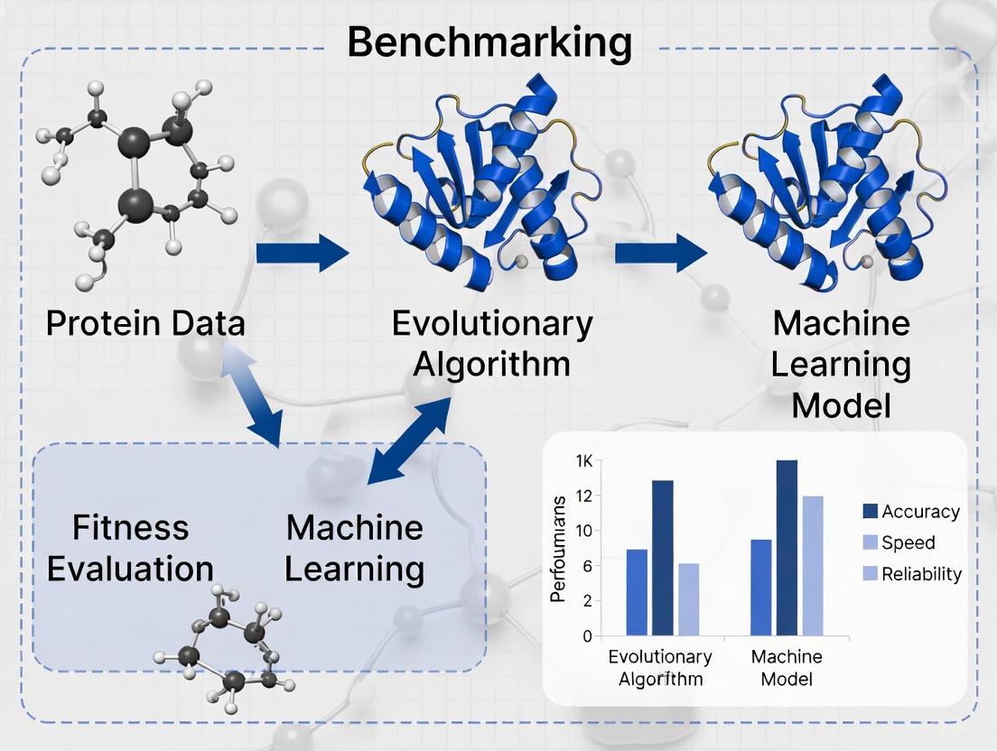
ML for Rapid Prediction vs. EA for De Novo Design and Optimization
The advent of sophisticated computational methods has revolutionized structural biology and protein engineering. Two dominant paradigms have emerged: machine learning (ML) for the rapid prediction of protein structures from sequences, and evolutionary algorithms (EA) for the de novo design and optimization of protein sequences for desired properties. This guide provides a objective comparison of these approaches, benchmarking their performance, outlining experimental protocols, and contextualizing their roles within a modern research workflow.
ML models, such as AlphaFold and ESMFold, have achieved remarkable accuracy in predicting protein structures by learning from vast datasets of known sequences and structures [11] [44]. In contrast, evolutionary algorithms excel at navigating the vast sequence space to solve inverse problems, such as finding sequences that fold into a target structure or optimizing for stability and function [8]. The following sections synthesize quantitative performance data and detailed methodologies to equip researchers with the information needed to select the appropriate tool for their specific application.
Performance Benchmarking and Quantitative Comparison
Directly comparing ML and EA is complex, as they are often applied to different problems—structure prediction versus sequence design. However, by examining their performance on related tasks and their computational footprints, meaningful comparisons can be drawn. The table below summarizes key performance indicators for leading ML models and EA approaches.
Table 1: Performance Benchmarking of ML Prediction Models
| Model | Primary Application | Key Metric | Performance | Computational Load | Notable Strengths |
|---|---|---|---|---|---|
| AlphaFold 2/3 [45] [11] [12] | Protein Structure & Complex Prediction | Global Distance Test (GDT) | >90 GDT on most CASP14 targets [11] | High (Requires significant GPU memory) [4] | Atomic accuracy; predicts complexes with ligands, DNA, RNA [45] |
| ESMFold [4] | Protein Structure Prediction | Predicted LDDP (pLDDT) | pLDDT >90 on some targets; variable on longer sequences [4] | Medium (Faster than AlphaFold, but high memory use) [4] | Very fast prediction; does not require multiple sequence alignments (MSAs) |
| OmegaFold [4] | Protein Structure Prediction | pLDDT | High pLDDT on short sequences (<400 aa) [4] | Medium (More efficient GPU use than ESMFold) [4] | Balanced speed, accuracy, and resource efficiency for shorter sequences |
| Boltz 2 [45] | Structure & Binding Affinity Prediction | Pearson Correlation (Affinity) | Pearson ~0.62 for binding affinity (comparable to FEP) [45] | High (with Boltz-steering for physical plausibility) [45] | Approaches FEP accuracy for binding affinity; 1000x more efficient [45] |
Table 2: Characteristics of Evolutionary Algorithm Approaches for Protein Design
| Aspect | Description | Performance & Characteristics |
|---|---|---|
| Core Function [8] | Inverse Protein Folding Problem (IFP) | Finds sequences that fold into a defined structure. |
| Algorithm Example [8] | Multi-Objective Genetic Algorithm (MOGA) | Optimizes for secondary structure similarity and sequence diversity simultaneously. |
| Key Strength [8] | Diversity Preservation | Searches deeper in sequence solution space, finding highly dissimilar sequences for the same structure. |
| Validation [8] | Tertiary Structure Prediction | Generated sequences are validated by predicting their 3D structure and comparing it to the original target. |
| Limitation | Relies on Predictive Tools | Dependent on fast, approximate structure predictors (like ML models) during optimization for feasibility [8]. |
Experimental Protocols and Workflows
A clear understanding of the underlying methodologies is crucial for their practical application and critical evaluation. This section details the standard protocols for both ML-based prediction and EA-driven design.
Protocol for ML-Based Protein Structure Prediction
The workflow for models like AlphaFold and ESMFold is largely automated but follows a consistent pipeline [11] [44].
- Input Preparation: The user provides the amino acid sequence of the target protein in FASTA format.
- Homology Search (MSA Generation): For models requiring it (e.g., AlphaFold), the first step is to search genetic databases to find homologous sequences and construct a Multiple Sequence Alignment (MSA). This step is bypassed in single-sequence methods like ESMFold [44].
- Neural Network Inference: The sequence (and MSA) is fed into the pre-trained deep learning model.
- Output and Confidence Estimation: The model outputs a 3D structure file (e.g., PDB format) alongside a per-residue confidence score (pLDDT), which estimates the local accuracy of the prediction [11].
Protocol for Evolutionary Algorithm-Based Protein Design
The EA workflow for the Inverse Folding Problem is an iterative optimization process [8].
- Problem Definition: The target protein structure (secondary or tertiary) is defined as the goal for the design process.
- Initialization: An initial population of random or seed-based amino acid sequences is generated.
- Fitness Evaluation: Each sequence in the population is evaluated using one or more fitness functions. A common multi-objective approach includes:
- Objective 1 (Similarity): Predicting the secondary structure of the generated sequence and measuring its similarity to the target secondary structure.
- Objective 2 (Diversity): Measuring the sequence diversity within the population to encourage exploration of the solution space [8].
- Selection, Crossover, and Mutation: Sequences with high fitness scores are selected to "reproduce." Their genetic information is combined (crossover) and randomly altered (mutation) to create a new generation of candidate sequences.
- Termination and Validation: The loop (steps 3-4) continues for a set number of generations or until convergence. The final best sequences are then validated by using a high-accuracy ML structure predictor (like AlphaFold) to confirm they fold into the intended tertiary structure [8].
The following diagram illustrates the logical workflow of a Multi-Objective Genetic Algorithm for inverse protein folding:
The Scientist's Toolkit: Essential Research Reagents and Materials
Successful computational research relies on a suite of software tools, databases, and hardware. The following table details key resources in the field.
Table 3: Key Research Reagents and Computational Tools
| Category | Item | Function & Description |
|---|---|---|
| Software & Models | AlphaFold Server / ColabFold [45] [4] | Web and local servers for running AlphaFold, providing free access to state-of-the-art structure prediction. |
| ESMFold / OmegaFold [4] | Alternative ML models for fast protein structure prediction, useful for high-throughput screening or validation. | |
| Rosetta [46] | A comprehensive software suite for molecular modeling, widely used for physics-based protein design and refinement. | |
| Databases | Protein Data Bank (PDB) [44] | Worldwide repository for experimentally determined 3D structures of proteins, nucleic acids, and complexes. Essential for training and validation. |
| AlphaFold Database [46] | Provides pre-computed AlphaFold structure predictions for over 200 million proteins, greatly expanding structural coverage. | |
| Experimental Validation | cDNA Display Proteolysis [7] | A high-throughput experimental method for measuring thermodynamic folding stability for hundreds of thousands of protein variants. |
| X-ray Crystallography / Cryo-EM [12] | Traditional gold-standard experimental methods for determining high-resolution protein structures. |
Leveraging Knowledge-Based Potentials and Energy Profiles for Fitness Evaluation
In the fields of structural biology and computational drug development, accurately evaluating the quality of protein structures is a critical challenge. The "fitness" of a protein model—its closeness to a biologically active native state—directly influences the reliability of downstream applications, from understanding disease mechanisms to drug design. This guide objectively compares two dominant computational philosophies for this task: knowledge-based potentials (KBPs) and modern machine learning (ML) protein folding tools. KBPs, rooted in statistical mechanics and evolutionary information, provide a physics-based lens for scoring and refining models. In contrast, ML methods like AlphaFold have revolutionized structure prediction. Framed within the broader thesis of benchmarking evolutionary algorithms against ML research, this article provides a comparative analysis of these approaches, supported by experimental data and detailed protocols for researchers.
Performance Benchmarking: Knowledge-Based Potentials vs. Machine Learning Methods
The selection of a fitness evaluation method involves trade-offs between interpretability, accuracy, resource requirements, and applicability. The following tables summarize the quantitative performance and characteristics of prominent methods.
Table 1: Comparative Performance on Standardized Tasks
| Method | Core Approach | Native State Recognition Rate (CASP Decoys) | Typical Application | Key Metric |
|---|---|---|---|---|
| BACH Potential [47] | Knowledge-based (Bayesian) | 58% (ranked #1) | Scoring model ensembles, discriminating native from decoys | Z-score, Normalized Rank |
| Profile-level Potentials [48] | Knowledge-based (Evolutionary profiles) | N/A (Significantly outperforms residue-level potentials) | Fold recognition, model refinement | Fraction Correctly Predicted (CP) |
| BCL::Score [49] | Knowledge-based (SSE-focused) | Enriches native-like models in 80-94% of cases | Topology evaluation from limited data | Enrichment of native-like models |
| AlphaFold 2 [12] | Deep Learning (Transformer) | >90 GDT on two-thirds of CASP14 targets | De novo structure prediction | Global Distance Test (GDT) |
| ESMFold [4] | Deep Learning (Transformer) | Varies with sequence length | Rapid tertiary structure prediction | Predicted LDDT (pLDDT) |
| OmegaFold [4] | Deep Learning (Transformer) | High accuracy on short sequences (<400 aa) | Accurate prediction for short sequences | pLDDT |
Table 2: Computational Resource Requirements
| Method | Hardware Requirements | Computational Speed | Scalability | Accessibility |
|---|---|---|---|---|
| Energetic Profile (CPE/SPE) [50] | Standard CPU | Fast (210-dimensional vector comparison) | Highly scalable to large datasets | Method described in literature |
| BACH Potential [47] | Standard CPU | Fast (1091-parameter function) | Suitable for high-throughput scoring | Method described in literature |
| 3D FCC HP EA [51] | High-performance CPU | Slower (iterative search and evaluation) | Limited by conformational search space | Custom implementation required |
| AlphaFold 2 [4] | High-end GPU (100-200 for training) | Minutes to hours per prediction [4] | Highly scalable with dedicated resources | Public server; open-source code |
| ESMFold [4] | A10 GPU | Very fast (e.g., 1 sec for 50 aa) [4] | Failed on sequences >1600 aa [4] | Public server; open-source code |
| OmegaFold [4] | A10 GPU | Fast, but slower than ESMFold (e.g., 3.66 sec for 50 aa) [4] | Handles sequences ~800 aa [4] | Public server; open-source code |
Experimental Protocols for Fitness Evaluation
To ensure reproducibility and provide a clear framework for benchmarking evolutionary algorithms against ML methods, we outline detailed protocols for two representative approaches: one based on a novel knowledge-based potential and another utilizing a deep learning model.
Protocol 1: Fitness Scoring with Knowledge-Based Energy Profiles
This protocol, adapted from the fast approach for structural analysis using energetic profiles, is designed for high-throughput comparison and fitness evaluation of protein models [50].
- Objective: To rapidly score and compare protein structures based on their compositional and structural energy profiles.
- Materials:
- Input Data: Protein sequences (for CPE) and/or 3D structures (for SPE) in PDB format.
- Knowledge-Based Potential: A pre-derived potential function, such as the distance-dependent potential used to generate 210 pairwise interaction types [50].
- Software: A computational environment (e.g., Python/R) capable of vector mathematics and, if working with structures, parsing PDB files.
- Procedure:
- Feature Vector Generation:
- For a given protein, compute the Compositional Profile of Energy (CPE) from its sequence using Eq. 7 from the original study [50]. This sums the estimated energy for each of the 210 possible amino acid pair types based on their frequency in the sequence.
- Alternatively, for a 3D structure, compute the Structural Profile of Energy (SPE). Using a knowledge-based potential, calculate the total energy contribution for each of the same 210 amino acid pair types based on their spatial interactions in the structure [50].
- This results in a 210-dimensional vector that serves as a unique energetic signature for the protein.
- Dissimilarity Calculation:
- To compare two proteins (e.g., a candidate model against a native reference or another model), compute the Manhattan distance between their respective 210-dimensional energy profile vectors [50].
- A smaller distance indicates higher structural and evolutionary similarity, and thus a fitter model.
- Feature Vector Generation:
- Analysis: The calculated distances can be used to cluster proteins, construct phylogenetic trees, or rank a pool of decoy models generated by an evolutionary algorithm, with lower energy profile distances indicating higher fitness.
Protocol 2: Fitness Assessment Using Machine Learning Models
This protocol leverages state-of-the-art deep learning models for structure prediction and intrinsic confidence scoring.
- Objective: To generate a 3D protein structure from its sequence and evaluate its local and global accuracy.
- Materials:
- Input Data: Amino acid sequence(s) in FASTA format.
- Software:
- Hardware: A computer with a modern GPU is recommended for running these models locally in a reasonable time frame [4].
- Procedure:
- Structure Prediction:
- Input the target sequence into the chosen ML tool (e.g., ColabFold, ESMFold server).
- Execute the prediction. The model will output atomic coordinates in PDB format.
- Fitness Evaluation via Confidence Metrics:
- Analyze the pLDDT score. This is a per-residue estimate of the model's local confidence on a scale from 0 to 100 [4]. A higher average pLDDT and more residues with high scores (>90) indicate a more reliable, fitter model.
- For global assessment, use the predicted TM-score or GDT. These metrics are often correlated with the pLDDT and provide a single score for the overall model quality, with higher values indicating a fitter model.
- Structure Prediction:
- Analysis: For benchmarking, the models generated by an evolutionary algorithm can be used as input to ML tools to obtain their pLDDT scores. Conversely, ML-generated models can be scored using knowledge-based potentials to compare the fitness assessments of both paradigms.
The logical workflow for selecting and applying these fitness evaluation methods is summarized in the diagram below.
Successful fitness evaluation relies on a suite of computational "reagents." The following table details key resources, their functions, and their relevance to this field.
Table 3: Key Research Reagent Solutions for Fitness Evaluation
| Resource Name | Type / Category | Primary Function in Fitness Evaluation | Relevance to Benchmarking |
|---|---|---|---|
| Knowledge-Based Potential [50] [47] [52] | Scoring Function | Derives an effective energy function from statistical analysis of known protein structures in the PDB to score decoy models. | The standard against which EA-generated models are scored for fitness; can be used as the objective function within an EA. |
| ASTRAL/SCOPe Database [50] | Benchmark Dataset | Provides curated datasets of protein domains with low sequence similarity for training and testing scoring functions. | Provides a gold-standard set of native structures and a source for generating decoys to test EA and ML methods. |
| CASP Decoy Sets [47] [12] | Benchmark Dataset | Provides challenging sets of protein models from the Critical Assessment of Structure Prediction, used for rigorous testing. | The ultimate test bed for benchmarking any new fitness evaluation method or prediction algorithm against state-of-the-art. |
| PDB (Protein Data Bank) | Primary Data Repository | The central repository for experimentally solved protein structures, serving as the source data for deriving knowledge-based potentials. | Essential for deriving KBPs and for providing the "true" native structures required for benchmarking. |
| HP Lattice Model [51] | Simplified Protein Model | A coarse-grained model that reduces complexity for fundamental studies of protein folding principles and algorithm development. | Often used as a test case for Evolutionary Algorithms due to its NP-hard nature and simplified conformational space [51]. |
| AlphaFold/ESMFold/OmegaFold [4] [12] | ML Prediction Tool | Provides high-accuracy reference structures and intrinsic confidence scores (pLDDT) for fitness assessment. | Serves as a high-accuracy baseline predictor; its output can be used as a fitness target or for validating EA results. |
| BCL::ScoreProtein [49] | Software Application | Implements a knowledge-based potential focused on secondary structure element packing for topology-level evaluation. | Useful for benchmarking EAs that work with limited data or SSE-restrained models, as is common in experimental biology. |
The field of computational protein structure prediction has been revolutionized by deep learning methods, most notably AlphaFold, which achieved unprecedented accuracy by leveraging deep neural networks and attention mechanisms on vast datasets of known protein structures [12] [53] [54]. However, evolutionary algorithms (EAs) continue to offer complementary strengths for specific protein modeling challenges, particularly for problems with sparse homologous sequence data or where global optimization against physical force fields is required. This case study provides a systematic benchmarking of EA-based approaches against machine learning (ML) alternatives, examining their respective methodologies, performance characteristics, and ideal application domains through quantitative comparison of experimental results.
The core distinction lies in their fundamental approaches: ML methods like AlphaFold excel at pattern recognition from evolutionary data, while EAs perform global optimization searches through conformational space. As one researcher noted following AlphaFold2's breakthrough, "It's the biggest 'machine learning in science' story that there has been," yet acknowledged that significant gaps remain in simulating protein dynamics and temporal changes [53]. These gaps represent opportunities where EAs maintain relevance in the computational biologist's toolkit.
Methodological Comparison: EA vs. ML Approaches
Evolutionary Algorithm Framework for Protein Structure Prediction
Evolutionary algorithms approach protein structure prediction as a global optimization problem, seeking to find the lowest-energy conformation for an amino acid sequence. The USPEX algorithm exemplifies this approach, implementing key components through specialized variation operators and fitness evaluation against physical force fields [55].
Key Experimental Protocol for EA-based Protein Structure Prediction:
- Initialization: Generate initial population of diverse protein conformations through random or fragment-based initialization methods.
- Fitness Evaluation: Calculate energy for each conformation using molecular mechanics force fields (e.g., Amber, Charmm, Oplsaal) implemented in packages like Tinker or Rosetta with REF2015 scoring function [55].
- Selection: Apply tournament or fitness-proportional selection to identify promising conformations for variation.
- Variation: Implement specialized variation operators including:
- Crossover: Exchange structural fragments between parent conformations
- Mutation: Introduce local structural perturbations through torsion angle adjustments
- Local Optimization: Apply gradient-based minimization to refine promising candidates
- Termination: Continue through generations until convergence criteria met (fitness stabilization or maximum generations reached).
USPEX has demonstrated particular effectiveness on small protein domains (up to 100 residues), successfully predicting tertiary structures with high accuracy for proteins lacking cis-proline residues in tests [55].
Deep Learning Framework for Protein Structure Prediction
In contrast to the optimization-focused EA approach, deep learning methods like AlphaFold employ pattern recognition on evolutionary data. AlphaFold2 utilizes an intricate attention-based architecture that processes multiple sequence alignments (MSAs) to infer spatial relationships between residues [12] [53].
Key Experimental Protocol for AlphaFold2-based Prediction:
- Input Representation: Generate multiple sequence alignments and paired representations from sequence databases using tools like Jackhmmer and HHblits.
- Evoformer Processing: Apply attention mechanisms to extract co-evolutionary patterns and refine residue-pair representations in multiple rounds of processing.
- Structure Module: Generate 3D atomic coordinates from processed representations using invariant point attention.
- Recycling: Iteratively refine the prediction through multiple passes of the network.
- Loss Calculation: Minimize difference between predicted and actual structures using frame-aligned point error and structural violation terms.
The AlphaFold2 method demonstrated remarkable accuracy in CASP14, achieving a global distance test (GDT) score above 90 for approximately two-thirds of proteins, representing a level of accuracy much higher than any previous method [12].
Addressing the MSA Dependency Limitation
A significant limitation of AlphaFold and similar ML approaches is their dependency on high-quality multiple sequence alignments. When few homologous sequences exist, prediction accuracy declines substantially [56]. Researchers have developed generative models like MSA-Augmenter to address this gap by creating novel protein sequences that supplement shallow MSAs using transformer architectures from natural language processing [56]. This hybrid approach demonstrates how ML techniques can evolve to address specific weaknesses while maintaining their core methodological approach.
Table 1: Core Methodological Differences Between EA and ML Approaches
| Aspect | Evolutionary Algorithms (USPEX) | Machine Learning (AlphaFold) |
|---|---|---|
| Primary Approach | Global optimization through population-based search | Pattern recognition from evolutionary data |
| Key Input | Amino acid sequence + physical force fields | Amino acid sequence + multiple sequence alignments |
| Core Mechanism | Variation, selection, inheritance | Attention mechanisms, neural networks |
| Energy/Scoring | Physical force fields (Amber, Charmm, Oplsaal) | Learned statistical potentials from training data |
| Output | 3D atomic coordinates | 3D atomic coordinates |
| Theoretical Basis | Thermodynamic hypothesis (minimum free energy) | Evolutionary coupling + structural conservation |
Experimental Benchmarking and Performance Comparison
Quantitative Performance Metrics
Direct comparison of EA and ML approaches reveals a complementary performance profile, with each demonstrating strengths under different conditions. USPEX has been tested on proteins up to 100 residues, finding structures with energy values comparable to or lower than Rosetta's Abinitio protocol when evaluated using the same force fields [55]. However, the study noted that "existing force fields are not sufficiently accurate for accurate blind prediction of protein structures without further experimental verification," highlighting a fundamental challenge for all physics-based approaches.
AlphaFold2 achieved a median Global Distance Test (GDT) score of 92.4 across all targets in CASP14, with many predictions approaching experimental accuracy [12]. This represents a transformative improvement over previous methods. The inclusion of metagenomic data in its training significantly improved prediction quality, with the system trained on a custom-built database of nearly 66 million protein families covering over 2.2 billion protein sequences [12].
Table 2: Performance Comparison on Standardized Benchmarks
| Method | Test Dataset | Accuracy Metric | Performance | Limitations |
|---|---|---|---|---|
| USPEX (EA) | 7 proteins (≤100 residues) | Potential energy relative to native | Comparable or lower energy than Rosetta Abinitio [55] | Limited to small proteins; force field inaccuracies |
| AlphaFold2 (ML) | CASP14 proteins | Global Distance Test (GDT) | >90 GDT for ~2/3 of proteins [12] | Performance declines with poor MSA quality |
| MSA-Augmenter + AF2 | CASP14 (low MSA targets) | GDT improvement | Significant accuracy improvement for shallow MSAs [56] | Computational overhead for sequence generation |
| Traditional EA | PhyloBench benchmark | Robinson-Foulds distance | Lower accuracy than distance methods [57] | Less accurate than ML/distance methods for phylogeny |
Performance on Low-Homology Targets
The MSA dependency of AlphaFold represents a particular challenge for proteins with few homologs. Experimental results demonstrate that for targets with fewer than ten homologous sequences, AlphaFold's performance degrades, sometimes failing to produce meaningful results [56]. This specific scenario represents an opportunity for EA approaches, which operate independently of evolutionary data.
Generative models that create synthetic MSAs have shown promise in bridging this gap, with MSA-Augmenter demonstrating improved prediction accuracy when supplementing shallow MSAs with generated sequences [56]. This hybrid approach illustrates how ML methodology is evolving to address its limitations while maintaining its core pattern-recognition paradigm.
Table 3: Essential Research Reagents and Computational Tools for Protein Structure Prediction
| Tool/Resource | Type | Primary Function | Application Context |
|---|---|---|---|
| USPEX | Evolutionary Algorithm | Global optimization of protein structures | Ab initio structure prediction without templates [55] |
| AlphaFold | Deep Neural Network | End-to-end structure prediction from sequence | High-accuracy prediction when quality MSAs available [12] |
| Rosetta | Modeling Suite | Protein structure modeling and design | Comparative modeling, de novo structure prediction [58] |
| Tinker | Molecular Dynamics | Protein structure relaxation and energy calculation | Force field evaluation and structure refinement [55] |
| MSA-Augmenter | Generative Model | Synthetic MSA generation for low-homology targets | Enhancing AlphaFold performance on difficult targets [56] |
| PhyloBench | Benchmarking Platform | Evaluation of phylogenetic inference methods | Benchmarking evolutionary relationships [57] |
| Protein Data Bank | Data Repository | Experimentally determined protein structures | Training data, template sources, validation [53] |
Integrated Workflows and Signaling Pathways in Protein Structure Prediction
The relationship between different protein structure prediction methods and their application contexts can be visualized as a decision pathway that researchers navigate based on their specific protein of interest and available data.
This benchmarking analysis reveals that evolutionary algorithms and machine learning approaches offer complementary strengths for protein structure prediction. While deep learning methods like AlphaFold have demonstrated superior accuracy for targets with rich evolutionary data, EAs maintain relevance for specific challenges including low-homology proteins, structure prediction with physical constraints, and applications where interpretability of the folding process is valuable.
The most promising future direction likely lies in hybrid approaches that leverage the strengths of both paradigms. As noted in recent surveys, "the incorporation of deep learning techniques into different steps of protein folding and design approaches represents an exciting future direction and should continue to have a transformative impact on both fields" [58]. The integration of physical constraints from EAs with the pattern recognition capabilities of ML, along with emerging protein language models that capture evolutionary information without explicit MSA construction, represents the next frontier in computational protein science.
For researchers and drug development professionals, this case study underscores the importance of maintaining a diverse computational toolkit. The selection of appropriate methods should be guided by the specific protein characteristics, available evolutionary data, and research objectives, with the understanding that methodological diversity remains essential for addressing the complex challenges of protein structure prediction.
Overcoming Computational Hurdles: Optimization and Troubleshooting for Scalable Protein Folding
The groundbreaking success of Machine Learning (ML) in predicting protein structures represents one of the most significant achievements in computational biology. Models like AlphaFold have demonstrated accuracies rivaling experimental methods, yet their operation often remains a "black box" [12]. This creates a fundamental tension between performance and interpretability: while these models deliver unprecedented results, the mechanistic reasoning behind their predictions can be opaque [9]. For researchers, scientists, and drug development professionals, this interpretability gap presents significant challenges in validating results, identifying failure modes, and generating novel biological insights beyond structure prediction alone.
The protein folding problem encompasses three distinct yet related challenges: the physical folding code (thermodynamic forces), the folding mechanism (kinetic pathways), and structure prediction (computational determination from sequence) [9]. ML approaches have predominantly addressed the third challenge, often sacrificing mechanistic interpretability for predictive accuracy. This article benchmarks contemporary ML-based protein folding tools through the critical lens of interpretability, providing experimental protocols and comparative analyses to guide methodological selection in research and development contexts.
Comparative Performance Benchmarking of Protein Folding Tools
Quantitative Performance Metrics Across Model Architectures
Independent benchmarking provides crucial insights into the practical performance characteristics of different protein folding approaches. The following comparison evaluates key computational metrics across leading ML-based protein folding tools, highlighting the critical trade-offs between accuracy, resource requirements, and operational efficiency.
Table 1: Runtime and Accuracy Comparison Across Protein Lengths [4]
| Sequence Length | Tool | Running Time (s) | PLDDT Score | CPU Memory (GB) | GPU Memory (GB) |
|---|---|---|---|---|---|
| 50 | ESMFold | 1 | 0.84 | 13 | 16 |
| 50 | OmegaFold | 3.66 | 0.86 | 10 | 6 |
| 50 | AlphaFold | 45 | 0.89 | 10 | 10 |
| 100 | ESMFold | 1 | 0.30 | 13 | 16 |
| 100 | OmegaFold | 7.42 | 0.39 | 10 | 7 |
| 100 | AlphaFold | 55 | 0.38 | 10 | 10 |
| 400 | ESMFold | 20 | 0.93 | 13 | 18 |
| 400 | OmegaFold | 110 | 0.76 | 10 | 10 |
| 400 | AlphaFold | 210 | 0.82 | 10 | 10 |
| 800 | ESMFold | 125 | 0.66 | 13 | 20 |
| 800 | OmegaFold | 1425 | 0.53 | 10 | 11 |
| 800 | AlphaFold | 810 | 0.54 | 10 | 10 |
| 1600 | ESMFold | Failed (OOM) | - | - | 24 |
| 1600 | OmegaFold | Failed (>6000) | - | - | 17 |
| 1600 | AlphaFold | 2800 | 0.41 | 10 | 10 |
Table 2: Architectural and Interpretability Features Comparison [59] [12] [60]
| Tool | Core Architecture | Parameters | Training Data | Interpretability Features | Key Limitations |
|---|---|---|---|---|---|
| AlphaFold 2 | Evoformer (Attention-based) with template integration | ~93 million | 170,000+ PDB structures + evolutionary databases | Confidence per-residue (pLDDT), predicted aligned error | Limited to single-chain proteins (original version) |
| AlphaFold 3 | Pairformer + Diffusion model | Not specified | Expanded to complexes (proteins, DNA, RNA, ligands) | pLDDT, confidence metrics for interactions | Restricted server access for non-commercial use |
| ESMFold | Transformer-based single-sequence method | Not specified | Evolutionary Scale Modeling | pLDDT scores, single-sequence processing | Lower accuracy on some intermediate-length proteins |
| OmegaFold | Deep learning with evolutionary algorithms | Not specified | Large-scale protein structure data | pLDDT, memory-efficient design | Performance degradation on longer sequences |
| SimpleFold | Flow-matching with general-purpose transformers | Up to 3 billion | 8.6M+ distilled structures + PDB data | Ensemble prediction capabilities, simplified architecture | Emerging methodology, less established than alternatives |
Performance Analysis and Practical Implications
The benchmarking data reveals distinct operational profiles for each tool. ESMFold demonstrates exceptional speed for shorter sequences (≤100 residues) but shows inconsistent accuracy metrics and substantial memory demands, failing on longer sequences (1600 residues) due to GPU memory exhaustion [4]. OmegaFold provides a balanced compromise with competitive accuracy and superior memory efficiency, particularly for shorter sequences (50-400 residues) where it achieves the best accuracy-to-resource ratio [4]. AlphaFold/ColabFold maintains consistent memory usage across all sequence lengths and delivers robust accuracy, particularly for shorter sequences, though at the cost of significantly longer runtimes [4].
For research applications requiring high-throughput screening of shorter protein sequences, OmegaFold's balance of accuracy, runtime, and memory efficiency makes it particularly suitable for production environments. For longer sequences or when highest accuracy is critical, AlphaFold's more computationally intensive approach remains preferable despite longer wait times. ESMFold offers advantages for rapid preliminary screening when sufficient GPU memory is available and some accuracy trade-offs are acceptable.
Experimental Protocols for Method Evaluation
Standardized Benchmarking Methodology
To ensure reproducible evaluation of protein folding tools, researchers should implement standardized experimental protocols. The following methodology outlines key considerations for rigorous benchmarking:
Hardware Configuration: Benchmarks should be conducted on systems with standardized GPU resources (e.g., A10 GPU with 24GB memory as referenced in comparative studies) [4]. CPU memory should be monitored throughout execution, with 16GB RAM minimum recommended.
Evaluation Metrics: Primary metrics should include:
- PLDDT (Predicted Local Distance Difference Test): Measures local confidence on a scale from 0-100, with higher scores indicating greater reliability [4] [12].
- Running Time: Total execution time from sequence input to structure output.
- Resource Utilization: Peak CPU, GPU memory, and GPU utilization during execution.
- Global Distance Test (GDT): Alternative accuracy metric used in CASP competitions, with scores above 90 considered highly accurate [12].
Dataset Selection: Benchmarks should include proteins of varying lengths (50-1600 residues) and structural classifications to evaluate tool performance across diverse scenarios. Standardized test sets from CASP (Critical Assessment of Structure Prediction) competitions provide excellent reference points [9].
Implementation Protocols for Specific Tools
AlphaFold Implementation: For optimal AlphaFold performance, utilize the full multiple sequence alignment (MSA) generation pipeline despite its computational cost, as this significantly impacts accuracy. The model produces per-residue confidence estimates (pLDDT) and predicted aligned error matrices that are essential for interpretability [12].
SimpleFold Protocol: Implementation requires specific steps for data preparation and processing. The recommended workflow includes:
- Data preparation from mmCIF files using
process_mmcif.pywith--use-assemblyflag - Structure tokenization via
process_structure.pyto convert processed targets into model inputs - Inference execution with step control (
--num_steps) and sample variation (--nsample_per_protein) parameters [59]
ESMFold Execution: Leverage ESMFold's single-sequence processing capability for rapid predictions without MSA generation. This provides significant speed advantages but may sacrifice accuracy for sequences with limited evolutionary information [4].
Visualizing Comparative Analysis Workflows
The following diagram illustrates a systematic workflow for comparative analysis of protein folding tools, highlighting key decision points and evaluation metrics essential for rigorous benchmarking.
Figure 1: Protein Folding Tools Comparative Analysis Workflow
Interpretability Methods for ML Models in Structural Biology
Interpretability Approaches and Their Applications
The "black box" problem in deep learning refers to the difficulty in understanding how models arrive at their predictions [61]. Several interpretability methods have been developed to address this challenge, each with distinct strengths and limitations for protein folding applications.
Table 3: ML Interpretability Methods and Applications [62] [63] [64]
| Method | Core Principle | Applications in Protein Folding | Key Limitations |
|---|---|---|---|
| LIME (Local Interpretable Model-agnostic Explanations) | Creates local linear approximations of complex models | Interpreting specific residue contributions to structural features | Instance-specific explanations, may not capture global model behavior |
| SHAP (SHapley Additive exPlanations) | Game theory approach to quantify feature importance | Identifying critical sequence regions influencing fold stability | Computationally intensive for large models and inputs |
| Saliency Maps | Visualizes input features that most influence outputs | Mapping sequence-structure relationships in predictions | May not reveal complex feature interactions |
| Activation Maximization | Identifies inputs that maximize neuron activations | Understanding learned representations in folding networks | Results may not be biologically interpretable |
| Model Distillation | Trains simpler, interpretable proxy models | Creating simplified versions of complex folding models | Potential loss of predictive accuracy |
Implementing Interpretability in Protein Folding Research
For researchers seeking to implement interpretability methods, the following approaches show particular promise:
Confidence Metric Integration: Tools like AlphaFold provide built-in confidence measures (pLDDT) that serve as foundational interpretability features. These should be routinely examined rather than focusing solely on predicted structures [12]. Residues with low pLDDT scores (<70) often indicate regions requiring experimental validation or alternative modeling approaches.
Comparative Interpretation with LIME: When analyzing specific structural features, LIME can help identify contributing residues by creating local explanations. For example, when a model predicts a particular beta-sheet formation, LIME can highlight which residues most strongly influence this prediction [64].
Feature Importance with SHAP: For understanding global sequence-structure relationships, SHAP values can quantify how different sequence features contribute to overall fold prediction. This is particularly valuable for identifying potential stability determinants or functional regions [64].
The following diagram illustrates how interpretability methods can be integrated into protein structure prediction workflows to enhance model transparency and insight generation.
Figure 2: ML Model Interpretability Pipeline for Protein Folding
Research Reagent Solutions for Protein Folding Studies
Table 4: Essential Research Resources for Protein Folding Investigations [59] [12] [9]
| Resource Category | Specific Tools/Databases | Primary Function | Access Considerations |
|---|---|---|---|
| Protein Structure Databases | Protein Data Bank (PDB) | Repository of experimentally determined structures | Publicly available, essential for training and validation |
| Evolutionary Databases | Big Fantastic Database (AlphaFold), AFDB, SwissProt | Multiple sequence alignments, evolutionary constraints | AlphaFold's custom database covers 2.2+ billion sequences |
| Software Frameworks | TensorFlow, PyTorch, JAX | ML model development and training | Open-source with varying production readiness |
| Specialized Protein Folding Tools | AlphaFold Server, ColabFold, SimpleFold, OmegaFold | Structure prediction from sequence | Varying access restrictions; AlphaFold 3 limited to server |
| Validation Metrics | PLDDT, GDT, TM-score | Assessment of prediction accuracy and quality | Standardized metrics enable cross-study comparisons |
| Experimental Validation | X-ray crystallography, Cryo-EM, NMR | Empirical structure determination | Expensive and time-consuming but essential for ground truth |
The benchmarking analysis presented reveals that contemporary ML-based protein folding tools exhibit distinct performance profiles across accuracy, computational efficiency, and interpretability dimensions. While AlphaFold variants generally lead in accuracy, alternatives like OmegaFold and ESMFold provide valuable trade-offs for specific application contexts, particularly when computational resources or throughput requirements are limiting factors.
The interpretability challenge remains significant, with even the most accurate models offering limited mechanistic insights into the fundamental principles governing protein folding. However, emerging methodologies like SimpleFold's flow-matching approach suggest promising directions for developing both accurate and architecturally transparent models [60]. For the research community, prioritizing interpretability alongside accuracy will be essential for transforming protein structure prediction from a powerful pattern-matching tool into a genuine source of biological insight.
As the field progresses, the integration of ML approaches with evolutionary algorithms and physics-based simulations may help bridge the interpretability gap while maintaining predictive performance. For drug development professionals and researchers, maintaining a diversified toolkit of protein folding methods—while carefully considering their respective interpretability limitations—remains the most prudent strategy for leveraging these transformative technologies in practical applications.
In computational biology, efficiently navigating vast and complex search spaces is a fundamental challenge. This is particularly true in two critical fields: evolutionary algorithms (EAs) for protein design and machine learning (ML) for protein structure prediction. Both disciplines grapple with the same core problem—an exponentially large universe of possible solutions. EAs for the Inverse Protein Folding Problem (IFP) search through a colossal space of amino acid sequences to find those that fold into a desired structure [8]. Meanwhile, ML folding methods like AlphaFold confront Levinthal's paradox: the astronomical number of possible conformations a protein chain could theoretically adopt, which is on the order of 10^300 for a typical protein [65].
The strategy for traversing this search space is what separates different computational approaches. EAs often employ population-based metaheuristics, iteratively evolving a set of candidate solutions through operations like crossover and mutation, guided by fitness functions [66] [8]. In contrast, modern ML predictors use deep learning architectures, such as attention-based neural networks, to learn the mapping from sequence to structure directly from evolutionary and physical data [12] [67]. This guide benchmarks these strategies, focusing on their convergence behavior, computational efficiency, and practical utility in accelerating discovery within biomedical research.
Methodological Comparison: EA vs. Modern ML Folders
Evolutionary Algorithms for Inverse Folding
The Inverse Folding Problem is at the heart of rational protein design. The objective is to find amino acid sequences that will fold into a predefined tertiary structure [8]. EAs address this by optimizing sequences towards a target, often using a multi-objective genetic algorithm (MOGA). A key advancement is the use of diversity-as-objective (DAO), which optimizes for both secondary structure similarity and sequence diversity simultaneously. This pushes the algorithm to explore deeper into the solution space rather than converging prematurely on a local optimum [8].
Typical EA Workflow for IFP:
- Initialization: A population of random amino acid sequences is generated.
- Evaluation: Each sequence is scored using a fitness function (e.g., secondary structure similarity to the target).
- Selection: The fittest sequences are selected to "parent" the next generation.
- Variation: New sequences are created through genetic operations:
- Crossover: Combining parts of two parent sequences.
- Mutation: Randomly changing amino acids in a sequence.
- Diversity Preservation: Techniques like DAO are applied to maintain a diverse gene pool.
- Iteration: Steps 2-5 repeat until a termination condition is met (e.g., a high-fitness sequence is found or a generation limit is reached) [66] [8].
Machine Learning for Structure Prediction
Modern protein folding tools address the forward problem—predicting a 3D structure from a sequence—using deep learning. They have redefined the state-of-the-art in accuracy and speed.
- AlphaFold2: Developed by DeepMind, it uses an "Evoformer" module, a transformer-based neural network. This architecture processes multiple sequence alignments (MSAs) and uses an attention mechanism to reason about the spatial relationships between amino acids that may be far apart in the sequence, effectively piecing the structure together like a jigsaw puzzle [12] [67]. Its iterative refinement process significantly reduces stereochemical violations in its predictions [12].
- ESMFold: This model leverages a protein language model trained on millions of sequences. It can predict structures directly from a single sequence, bypassing the need for computationally expensive MSAs. This makes it exceptionally fast, though sometimes at a slight cost to accuracy compared to AlphaFold2 [4] [67].
- OmegaFold: Another deep learning model, OmegaFold aims for high accuracy without relying on MSAs. It is recognized for its balance of accuracy, speed, and memory efficiency, making it particularly suitable for shorter sequences and production environments [4].
The table below summarizes a comparative benchmark of these ML methods.
Table 1: Benchmarking ML Protein Folding Tools on an A10 GPU [4]
| Sequence Length | Method | Running Time (s) | PLDDT Accuracy | GPU Memory |
|---|---|---|---|---|
| 50 | ESMFold | 1 | 0.84 | 16 GB |
| OmegaFold | 3.66 | 0.86 | 6 GB | |
| AlphaFold (ColabFold) | 45 | 0.89 | 10 GB | |
| 400 | ESMFold | 20 | 0.93 | 18 GB |
| OmegaFold | 110 | 0.76 | 10 GB | |
| AlphaFold (ColabFold) | 210 | 0.82 | 10 GB | |
| 800 | ESMFold | 125 | 0.66 | 20 GB |
| OmegaFold | 1425 | 0.53 | 11 GB | |
| AlphaFold (ColabFold) | 810 | 0.54 | 10 GB |
Visualizing Workflow Divergence
The following diagram illustrates the fundamental differences in how EAs and modern ML folders navigate the search space to arrive at a solution.
Convergence Benchmarking: Performance and Applications
Quantitative Performance Metrics
The performance gap between traditional EA methods and modern ML folders is significant, primarily in terms of accuracy and computational cost. AlphaFold2's achievement of a median Global Distance Test (GDT) score above 90 in the CASP14 competition marked a paradigm shift, as a score above 90 is considered comparable to experimental methods [12]. EAs for inverse folding lack a direct equivalent to the GDT score but are typically validated by comparing the tertiary structures of their designed sequences to the original target, a process that often requires subsequent structure prediction [8].
Table 2: Comparative Analysis of Optimization Strategies
| Feature | Evolutionary Algorithms (for IFP) | ML Folders (e.g., AlphaFold2) |
|---|---|---|
| Primary Goal | Find sequences for a target structure [8] | Predict structure for a given sequence [12] |
| Core Mechanism | Population-based stochastic search [66] | Deep learning & attention networks [12] |
| Key Strength | Designs novel sequences; explains solution space [8] | Unprecedented prediction accuracy & speed [67] |
| Convergence Metric | Fitness score (e.g., structure similarity) [8] | GDT_TS, PLDDT [4] [12] |
| Typical Runtime | Highly variable; can be long [8] | Seconds to minutes for a single prediction [4] |
| Search Strategy | Explores sequence space via genetic operations [8] | Direct mapping via trained neural network [12] |
Experimental Validation Protocols
Validating the outputs of these algorithms requires distinct experimental pathways.
Validating EA-Designed Sequences:
- Inverse Folding: A MOGA is run to produce a set of high-fitness, diverse amino acid sequences predicted to fold into the target structure [8].
- Tertiary Structure Prediction: The designed sequences are fed into a high-accuracy structure predictor like AlphaFold2 to generate their predicted 3D models [8] [68].
- Structure Comparison: The predicted model is compared to the original target structure using metrics like Root-Mean-Square Deviation (RMSD) and Template-Modeling (TM) score to confirm the design's success [8].
Validating ML-Predicted Structures:
- Blind Prediction: In competitions like CASP, predictors forecast structures for proteins with solved but unpublished experimental structures [67].
- Experimental Comparison: The predicted model is compared against the ground-truth experimental data (from X-ray crystallography, cryo-EM, etc.) using the GDT_TS and PLDDT metrics [4] [12].
- Physical Realism: The structure is also checked for stereochemical violations (e.g., unrealistic bond lengths/angles) using tools like MolProbity [12].
The Scientist's Toolkit: Essential Research Reagents
Table 3: Key Resources for Computational Protein Research
| Item / Resource | Function in Research |
|---|---|
| AlphaFold Database | Provides free, immediate access to over 200 million predicted protein structures, serving as a foundational resource for hypothesis generation and validation [12] [65]. |
| Protein Data Bank (PDB) | The global repository for experimentally determined 3D structures of proteins, nucleic acids, and complex assemblies. Serves as the primary source of ground-truth data for training and testing algorithms [12] [67]. |
| Multiple Sequence Alignments (MSAs) | Collections of evolutionarily related protein sequences. Critical for algorithms like AlphaFold2 to infer distance constraints between residues based on co-evolution [12] [67]. |
| CASP Competition | A biennial blind community experiment that objectively assesses the state-of-the-art in protein structure prediction, providing a standardized benchmark for new methods [12] [67]. |
| Genetic Algorithm Framework | Software libraries (e.g., in Python or R) that enable the implementation of custom EA optimizations, such as for multi-objective inverse folding projects [66] [8]. |
The benchmarking of EA and ML strategies reveals a landscape of powerful complementarity rather than outright superiority of one approach. ML folding tools, led by AlphaFold2, have achieved dominant performance in the forward problem of structure prediction, offering breathtaking speed and accuracy that has democratized structural biology [67] [65]. Meanwhile, EAs remain highly relevant for the inverse problem of protein design, where the goal is to explore the vast sequence space to discover novel proteins that fulfill a predefined structural or functional role [8] [68].
The future of navigating biological search spaces lies in convergence. EA principles of diversity-preservation and multi-objective optimization can inform the development of more robust ML models [69]. Conversely, fast, approximate ML folders can be integrated into EA fitness evaluation loops to rapidly assess candidate sequences, creating powerful hybrid pipelines. This synergistic approach, leveraging the exploratory power of EAs and the predictive precision of ML, will ultimately provide researchers and drug developers with the most advanced toolkit to accelerate the design of new therapeutics and enzymes, pushing the boundaries of computational biology.
In the rapidly advancing field of protein structure prediction, computational resources represent a significant practical constraint for researchers and drug development professionals. The groundbreaking success of machine learning (ML) models like AlphaFold2 has democratized access to accurate protein folding, yet the computational cost of these models varies dramatically. This guide provides an objective performance comparison of leading protein folding algorithms by synthesizing empirical data on their runtime and memory characteristics. Framed within a broader thesis on benchmarking methodologies, this analysis extends principles from evolutionary algorithm runtime analysis—where the efficiency of searching vast combinatorial spaces is rigorously quantified—to the domain of ML-based protein folding. Understanding these computational profiles is essential for laboratories to select the right tool that balances prediction accuracy with available infrastructure, thereby optimizing research throughput and cost.
Key Protein Folding Models and Their Computational Profiles
The landscape of protein folding tools is diverse, with each model employing a distinct architectural approach that directly influences its computational demands. The following models are central to current research and development efforts.
AlphaFold2/ColabFold: Developed by DeepMind, AlphaFold2 represents a seminal advancement in the field. It employs a complex architecture that integrates an Evoformer for processing evolutionary data and a structure module to generate 3D atomic coordinates. Its operation requires generating Multiple Sequence Alignments (MSAs), which is often the most computationally intensive step. ColabFold is a popular reimplementation that offers enhanced accessibility and includes optimizations like the use of MMseqs2 for faster MSA generation, making it a widely used benchmark for comparison [70].
ESMFold: A product of Meta's FAIR team, ESMFold is an end-to-end single-sequence protein language model based on the ESM-2 transformer architecture. Its key innovation is bypassing the need for explicit MSAs, instead deriving evolutionary insights directly from the sequence via its pretrained language model. This architectural choice makes it exceptionally fast, particularly for shorter sequences, though it can require more GPU memory than other models [4] [35].
OmegaFold: This deep learning model is designed to predict protein structures with high accuracy without relying on MSAs or database homology. Its efficiency stems from a data-driven approach that learns patterns from known protein structures. OmegaFold is often noted for its balance of accuracy and resource efficiency, especially on shorter sequences, making it a strong candidate for production environments with limited resources [4].
OpenFold: Conceived as a fully open-source trainable replica of AlphaFold2, OpenFold is optimized for execution on widely available GPUs. It uses PyTorch and incorporates several memory and speed optimizations, such as low-memory attention and FlashAttention. These features allow it to handle very long protein sequences (up to 4,600 residues) on a single A100 GPU, offering a compelling blend of performance and cost-effectiveness [70].
SimpleFold: Introduced by Apple, SimpleFold challenges the reliance on complex, domain-specific architectures. It employs a standard flow-matching objective and uses general-purpose transformer layers with adaptive layers, forgoing expensive modules like triangle attention. As a generative model, it also shows strong performance in ensemble prediction, providing a simplified yet powerful alternative [60].
Empirical benchmarking reveals clear trade-offs between speed, accuracy, and resource consumption across different protein folding tools. The data below, synthesized from independent benchmarks, provides a quantitative basis for comparison. All runtime and memory data was collected using an A10 GPU unless otherwise specified [4].
Runtime and PLDDT Score Comparison
Table 1: Comparative runtime (in seconds) and accuracy (PLDDT score) across different protein sequence lengths.
| Sequence Length | ESMFold Runtime (s) | ESMFold PLDDT | OmegaFold Runtime (s) | OmegaFold PLDDT | AlphaFold/ColabFold Runtime (s) | AlphaFold/ColabFold PLDDT |
|---|---|---|---|---|---|---|
| 50 | 1 | 0.84 | 3.66 | 0.86 | 45 | 0.89 |
| 100 | 1 | 0.30 | 7.42 | 0.39 | 55 | 0.38 |
| 200 | 4 | 0.77 | 34.07 | 0.65 | 91 | 0.55 |
| 400 | 20 | 0.93 | 110 | 0.76 | 210 | 0.82 |
| 800 | 125 | 0.66 | 1425 | 0.53 | 810 | 0.54 |
| 1600 | Failed (OOM) | Failed | Failed (>6000) | Failed | 2800 | 0.41 |
System Memory and GPU Memory Usage
Table 2: Comparative memory usage (in GB) across different protein folding models [4].
| Model | CPU Memory (GB) | GPU Memory (GB) |
|---|---|---|
| ESMFold | 13 | 16-24* |
| OmegaFold | 10 | 6-17* |
| AlphaFold/ColabFold | 10 | 10 |
Note: GPU memory usage for ESMFold and OmegaFold can increase with longer sequence lengths, as indicated in Table 1.
Performance on AWS G4dn Instances
A separate benchmark on AWS g4dn.xlarge instances (T4 GPU) compared OpenFold and AlphaFold on 32 monomer proteins. OpenFold generated predictions 90% faster than AlphaFold on average, with a mean difference in prediction accuracy (GDT_TS) of less than 1% [70].
Detailed Experimental Protocols and Methodologies
To ensure the reproducibility of the comparative data and facilitate future benchmarking, this section outlines the key experimental methodologies employed in the cited studies.
Benchmarking Protein Folding Models on A10 GPU
The comparative data in Tables 1 and 2 was generated using a standardized benchmarking protocol [4].
- Hardware Setup: All models were executed on a machine equipped with a g5.2xlarge A10 GPU.
- Performance Metrics: Two primary parameters were assessed: 1) Running Time: The total time taken to predict the structure from a given protein sequence. 2) PLDDT Score: A per-residue estimate of confidence, on a scale from 0 to 1.
- Memory Assessment: Both CPU system memory and GPU memory usage were monitored during the execution of the prediction.
- Test Sequences: A range of protein sequences of different lengths (from 50 to 1600 residues) were used to evaluate performance across a spectrum of realistic scenarios.
AWS Batch Folding Architecture for OpenFold and AlphaFold
The performance comparison between OpenFold and AlphaFold on AWS was conducted using a scalable cloud-based workflow [70].
- Infrastructure Provisioning: The AWS Batch Architecture for Protein Folding and Design was deployed via an AWS CloudFormation template, which provisioned necessary compute, storage, and container resources.
- Instance Configuration: Folding jobs for both algorithms were executed on g4dn.xlarge Amazon EC2 instances, each equipped with 4 vCPUs, 16 GiB of memory, and a single T4 GPU.
- Data and Pre-processing: The study used 32 monomer proteins from the CAMEO dataset. MSAs were pre-computed for all targets using JackHMMER against the full BFD database to ensure a consistent starting point.
- Accuracy Validation: Predictions from both OpenFold and AlphaFold were compared against experimentally determined structures from the RCSB Protein Data Bank. The Template Modeling Score (TMScore) tool was used to calculate the GDT_TS metric, which quantifies structural similarity.
Connecting Evolutionary Algorithm Principles to Protein Folding Benchmarking
The theoretical foundation of benchmarking computational efficiency has deep roots in the analysis of evolutionary algorithms (EAs). Runtime analysis, a core subfield of evolutionary computation, provides a rigorous framework for understanding how the performance of iterative search algorithms scales with problem size and complexity. This involves deriving bounds on the expected runtime—the number of fitness evaluations until an optimal solution is found—for EAs on canonical problems like pseudo-Boolean functions and permutation-based problems [71] [72].
This principled approach to performance evaluation directly informs the benchmarking of ML-based protein folding. The search for a protein's native structure from its amino acid sequence is a high-dimensional combinatorial optimization problem. Just as runtime analysis quantifies an EA's efficiency in navigating a fitness landscape, our comparative analysis quantifies how effectively different ML models traverse the conformational space of proteins. Furthermore, concepts like maintaining diversity in a population of candidate solutions—a well-studied challenge in EAs—find parallels in the exploration strategies of different folding architectures [73]. By adopting the rigorous, quantitative mindset of evolutionary algorithm analysis, we can move beyond mere empirical comparisons to develop a more fundamental understanding of what makes a protein folding model computationally efficient.
Successful and efficient protein structure prediction relies on a suite of computational tools and data resources. The following table details key components of the modern computational biologist's toolkit.
Table 3: Essential resources for computational protein folding research.
| Resource Name | Type | Primary Function | Key Application |
|---|---|---|---|
| JackHMMER | Software Tool | Generates Multiple Sequence Alignments (MSAs) by searching protein sequence databases. | Identifying evolutionary related sequences; essential first step for MSA-dependent folders like AlphaFold [70]. |
| MMseqs2 | Software Tool | Rapid, sensitive protein sequence searching and clustering. | Can be used as a faster alternative to JackHMMER for MSA generation, especially in pipelines like ColabFold [70]. |
| UniRef90/BDD | Database | Clustered sets of protein sequences from UniProt. | Primary databases for MSA generation, providing evolutionary context [70]. |
| PDB70 | Database | Database of profile HMMs built from the PDB. | Used for template-based modeling in some folding pipelines [70]. |
| AWS Batch | Cloud Service | Orchestrates and scales batch computing jobs. | Manages the submission and execution of thousands of folding jobs across scalable EC2 instance fleets [70]. |
| FSx for Lustre | Cloud Storage | High-performance file system. | Provides low-latency access to large reference datasets (e.g., UniRef90) for folding workflows on AWS [70]. |
| PyTorch | Framework | Open-source machine learning library. | The underlying framework for models like ESMFold and OpenFold, enabling model training and inference [70] [35]. |
The computational profiling of leading protein folding models reveals that there is no single "best" tool for all scenarios. The optimal choice is a function of the researcher's specific constraints regarding protein length, computational budget, and accuracy requirements.
- For short sequences (under ~400 residues) where computational efficiency is paramount, OmegaFold presents a strong option, offering a superior balance of accuracy, speed, and lower memory usage [4].
- For scenarios demanding the highest possible accuracy and where longer runtimes are acceptable, AlphaFold/ColabFold remains a gold standard, though it is the slowest option in this comparison [4] [70].
- For high-throughput screening of many proteins, particularly shorter sequences, ESMFold's exceptional speed is a major advantage, though users must be prepared for its higher GPU memory demands [4].
- For a well-balanced, open-source alternative that is optimized for modern cloud GPUs, OpenFold is highly recommended, offering near-AlphaFold accuracy with significantly faster runtimes [70].
Ultimately, managing computational resources in protein folding research requires a nuanced understanding of the trade-offs inherent in each model. By leveraging the empirical data and methodologies outlined in this guide, research teams can make informed decisions that accelerate discovery while responsibly managing their computational infrastructure.
The accurate computational prediction of protein structures has been revolutionized by machine learning (ML), with tools like AlphaFold achieving unprecedented accuracy on many targets. However, significant challenges remain for specific protein classes, notably intrinsically disordered regions (IDRs) and large multi-domain proteins. These targets represent a critical frontier in structural biology. Disordered regions, which lack a fixed three-dimensional structure, are abundant in eukaryotic proteomes and play vital roles in cell signaling and regulation [74]. Multi-domain proteins, which constitute the majority of proteins in nature, pose a folding challenge due to the complex interplay between independently folding domains and the linker regions that connect them [75] [76]. This guide provides an objective comparison of the performance of leading ML-based protein folding methods on these challenging targets, framing the analysis within a broader thesis on benchmarking against evolutionary and physical algorithms.
Performance Comparison on Disordered Regions and Multi-Domain Proteins
Quantitative Performance Metrics
The following tables summarize key performance metrics for leading protein folding models, highlighting their capabilities and limitations.
Table 1: Overall Model Characteristics and Performance on Disordered Regions
| Model | Approach to Disordered Regions | Reported Strengths | Reported Limitations |
|---|---|---|---|
| AlphaFold2/3 | Predicts per-residue confidence (pLDDT); low confidence often indicates disorder [44] [9]. | High accuracy on structured regions; low pLDDT scores can correctly hint at disorder [9]. | Does not directly model the structural ensemble of disordered proteins; treats low confidence as an uncertainty metric [74] [9]. |
| ESMFold | Leverages a protein language model; less reliant on homologous sequences [4]. | Fast prediction times; effective on sequences with few homologs [4]. | Generally lower accuracy than AlphaFold on structured domains, which may affect the interpretation of flanking disordered regions [4]. |
| OmegaFold | Designed for high accuracy without MSAs [4]. | Balanced accuracy and resource usage, especially on shorter sequences [4]. | Like others, it predicts a single structure rather than an ensemble for disordered regions [4]. |
| SimpleFold | Uses a standard transformer architecture with a flow-matching objective [60]. | Challenges the need for complex, domain-specific architectures; demonstrates strong ensemble prediction capability [60]. | A relatively new approach; broader community validation on disordered regions is ongoing [60]. |
Table 2: Performance and Resource Usage on Multi-Domain and Long Sequences
| Model | Performance on Long Sequences (>800 residues) | CPU Memory Usage | GPU Memory Usage |
|---|---|---|---|
| ESMFold | Failed on a 1600-residue sequence (out of GPU memory) [4]. | ~13 GB [4] | 16-24 GB (increases with sequence length) [4]. |
| OmegaFold | Failed on a 1600-residue sequence (excessive runtime) [4]. | ~10 GB [4] | 6-17 GB (increases with sequence length) [4]. |
| AlphaFold (ColabFold) | Successfully processed a 1600-residue sequence in ~2800 seconds [4]. | ~10 GB [4] | ~10 GB (consistent across lengths) [4]. |
Key Experimental Protocols in Benchmarking Studies
The comparative data presented in this guide are derived from standardized benchmarking experiments. Understanding the underlying methodologies is crucial for interpreting the results.
- Benchmarking Method for Runtime/Accuracy: One key study evaluated ESMFold, OmegaFold, and AlphaFold (via ColabFold) on a g5.2xlarge A10 GPU instance. The models were run on protein sequences of varying lengths (50, 100, 200, 400, 800, and 1600 residues). Performance was assessed based on Running Time (seconds), PLDDT Accuracy (a score from 0-1 where 1 is a perfect prediction), and memory usage on both CPU and GPU [4].
- Principles of Multi-Domain Protein Folding: Experimental studies, often using single-molecule techniques like optical tweezers, have revealed that multi-domain proteins often fold co-translationally. As the polypeptide chain emerges from the ribosome, individual domains can fold sequentially, which helps prevent inter-domain misfolding and aggregation. The high local concentration enforced by covalent linkage of domains strongly promotes inter-domain interactions and is a key factor in their stability and function [76].
- Analysis of Disordered Regions: The propensity for intrinsic disorder is encoded in the amino acid sequence, typically characterized by a low content of bulky hydrophobic amino acids and a high proportion of polar and charged residues. Disordered regions are highly dynamic and can adopt a structural ensemble rather than a single conformation. Their biological roles often involve functioning as flexible linkers, molecular switches, or in forming "fuzzy complexes" where they retain conformational freedom even when bound to a partner [74].
Visualizing Complex Folding Landscapes and Workflows
Multi-Domain Protein Folding and Interactions
The diagram below illustrates the folding pathways and interactions in multi-domain proteins.
Multi-Domain Folding Pathways
Experimental Workflow for Folding Analysis
This diagram outlines a general workflow for benchmarking protein folding methods, incorporating experimental validation.
Folding Method Benchmarking Workflow
Table 3: Key Research Reagents and Computational Tools
| Tool/Reagent | Function/Description | Relevance to Challenging Targets |
|---|---|---|
| Optical Tweezers | A single-molecule force spectroscopy technique that allows precise manipulation and measurement of folding dynamics. | Ideal for dissecting the energetics and kinetics of individual domains within a multi-domain protein without ensemble averaging [76]. |
| Nuclear Magnetic Resonance (NMR) | A high-resolution method for studying protein structure and dynamics in solution. | Can provide atomic-level details on flexible, disordered regions and transient structural elements that are invisible to crystallography [74]. |
| ColabFold | A popular, accessible server that combines AlphaFold2 with fast homology search (MMseqs2). | Enables researchers to run state-of-the-art structure predictions without extensive computational resources; robust for long sequences [4]. |
| pLDDT Score | A per-residue confidence score (0-100) output by AlphaFold. | Low scores (<70) are a strong computational indicator of intrinsic disorder or high flexibility [44] [9]. |
| DISOPRED2 | A bioinformatics tool for predicting disordered regions from amino acid sequence. | Used to identify and characterize intrinsically disordered proteins and regions (IDPs/IDRs) prior to experimental studies [74]. |
Current ML-based protein folding methods have dramatically advanced the field, but a performance gap remains for intrinsically disordered regions and large multi-domain proteins. While tools like AlphaFold excel at predicting structured domains and can infer disorder through low confidence scores, they do not natively predict the conformational ensembles that characterize these dynamic systems [74] [9]. On long, multi-domain sequences, resource constraints become a significant bottleneck, with some models failing entirely on very large proteins [4]. The future of folding research on these challenging targets lies in the development of methods that explicitly model ensembles and dynamics, such as the flow-matching approach of SimpleFold [60], and in the closer integration of computational predictions with experimental data from biophysical techniques tailored to resolve heterogeneity and complexity.
The prediction of a protein's three-dimensional structure based solely on its amino acid sequence represents one of the most challenging problems in computational biology and biophysics [9]. This challenge, known as the protein folding problem, is fundamentally important because a protein's structure ultimately determines its biological function [15] [9]. For decades, researchers have approached this problem through two distinct computational paradigms: evolutionary algorithms (EAs) grounded in biophysical principles and, more recently, machine learning (ML) methods trained on vast structural databases [15] [9] [12]. Evolutionary algorithms simulate the folding process as a search for low-energy conformations, often using simplified models to make the problem computationally tractable [15] [77]. In contrast, modern ML approaches, epitomized by AlphaFold, learn the mapping from sequence to structure directly from experimental data [9] [12]. This guide provides a comparative benchmark of these methodologies, with a special focus on emerging hybrid strategies that integrate EA-driven search with ML-based fitness prediction. We present structured experimental data and detailed protocols to assist researchers in selecting and implementing appropriate algorithms for protein structure prediction, particularly within drug discovery and basic research contexts.
Quantitative Benchmarking of Modern Protein Folding AI
Performance benchmarking reveals significant differences in the computational efficiency and prediction accuracy of modern protein structure prediction algorithms. The table below summarizes a comparative study of three leading ML-based methods—ESMFold, OmegaFold, and AlphaFold (via ColabFold)—evaluated on an A10 GPU system, measuring running time and accuracy (PLDDT score) across varying protein sequence lengths [4].
Table 1: Performance Comparison of ML-Based Protein Folding Algorithms on A10 GPU
| Sequence Length | Metric | ESMFold | OmegaFold | AlphaFold (ColabFold) |
|---|---|---|---|---|
| 50 | Running Time (s) | 1 | 3.66 | 45 |
| PLDDT Score | 0.84 | 0.86 | 0.89 | |
| 100 | Running Time (s) | 1 | 7.42 | 55 |
| PLDDT Score | 0.30 | 0.39 | 0.38 | |
| 200 | Running Time (s) | 4 | 34.07 | 91 |
| PLDDT Score | 0.77 | 0.65 | 0.55 | |
| 400 | Running Time (s) | 20 | 110 | 210 |
| PLDDT Score | 0.93 | 0.76 | 0.82 | |
| 800 | Running Time (s) | 125 | 1425 | 810 |
| PLDDT Score | 0.66 | 0.53 | 0.54 |
The data indicates a clear trade-off between speed and accuracy. ESMFold demonstrates superior speed for shorter sequences but exhibits variable accuracy [4]. OmegaFold shows a favorable balance for shorter sequences (up to length 400), offering good accuracy with reasonable resource consumption, making it potentially suitable for production environments with limited resources [4]. AlphaFold, while generally slower, consistently achieves high accuracy, particularly for shorter sequences, but requires significant computational resources [4]. This benchmarking data is crucial for researchers to select the appropriate tool based on their specific protein of interest and available computational infrastructure.
Experimental Protocols: EA and ML Methodologies
Evolutionary Algorithm Protocol for Lattice Folding
Evolutionary algorithms for protein folding often utilize simplified models to make the vast conformational search feasible. The following protocol is adapted from research on the 3D Face-Centered Cubic (FCC) HP model [15].
- Objective: To find the optimal conformation of a protein sequence on a 3D FCC lattice that minimizes the free energy, typically defined by maximizing hydrophobic (H-H) contacts.
- Lattice Model: The 3D FCC lattice is used, where each point has 12 neighbors. This model offers high packing density and avoids the parity problem of cubic lattices, producing conformations closer to real structures [15].
- Initialization: Generate an initial population of random self-avoiding walks (SAWs) that represent possible conformations of the protein chain on the lattice.
- Fitness Evaluation: The fitness of a conformation is calculated as the number of topological H-H contacts. A contact is defined when two hydrophobic residues are non-adjacent in the chain but occupy neighboring lattice points.
- Genetic Operators:
- Crossover: Implement a lattice rotation-based crossover. Parent conformations are aligned by rotating lattice segments to facilitate productive recombination of structural motifs [15].
- Mutation: Employ a combination of local move sets to create new conformations:
- K-site Move: A segment of consecutive residues of length K is selected and replaced with a new, randomly generated conformation for that segment [15].
- Generalized Pull Move: A local deformation that "pulls" a chain segment to a new position, preserving the self-avoiding walk property and enabling efficient local search [15].
- Selection: Utilize a selection mechanism (e.g., tournament selection) that favors conformations with higher fitness (more H-H contacts). A twin-removal strategy is often incorporated to maintain population diversity and prevent premature convergence [15].
- Termination: The algorithm iterates until a predetermined number of generations is reached, a solution with satisfactory fitness is found, or population convergence is detected.
Machine Learning Protocol for Structure Prediction
Modern ML methods like AlphaFold2 have revolutionized protein structure prediction by leveraging deep learning on known structures [9] [12].
- Objective: To predict the 3D coordinates of all heavy atoms in a protein from its amino acid sequence.
- Training Data: The model is trained on a vast dataset of experimentally determined protein structures from the Protein Data Bank (PDB), which contains over 170,000 structures, combined with evolutionary information from multiple sequence alignments (MSAs) [12].
- Input Features: The primary inputs include:
- The target amino acid sequence.
- MSAs derived from homologous sequences.
- Templates of known structures from the PDB (though AlphaFold2 can achieve high accuracy without them).
- Core Architecture (AlphaFold2):
- The system is an end-to-end deep learning model based on an "Evoformer" architecture, a specialized transformer network [12].
- The Evoformer processes the inputs and iteratively refines two sets of representations: a pair-wise distance map between residues and a set of single residue representations [12].
- This refinement uses an attention mechanism to reason about spatial and evolutionary relationships simultaneously.
- Output: The model outputs a 3D structure, typically represented as atomic coordinates. A key output is the per-residue pLDDT (predicted Local Distance Difference Test) score, which estimates the confidence of the prediction on a scale from 0 to 100 [4] [12].
- Physical Refinement: A final refinement step applies a lightweight energy minimization using a physical force field (like AMBER) to correct minor stereochemical violations, such as unrealistic bond lengths or angles [12].
Diagram 1: Machine Learning Prediction Workflow (simplified from AlphaFold2)
A Hybrid Framework: Integrating EA Search with ML Fitness
The integration of Evolutionary Algorithms and Machine Learning represents a promising frontier for tackling complex structural biology problems beyond the scope of current ML methods alone. A hybrid framework leverages the exploratory power of EA with the predictive accuracy of ML.
- ML as a Fitness Predictor: The most straightforward integration uses a fast, trained ML model to replace the traditional physics-based or simplified energy function for evaluating candidate structures within the EA. This can guide the evolutionary search more accurately toward native-like conformations without the cost of full atomic simulations.
- EA for Refinement and Exploration: EAs can be deployed to refine ML-predicted structures, especially in low-confidence regions indicated by a low pLDDT score. Furthermore, EAs are exceptionally well-suited for exploring conformational states that are underrepresented in the PDB, such as intermediate folding states, misfolded structures, or conformations of designed proteins with no natural homologs [9].
- Handling Complex Systems: While ML models like AlphaFold3 have expanded to predict protein complexes with DNA, RNA, and ligands, EAs remain robust and can handle arbitrary energy functions or complex multi-molecule systems without being constrained by the training data distribution [15] [12]. This makes hybrid models particularly valuable for de novo drug design and modeling intricate biological pathways.
Diagram 2: Evolutionary Algorithm Folding Workflow
The Scientist's Toolkit: Essential Research Reagents
Table 2: Key Software and Data Resources for Protein Folding Research
| Resource Name | Type | Primary Function | Relevance to EA/ML Research |
|---|---|---|---|
| Protein Data Bank (PDB) | Database | Repository for experimentally determined 3D structures of proteins, nucleic acids, and complex assemblies. | Serves as the ground-truth dataset for training ML models like AlphaFold and for validating EA predictions [9] [12]. |
| Critical Assessment of Structure Prediction (CASP) | Benchmarking Initiative | A community-wide, blind competition to objectively assess the state-of-the-art in protein structure prediction. | Provides the standard benchmark (e.g., GDT_TS score) for comparing the performance of new EA, ML, and hybrid methods against established tools [9] [12]. |
| AlphaFold Protein Structure Database | Database | A vast public database containing pre-computed AlphaFold predictions for over 200 million proteins [12]. | Offers instant access to high-accuracy predictions for most known proteins, which can be used as starting points for EA refinement or as a baseline for comparison. |
| HP Lattice Model | Computational Model | A simplified model that classifies amino acids as Hydrophobic (H) or Polar (P) and folds the chain onto a discrete lattice. | A standard and tractable testing ground for developing and benchmarking new EA strategies and genetic operators before applying them to all-atom models [15]. |
| Rosetta | Software Suite | A comprehensive software suite for macromolecular modeling, including de novo structure prediction and design. | Represents a powerful alternative approach that combines fragment assembly with Monte Carlo search and physical energy functions; useful for comparative studies [9]. |
Rigorous Benchmarking: Validating and Comparing EA and ML Performance on Key Metrics
The accurate prediction of protein structures from amino acid sequences remains a cornerstone challenge in structural bioinformatics. To objectively measure progress and compare the performance of diverse computational methods—from evolutionary algorithms (EAs) to modern machine learning (ML) systems—the field relies on rigorous, community-established benchmarking frameworks. These frameworks are built upon standardized datasets and evaluation metrics that allow for a fair comparison of different methodological paradigms. Initiatives like the Critical Assessment of protein Structure Prediction (CASP) and the Critical Assessment of Intrinsic Disorder (CAID) provide blind testing environments where predictors are tested on proteins with recently solved, previously unpublished structures [78]. For researchers and drug development professionals, understanding this landscape is crucial for selecting appropriate tools and interpreting their results confidently. This guide details the key components of this framework, enabling a direct comparison of traditional algorithms against modern AI-driven research.
Critical Benchmarking Datasets
Standardized, high-quality datasets are the foundation of any robust benchmarking framework. They allow for the reproducible training, testing, and comparison of protein structure prediction methods.
The Critical Assessment of Protein Structure Prediction (CASP)
CASP is a community-wide, double-blind experiment that has been held every two years since 1994. It is the gold standard for assessing the state of the art in protein structure prediction [78].
- Objective: CASP evaluates the performance of protein structure prediction methods on targets whose experimental structures have been recently solved but not yet published. This ensures a truly blind and objective assessment [78].
- Role in Benchmarking: The CASP test set is widely used for benchmarking 1D, 2D, and 3D prediction tasks, including secondary structure and solvent accessibility. Its targets are known for including challenging sequences with low homology to known structures, providing a rigorous test of a method's generalizability [78]. Recent competitions, such as CASP15 and CASP16, have dedicated specific categories to assessing methods for estimating the accuracy of predicted protein complex (multimer) structures [79].
The Critical Assessment of Intrinsic Disorder (CAID)
As the importance of intrinsically disordered regions (IDRs) became apparent, CAID was established as a specialized benchmarking initiative analogous to CASP.
- Objective: CAID is dedicated to benchmarking computational tools for predicting IDRs in proteins [80] [78].
- Data Sources and Quality: CAID uses high-quality, experimentally validated annotations for disordered regions from the manually curated DisProt database as its gold standard. To ensure a reliable benchmark, datasets are defined using DisProt annotations for disordered regions and Protein Data Bank (PDB) annotations for structured regions, while explicitly excluding regions without experimental data [78].
Beyond CASP and CAID, other datasets play crucial roles in training and evaluation.
- PSBench: A recently introduced, large-scale benchmark suite focused on protein complexes. It incorporates over one million structural models from CASP15 and CASP16, labeled with multiple quality scores. It is designed to facilitate the development of model accuracy estimation (EMA) methods [31] [79].
- Specialized Databases: Resources like MobiDB (which combines experimental and computational annotations of IDRs) and the Protein Ensemble Database (PED) (which focuses on structural ensembles of IDRs) provide critical data for understanding protein dynamics and disorder [78].
Table 1: Key Datasets for Benchmarking Protein Structure Prediction
| Dataset/Resource | Primary Focus | Description & Utility | Notable Features |
|---|---|---|---|
| CASP [78] | Protein Structure Prediction | Community-wide blind assessment of 3D structure prediction methods. | Provides targets of varying difficulty; the standard for judging predictive accuracy. |
| CAID [80] [78] | Intrinsic Disorder Prediction | Blind assessment of IDR prediction tools. | Uses DisProt as a manually curated, experimental gold standard. |
| PSBench [31] [79] | Protein Complexes & EMA | Large-scale benchmark with over 1 million labeled models for training and testing Model Quality Assessment (EMA) methods. | Includes models from CASP15/16; offers 10 complementary quality scores per model. |
| DisProt [78] | Intrinsic Disorder | Manually curated database of experimentally validated IDRs. | Serves as the reference dataset for CAID benchmarks. |
| MobiDB [78] | Intrinsic Disorder | Resource combining experimental and computational IDR annotations. | Offers broader sequence coverage than DisProt, suitable for large-scale analysis. |
| ACPro [3] | Folding Kinetics | Curated database of verified experimental protein folding rate constants. | Useful for benchmarking models that predict folding kinetics and stability. |
Essential Evaluation Metrics
A method's predictive performance is quantified using a suite of metrics, each designed to measure a different aspect of structural accuracy.
Global and Local Structure Metrics
These metrics evaluate the overall topological similarity and per-residue accuracy of a predicted model compared to the experimental structure.
- Template Modeling Score (TM-score) & predicted TM-score (pTM): The TM-score measures the global fold accuracy of a model, with a value above 0.5 indicating a model with the correct topology. Its predicted equivalent, pTM, is used in tools like AlphaFold-Multimer to evaluate the overall structure of a complex [81].
- Root-Mean-Square Deviation (RMSD): Measures the average distance between equivalent atoms in superimposed structures. Lower values indicate higher accuracy, though it can be sensitive to local errors in otherwise correct global folds [31].
- predicted Local Distance Difference Test (pLDDT): A per-residue confidence score that estimates the reliability of the local structure. pLDDT ranges from 0-100, with higher values indicating higher confidence. It is also used as a proxy for predicting intrinsic disorder, with low pLDDT scores often corresponding to disordered regions [80] [4].
Protein Complex-Specific Metrics
Predicting the structure of multi-chain complexes requires specialized metrics to evaluate the interfaces between subunits.
- Interface predicted TM-score (ipTM): A key metric from AlphaFold-Multimer that measures the accuracy of the predicted relative positions of subunits in a complex. An ipTM score > 0.8 represents a high-confidence, high-quality prediction, while a score < 0.6 suggests a likely failed prediction. The range of 0.6-0.8 is a grey zone [81].
- DockQ Score: A composite score for evaluating protein-protein docking models, which is also used in benchmarks like PSBench to assess the quality of protein complex interfaces (listed as
dockq_wave) [31]. - Interface Contact Score (ICS): Measures the accuracy of the specific residue-residue contacts at the interface between protein chains [31].
Table 2: Key Metrics for Evaluating Predicted Protein Structures
| Metric | Scale | What It Measures | Interpretation |
|---|---|---|---|
| TM-score / pTM [81] | 0-1 | Global fold similarity. | > 0.5: Correct fold. < 0.5: Likely incorrect fold. |
| RMSD [31] | Ångströms (Å) | Average atomic distance between superimposed models. | Lower is better. Sensitive to local errors. |
| pLDDT [4] | 0-100 | Per-residue local confidence. | ~90: High confidence. < 50: Very low confidence/Often disordered. |
| ipTM [81] | 0-1 | Interface quality in complexes. | > 0.8: High confidence. < 0.6: Likely failed. |
| DockQ [31] | 0-1 | Quality of protein-protein interfaces. | Higher is better. Used for complex assessment. |
Experimental Protocols for Benchmarking
Adherence to standardized protocols is critical for ensuring that benchmark results are consistent, comparable, and meaningful.
The CASP and CAID Blind Assessment Protocol
The core methodology for the most authoritative benchmarks involves a strict double-blind process.
- Target Selection: Organizers select proteins whose experimental structures have been recently determined but not yet published [78].
- Sequence Release: Only the amino acid sequences of these target proteins are released to the prediction teams [78].
- Model Prediction: Participants submit their predicted structures within a defined timeframe without access to the true experimental structure [78].
- Independent Evaluation: The organizers compare the submitted models against the experimental reference structures using a standardized set of metrics. The results are then presented and discussed at a public meeting [78].
Protocol for Comparative Studies of ML Tools
Independent comparative studies, such as those benchmarking AI models like AlphaFold, ESMFold, and OmegaFold, follow a different, yet still critical, methodology.
- Dataset Curation: A set of protein sequences of varying lengths is selected to test performance across different scales [4].
- Uniform Execution Environment: All tools are run on identical hardware (e.g., a specific GPU model) to ensure a fair comparison of computational efficiency [4].
- Multi-Dimensional Evaluation: Each tool is evaluated not just on accuracy (e.g., via pLDDT), but also on running time, CPU memory, and GPU memory usage. This provides a holistic view of performance suitable for different research constraints [4].
Table 3: Essential Resources for Protein Structure Prediction Research
| Resource / Reagent | Function / Utility | Relevance to Benchmarking |
|---|---|---|
| AlphaFold DB [82] | Database of over 200 million pre-computed protein structure predictions. | Provides immediate access to models for analysis; a baseline for comparison. |
| PSBench GitHub Repo [31] | Code, datasets, and scripts for benchmarking Model Quality Assessment (EMA) methods. | Standardized environment for developing and testing new EMA methods. |
| OpenStructure [31] | Software suite for structural bioinformatics. | Used in benchmarks like PSBench for calculating quality scores and analyzing models. |
| DisProt & MobiDB [78] | Specialized databases for intrinsically disordered proteins (IDPs). | Essential for training and testing disorder predictors, as used in CAID. |
| UniProtKB [78] | Comprehensive repository of protein sequence and functional information. | A primary source for obtaining sequences for prediction and functional annotation. |
Benchmarking Ecosystem Relationships
The diagram below illustrates the logical relationships and workflow between the key datasets, assessment initiatives, and evaluation processes in the protein structure prediction benchmarking ecosystem.
The prediction of protein three-dimensional structures from amino acid sequences has been revolutionized by deep learning methods such as AlphaFold2, RoseTTAFold, and ESMFold [83] [12] [11]. As these computational models increasingly supplement experimental methods like X-ray crystallography and cryo-electron microscopy, robust benchmarking metrics have become essential for evaluating prediction accuracy [83]. The Critical Assessment of Protein Structure Prediction (CASP) experiments serve as the gold-standard benchmark for comparing the performance of different prediction methods [83] [12]. This review provides a comprehensive analysis of three fundamental metrics—pLDDT, RMSD, and GDT_TS—used to evaluate the accuracy of protein structure predictions, with a focus on their interpretation, strengths, and limitations in benchmarking evolutionary algorithms against machine learning-based protein folding research.
Core Metrics for Accuracy Assessment
pLDDT (Predicted Local Distance Difference Test)
pLDDT is a per-residue confidence score estimated by AlphaFold2 that measures the local reliability of a predicted structure [84]. Ranging from 0 to 100, it indicates the predicted quality of individual amino acid residues in a protein structure [83] [84].
- Scores above 90: Considered highly reliable [83]
- Scores between 70-90: Represent confident predictions [83]
- Scores between 50-70: Should be interpreted with caution [83]
- Scores below 50: Indicate low-confidence regions that may be unstructured [83]
pLDDT is particularly valuable for identifying structurally ambiguous regions and assessing intra-domain confidence, allowing researchers to determine which parts of a prediction can be trusted for downstream applications [83] [84].
RMSD (Root Mean Square Deviation)
RMSD quantifies the average distance between corresponding atoms in two superimposed protein structures, typically measured in Ångströms (Å) [84]. A lower RMSD indicates greater similarity between the predicted and experimental structures [84].
While RMSD is widely used, it has significant limitations for evaluating flexible proteins. Traditional RMSD calculations can be skewed by mobile regions such as loops and hinged domains, where even correct predictions may display high RMSD values due to natural flexibility [85]. To address this, modified approaches like Gaussian-weighted RMSD (wRMSD) have been developed, which assign higher weight to static regions and lower weight to flexible areas, providing a more nuanced assessment of prediction quality [85].
GDT_TS (Global Distance Test Total Score)
GDT_TS was developed to overcome limitations of RMSD and provides a more robust measure of global structural similarity [86]. The metric calculates the largest set of alpha carbon atoms in a model structure that fall within defined distance cutoffs (1, 2, 4, and 8 Å) of their positions in the experimental structure after optimal superposition [86]. The results are averaged and reported as a percentage from 0 to 100, with higher scores indicating better accuracy [86].
GDTTS is less sensitive to outlier regions than RMSD and has become a major assessment criterion in CASP experiments [86]. Variations include GDTHA (High Accuracy) which uses stricter distance cutoffs, and GDC (Global Distance Calculation) scores that evaluate side-chain positioning [86].
Table 1: Key Protein Structure Assessment Metrics
| Metric | Full Name | Scale/Range | Interpretation | Primary Application |
|---|---|---|---|---|
| pLDDT | Predicted Local Distance Difference Test | 0-100 | Higher scores indicate higher confidence | Per-residue local accuracy assessment [83] [84] |
| RMSD | Root Mean Square Deviation | 0 Å and above | Lower values indicate better fit | Overall structural similarity [84] |
| GDT_TS | Global Distance Test Total Score | 0-100% | Higher percentages indicate better accuracy | Global fold recognition assessment [86] |
Comparative Performance Across Prediction Methods
AlphaFold2 Breakthrough Accuracy
AlphaFold2 demonstrated remarkable performance in CASP14, achieving a median backbone accuracy of 0.96 Å RMSD at 95% residue coverage, significantly outperforming other methods which had a median accuracy of 2.8 Å [11]. In terms of GDT_TS scores, AlphaFold2 scored above 90 for approximately two-thirds of proteins in CASP14, a substantial improvement over previous methods [12]. The all-atom accuracy of AlphaFold2 was 1.5 Å RMSD compared to 3.5 Å for the best alternative method [11].
Benchmarking Other Deep Learning Approaches
While AlphaFold2 sets the standard, other deep learning methods show varying performance profiles:
- RoseTTAFold: Integrates deep learning with energy-based refinement, showing strong performance particularly for protein-protein interactions [83]
- ESMFold: Leverages protein language models for rapid prediction, enabling large-scale metagenomic protein structure determination [83]
- trRosetta: Uses transform-restrained Rosetta for prediction, balancing accuracy with computational efficiency [83]
Table 2: Comparative Performance of Protein Structure Prediction Tools
| Method | Key Features | Reported GDT_TS Ranges | Strengths | Limitations |
|---|---|---|---|---|
| AlphaFold2 | Evoformer architecture, end-to-end learning [11] | >90 for 2/3 of CASP14 targets [12] | High accuracy, reliable confidence measures [12] [11] | Computational intensity, template dependence |
| RoseTTAFold | Three-track architecture, homology modeling [83] | Varies by target difficulty | Good for complexes, faster than AF2 [83] | Lower accuracy than AF2 for single chains |
| ESMFold | Protein language model, single forward pass [83] | Lower than AF2 but faster | High speed, suitable for metagenomics [83] | Reduced accuracy for novel folds |
| ColabFold | MMseqs2 integration, accelerated MSA [83] | Comparable to AF2 with faster MSA | Accessibility, reduced compute requirements [83] | Dependent on AF2 architecture |
Integrated Pipelines: AlphaMod Case Study
The AlphaMod pipeline demonstrates how integrating multiple approaches can enhance prediction quality. By combining AlphaFold2 with MODELLER for template-based modeling, AlphaMod achieved an 11-34% improvement in GDTTS scores over standalone AlphaFold2 for certain targets [87]. The pipeline employs a composite BORDASCORE that incorporates pLDDT and QMEANDisCo metrics to select optimal models without reference structures, showing strong correlation with GDTTS (ρ=0.78 for pLDDT) [87].
Experimental Protocols for Method Benchmarking
CASP Evaluation Framework
The Critical Assessment of Protein Structure Prediction (CASP) provides the standard experimental protocol for benchmarking protein structure prediction methods [83] [12]. This biannual blind assessment uses recently solved structures not yet published in the Protein Data Bank to ensure unbiased evaluation [12] [11]. The standard protocol involves:
- Target Selection: Recently determined experimental structures with no public availability [11]
- Structure Prediction: Participants submit predicted structures for target sequences [12]
- Accuracy Assessment: Predictions evaluated against experimental structures using GDT_TS, RMSD, and other metrics [86]
- Statistical Analysis: Results aggregated and compared across methods [83]
Standardized Assessment Workflow
The following diagram illustrates the generalized experimental workflow for benchmarking protein structure prediction methods:
Diagram 1: Protein Structure Prediction Benchmarking Workflow (76 characters)
Implementation Considerations
When benchmarking protein structure prediction methods, several technical factors significantly impact results:
- Multiple Sequence Alignment Depth: The quality and depth of MSAs directly affect prediction accuracy, particularly for evolutionary covariance estimation [12]
- Template Availability: Methods perform differently when homologous structures are available versus ab initio prediction [83]
- Computational Resources: Variation in GPU/TPU availability and processing time can influence model selection and refinement iterations [12]
- Recycling Iterations: Increasing the number of recycling steps in AlphaFold2 improves accuracy but requires more computation [84]
Table 3: Key Resources for Protein Structure Prediction Research
| Resource | Type | Function | Access |
|---|---|---|---|
| AlphaFold DB | Database | >214 million predicted structures [84] | Public |
| Protein Data Bank | Database | Experimentally determined structures [84] | Public |
| ColabFold | Software | Accelerated AF2 with MMseqs2 [83] | Public |
| Robetta | Web Server | Protein structure prediction service [83] | Public |
| CAMEO | Platform | Continuous automated model evaluation [83] | Public |
| UniProt | Database | Protein sequences and functional annotation [84] | Public |
| Pfam | Database | Protein families and domains [83] | Public |
The benchmarking of protein structure prediction methods requires a multifaceted approach combining complementary metrics. pLDDT provides crucial per-residue confidence estimates, GDT_TS delivers robust global accuracy assessment, and RMSD offers intuitive structural similarity measurement, despite its limitations with flexible regions [83] [86] [84]. While AlphaFold2 currently sets the standard for prediction accuracy, integrated pipelines like AlphaMod demonstrate that combining deep learning with traditional modeling approaches can yield further improvements [87]. As the field advances toward predicting more complex biological assemblies and characterizing conformational dynamics, continued refinement of these benchmarking metrics and protocols will remain essential for driving progress in computational structural biology.
This guide provides an objective performance comparison of modern machine learning-based protein structure prediction tools, focusing on computational efficiency metrics critical for research and development in drug discovery.
Performance Comparison Tables
The following tables summarize key performance metrics for major protein folding tools, based on experimental benchmarks.
Running Time Comparison (Seconds)
| Sequence Length | ESMFold [4] | OmegaFold [4] | AlphaFold (ColabFold) [4] | FastFold (Optimized) [88] |
|---|---|---|---|---|
| 50 | 1 | 3.66 | 45 | - |
| 100 | 1 | 7.42 | 55 | - |
| 200 | 4 | 34.07 | 91 | - |
| 400 | 20 | 110 | 210 | - |
| 800 | 125 | 1425 | 810 | - |
| 1600 | Failed (OOM) | Failed (>6000) | 2800 | - |
| 2000 | - | - | - | ~600 (4xA100) |
| 10000 | - | - | - | Supported (A100) |
GPU Memory Consumption (GB)
| Sequence Length | ESMFold [4] | OmegaFold [4] | AlphaFold (ColabFold) [4] | FastFold (Optimized) [88] |
|---|---|---|---|---|
| 50 | 16 | 6 | 10 | - |
| 100 | 16 | 7 | 10 | - |
| 200 | 16 | 8.5 | 10 | - |
| 400 | 18 | 10 | 10 | - |
| 800 | 20 | 11 | 10 | - |
| 1200 | - | - | - | 5 (vs. 16 original) |
| 1600 | 24 (Failed) | 17 (Failed) | 10 | - |
Accuracy Comparison (PLDDT Score)
| Sequence Length | ESMFold [4] | OmegaFold [4] | AlphaFold (ColabFold) [4] |
|---|---|---|---|
| 50 | 0.84 | 0.86 | 0.89 |
| 100 | 0.30 | 0.39 | 0.38 |
| 200 | 0.77 | 0.65 | 0.55 |
| 400 | 0.93 | 0.76 | 0.82 |
| 800 | 0.66 | 0.53 | 0.54 |
| 1600 | Failed | Failed | 0.41 |
Experimental Protocols and Methodologies
Benchmarking Environment Specifications
The primary comparative data was obtained from controlled benchmarks running on a g5.2xlarge AWS instance equipped with an NVIDIA A10 GPU (24GB VRAM). All models were tested using identical protein sequences across varying lengths to ensure consistent comparison. The software environment utilized Python-based inference scripts with model-specific Docker containers, ensuring optimal configuration for each tool [4].
Performance Evaluation Metrics
- Running Time: Measured from sequence input to complete 3D structure output, including all processing steps
- GPU Memory: Peak memory consumption during inference measured using NVIDIA System Management Interface (nvidia-smi)
- Accuracy Assessment: PLDDT (Predicted Local Distance Difference Test) scores ranging from 0-1, where higher values indicate better accuracy [4]
- Sequence Length Handling: Tests conducted across 50-1600 residue lengths, covering approximately 90% of natural proteins [88]
Optimization Methodologies
FastFold employs several advanced optimization techniques that explain its superior performance with long sequences:
- Fine-grained memory management: Reduces peak memory usage by 40% through optimized chunking technology
- Memory sharing technology: Implements in-place operations to avoid memory copying, reducing overhead by up to 50%
- Dynamic axial parallelism: Distributes computation along sequence dimension with efficient AlltoAll communication
- GPU kernel optimization: Uses operator fusion and custom implementations of LayerNorm and Fused Softmax [88]
MMseqs2-GPU addresses the Multiple Sequence Alignment (MSA) bottleneck:
- Gapless prefiltering algorithm: GPU-optimized implementation achieving 177x speedup over CPU-based JackHMMER
- CUDA-accelerated alignment: Parallel processing across thousands of GPU cores using optimized Smith-Waterman-Gotoh variants
- Multi-GPU support: Distributed computation across multiple GPUs for additional scalability [89]
Workflow and System Architecture
End-to-End Protein Structure Prediction Pipeline
Model Architecture Comparison
The Scientist's Toolkit: Essential Research Reagents
| Tool/Solution | Function | Performance Characteristics |
|---|---|---|
| ESMFold [4] [90] | Ultra-fast structure prediction | 10x faster than AlphaFold2, best for high-throughput screening |
| OmegaFold [4] | Accurate short-sequence prediction | Superior PLDDT on sequences <400 residues, memory efficient |
| AlphaFold2/ColabFold [4] [12] | Gold standard accuracy | Highest accuracy, extensive database support, slower inference |
| FastFold [88] | Long-sequence specialist | Enables 10,000+ residue folding, 5x acceleration over AlphaFold2 |
| MMseqs2-GPU [89] | Accelerated MSA generation | 177x faster MSA vs CPU methods, eliminates major bottleneck |
| OpenFold [91] [92] | Open-source AlphaFold2 replica | Training flexibility, good for custom model development |
| NVIDIA RTX PRO 6000 [91] | High-memory inference accelerator | 96GB HBM enables large protein complexes and ensembles |
Key Performance Insights
- Short Sequences (<400 residues): OmegaFold provides the best accuracy/memory tradeoff [4]
- Medium Sequences (400-1200 residues): ESMFold offers the fastest inference for large-scale studies [4] [90]
- Long Sequences (>1200 residues): FastFold is the only solution capable of handling extremely long sequences efficiently [88]
- Budget-Constrained Research: Optimized models like FastFold enable consumer GPU usage (5GB for 1200 residues) [88]
- Production Deployment: NVIDIA RTX PRO 6000 with OpenFold provides optimal throughput for enterprise-scale research [91]
The ability to accurately predict the three-dimensional structure of proteins from their amino acid sequence is a cornerstone of structural biology, with profound implications for understanding disease and designing new therapeutics. For researchers working with novel genes and de novo designed sequences, a critical challenge persists: how do state-of-the-art structure prediction tools perform when confronted with sequences that have no evolutionary homologs or are entirely new creations? These "non-native" sequences lack the evolutionary history that many machine learning (ML) models leverage, pushing these tools to their functional limits [93] [9].
This guide provides an objective comparison of leading protein folding models, focusing on their performance on novel and de novo sequences. We synthesize published benchmarking data and experimental methodologies to help researchers and drug development professionals select the appropriate tool for pioneering work in synthetic biology and rational protein design, where sequences often diverge from natural evolutionary patterns.
Performance Comparison on Non-Native and Short Sequences
Independent benchmarking provides crucial insights into how different models handle sequences of varying lengths and novelty. The following data, derived from controlled tests, highlights the trade-offs between accuracy, speed, and resource consumption.
Table 1: Benchmarking Results for Protein Folding Tools on Variable-Length Sequences
| Sequence Length | Tool | Running Time (s) | pLDDT Accuracy | GPU Memory (GB) | CPU Memory (GB) |
|---|---|---|---|---|---|
| 50 | ESMFold | 1 | 0.84 | 16 | 13 |
| OmegaFold | 3.66 | 0.86 | 6 | 10 | |
| AlphaFold (ColabFold) | 45 | 0.89 | 10 | 10 | |
| 100 | ESMFold | 1 | 0.30 | 16 | 13 |
| OmegaFold | 7.42 | 0.39 | 7 | 10 | |
| AlphaFold (ColabFold) | 55 | 0.38 | 10 | 10 | |
| 400 | ESMFold | 20 | 0.93 | 18 | 13 |
| OmegaFold | 110 | 0.76 | 10 | 10 | |
| AlphaFold (ColabFold) | 210 | 0.82 | 10 | 10 | |
| 800 | ESMFold | 125 | 0.66 | 20 | 13 |
| OmegaFold | 1425 | 0.53 | 11 | 10 | |
| AlphaFold (ColabFold) | 810 | 0.54 | 10 | 10 |
Source: Adapted from 310.ai Benchmarking Study [4]
Performance Analysis:
- OmegaFold demonstrates superior accuracy on shorter sequences (lengths 50-400), achieving the highest pLDDT score of 0.86 for a 50-residue sequence. Its relatively low GPU memory requirement (6 GB) also makes it a cost-effective choice for environments with limited computational resources [4].
- ESMFold excels in speed, processing a 50-residue sequence in approximately one second. It also shows high accuracy (pLDDT=0.93) on the 400-residue sequence, though its performance can be inconsistent, as seen in the low score for the 100-residue sequence. Its high GPU memory consumption can be a limiting factor [4].
- AlphaFold (via ColabFold) consistently maintains stable GPU memory usage (10 GB across all lengths) but is significantly slower, especially on shorter sequences. Its accuracy is competitive but does not consistently outperform the others enough to justify the long run times for shorter sequences [4].
For researchers focusing on short, novel peptides or designed protein fragments, OmegaFold offers the best balance of accuracy and resource efficiency. For high-throughput screening where speed is critical, ESMFold is advantageous, provided its variable accuracy is acceptable for the application.
Methodologies for Benchmarking and Validation
Standardized Experimental Protocols
To ensure fair and reproducible comparisons, benchmarking studies typically follow a structured workflow. The core protocol involves running each tool on a curated set of protein sequences with known structures but excluding these structures from the models' training data. Performance is then quantified using key metrics [4] [94].
The primary metric is the predicted Local Distance Difference Test (pLDDT), a per-residue estimate of the model's confidence on a scale from 0 to 1. A higher pLDDT indicates a more reliable prediction [4] [94]. The Global Distance Test (GDT) is another key metric, measuring the overall similarity between the predicted and experimental structures, with a score of 100 representing a perfect match [12]. In the Critical Assessment of protein Structure Prediction (CASP) competition, AlphaFold2 achieved a median GDT score of over 90 for two-thirds of its predictions, a accuracy level comparable to experimental methods [12] [94].
The Scientist's Toolkit: Essential Research Reagents
Table 2: Key Resources for Protein Folding Research
| Resource Name | Type | Primary Function in Research |
|---|---|---|
| Protein Data Bank (PDB) | Database | Central repository for experimentally-determined 3D structures of proteins, used for model training and validation [13] [9]. |
| SCOP / SCOP2 | Database | Hierarchical database providing detailed structural and evolutionary relationships between known protein structures [13]. |
| CATH | Database | A alternative hierarchical classification of protein domain structures based on Class, Architecture, Topology, and Homology [13]. |
| ColabFold | Software Platform | A cloud-based system that provides accessible, web-based interfaces for running both AlphaFold2 and RoseTTAFold without local installation [94]. |
| RoseTTAFold | Software Tool | An academic-developed deep learning-based protein structure prediction tool that uses a three-track neural network architecture [94]. |
Architectural Divergence: Implications for Novel Sequence Handling
The performance differences between tools stem from their underlying architectures and training data strategies, which become critically important for novel sequences.
AlphaFold's Evoformer and End-to-End Design: AlphaFold2 employs a complex architecture built around the Evoformer module, which uses an attention mechanism to reason about spatial relationships and the constraints placed by the protein's sequence. It is an end-to-end model that was trained on structures from the PDB and leverages vast multiple sequence alignments (MSAs) to infer evolutionary constraints [12] [94]. While this makes it highly accurate for natural proteins, its performance can be affected for de novo sequences that lack evolutionary context.
The "Simpler" Generative Approach of SimpleFold: In contrast, Apple's SimpleFold challenges the need for complex, domain-specific architectures. It employs a standard transformer model trained with a generative flow-matching objective on a massive dataset of over 8.6 million distilled protein structures. This architecture does not rely on components like triangle attention, which may allow it to generalize differently to sequences without evolutionary precursors [60].
ESMFold's Language Model Foundation: Meta's ESMFold is based on a large language model that was pre-trained on millions of protein sequences. It can often generate predictions from a single sequence without the need for explicit multiple sequence alignments, potentially offering a speed and simplicity advantage for novel sequences that have few homologs for an MSA to be built [4] [94].
The benchmarking data reveals that no single tool is universally superior for all types of novel sequences. The choice depends heavily on the specific research context: OmegaFold is optimal for short sequences due to its accuracy and efficiency; ESMFold is ideal for rapid, high-throughput screening of longer sequences; while AlphaFold remains a robust choice for detailed analysis where computational resources are less constrained.
The field is rapidly evolving. The recent advent of AlphaFold3, which expands prediction capabilities to protein complexes with DNA, RNA, and ligands, and the development of generative models like SimpleFold, signal a shift from pure structure prediction to functional design [12] [60]. For researchers benchmarking evolutionary algorithms, this underscores the need to test against these latest ML models, focusing on the challenging frontier of de novo sequences that truly probe a model's understanding of the physical principles of protein folding, beyond pattern matching in evolutionary data.
The prediction of protein structures from amino acid sequences represents a cornerstone challenge in computational biology, with profound implications for understanding biological functions and accelerating drug discovery. For decades, two distinct computational philosophies have evolved to address this challenge: evolutionary algorithms (EAs) grounded in physicochemical principles and population-based search, and machine learning (ML) approaches that leverage statistical patterns from known protein structures. EAs operate through iterative generation and selection of candidate solutions, mimicking natural evolution to explore the vast conformational space of protein structures. In contrast, ML methods, particularly deep learning, construct sophisticated models trained on large datasets of known protein sequences and structures to predict novel configurations. This guide provides a comprehensive, scenario-based comparison of these approaches, equipping researchers with the practical knowledge to select the optimal methodology for their specific protein folding research requirements.
Technical Foundations: How EA and ML Approaches Work
Evolutionary Algorithms in Protein Science
Evolutionary algorithms address protein folding as a global optimization problem, seeking to find the lowest-energy conformation by exploring the protein's conformational space. These methods employ a population of candidate structures that undergo iterative selection, recombination (crossover), and mutation operations, guided by a fitness function typically based on empirical force fields or knowledge-based statistical potentials. The EvoFold protocol, for instance, demonstrated that real-value encoding of dihedral angles and multipoint crossover operators significantly enhanced performance for polyalanine sequences and real proteins like met-enkephalin [95]. These algorithms are considered ab initio methods, as they theoretically require only the amino acid sequence and physicochemical principles, without direct reliance on databases of known structures [44]. Their strength lies in comprehensively exploring conformational spaces, making them particularly valuable for proteins with no structural homologs.
Machine Learning Revolution in Structure Prediction
Modern ML approaches for protein folding have diverged from physical principles, instead learning the mapping between sequence and structure from vast datasets of known proteins. AlphaFold2 established a new paradigm through its novel Evoformer architecture—a transformer-based neural network that processes multiple sequence alignments (MSAs) and residue pair representations through attention mechanisms and triangular multiplicative updates to enforce spatial constraints [11]. This system directly predicts 3D coordinates of all heavy atoms through a structure module that employs iterative refinement, achieving unprecedented accuracy competitive with experimental methods [11]. Subsequent innovations like SimpleFold further demonstrate that general-purpose transformer architectures trained with flow-matching generative objectives can achieve state-of-the-art performance without domain-specific components like MSAs or pair representations [96]. These ML methods excel at leveraging evolutionary information and patterns learned from the Protein Data Bank to achieve atomic accuracy.
Comparative Performance Benchmarking
Quantitative Performance Metrics Across Methods
Table 1: Computational Performance Across Protein Folding Methods
| Method | Type | Approach | 50-residue Time (s) | 50-residue PLDDT | 400-residue Time (s) | 400-residue PLDDT | GPU Memory Use |
|---|---|---|---|---|---|---|---|
| OmegaFold | ML | Deep Learning | 3.66 | 0.86 | 110 | 0.76 | Moderate (10-11GB) |
| ESMFold | ML | Transformer-based | 1.0 | 0.84 | 20 | 0.93 | High (13-18GB) |
| AlphaFold (ColabFold) | ML | Evoformer | 45 | 0.89 | 210 | 0.82 | Efficient (~10GB) |
| SimpleFold-100M | ML | Flow-matching | N/A | Competitive | N/A | ~90% of 3B model | Very Efficient |
| Evolutionary Algorithms | EA | Ab Initio | Days-Weeks | Variable | Impractical | Low-Medium | Minimal |
Table 2: Performance Across Protein Lengths and Resource Requirements
| Method | Short Sequence Performance | Long Sequence Handling | Computational Demand | Primary Strength |
|---|---|---|---|---|
| OmegaFold | High accuracy (PLDDT: 0.86) | Good up to ~800 residues | Moderate | Balanced speed/accuracy |
| ESMFold | Fast but lower accuracy | Fails beyond 1600 residues | High GPU memory | Inference speed |
| AlphaFold | Highest accuracy (PLDDT: 0.89) | Robust across all lengths | High | Overall accuracy |
| SimpleFold | Competitive | Excellent with large models | Scalable options | Architectural simplicity |
| Evolutionary Algorithms | Limited by search space | Theoretically possible | Extreme CPU time | Physical principles |
Recent benchmarking studies reveal distinct performance profiles across leading ML-based protein folding methods. For shorter sequences (50 residues), OmegaFold achieves an excellent balance of accuracy (PLDDT=0.86) and reasonable speed (3.66 seconds), while ESMFold provides the fastest inference (1.0 second) with slightly reduced accuracy (PLDDT=0.84) [4]. AlphaFold delivers the highest accuracy (PLDDT=0.89) for short sequences but requires significantly longer computation times (45 seconds) [4]. For medium-length proteins (400 residues), ESMFold emerges as particularly efficient, maintaining high accuracy (PLDDT=0.93) with relatively short runtimes (20 seconds), whereas OmegaFold and AlphaFold require 110 and 210 seconds respectively [4]. Evolutionary algorithms remain computationally intensive for all but the smallest proteins, requiring days to weeks of computation while typically achieving lower accuracy than modern ML methods.
Memory Efficiency and Hardware Requirements
ML methods exhibit substantially different resource profiles, with important implications for deployment. ESMFold demonstrates the highest GPU memory consumption, requiring 16-18GB for 400-residue proteins and failing at 1600 residues due to memory constraints [4]. In contrast, OmegaFold and AlphaFold show more moderate and consistent memory usage patterns, with AlphaFold maintaining approximately 10GB across various protein lengths [4]. The newer SimpleFold architecture offers particularly favorable scaling, with a 100M parameter model recovering approximately 90% of the performance of their largest 3B parameter model while remaining efficient enough for inference on consumer-level hardware [96]. Evolutionary algorithms typically require minimal GPU resources but demand substantial CPU computation time and memory for storing population states and energy calculations.
Scenario-Based Decision Framework
Method Selection Guide for Research Objectives
Diagram 1: Decision Framework for EA vs. ML Protein Folding Approaches
Detailed Application Scenarios
Scenario 1: High-Accuracy Structure Prediction for Proteins with Known Homologs
Recommended Approach: ML methods (AlphaFold or OmegaFold) When predicting structures for proteins with homologs in databases, ML approaches leveraging multiple sequence alignments (MSAs) significantly outperform other methods. AlphaFold's Evoformer architecture specifically designs information exchange between MSA and pair representations, enabling it to achieve atomic accuracy (median backbone accuracy: 0.96Å) competitive with experimental methods [11]. The system's iterative refinement process (recycling) and novel loss functions that emphasize orientational correctness contribute to its exceptional performance for these targets [11]. In such scenarios, the computational investment required by AlphaFold (45 seconds for 50 residues; 210 seconds for 400 residues) is justified by the resulting accuracy (PLDDT: 0.89 for short sequences) [4].
Scenario 2: Orphan Proteins with No Known Homologs
Recommended Approach: ESMFold or EA methods For orphan proteins lacking evolutionary relatives, MSA-dependent methods like AlphaFold face limitations. ESMFold leverages transformer-based protein language models that capture evolutionary patterns from single sequences, effectively addressing this "twilight zone" problem [4]. Its architectural strength enables accurate tertiary structure prediction even without homologous sequences. Evolutionary algorithms provide an alternative ab initio approach for these challenging targets, as they rely solely on physicochemical principles rather than evolutionary information [95]. While typically lower in accuracy, EAs offer the advantage of providing physics-based folding pathways, which can yield valuable insights into folding mechanisms.
Scenario 3: High-Throughput Screening or Resource-Constrained Environments
Recommended Approach: ESMFold or SimpleFold When computational efficiency is paramount—such as in large-scale virtual screening or when using consumer-grade hardware—streamlined ML architectures offer the best balance of speed and accuracy. ESMFold provides the fastest inference times (1.0 second for 50 residues; 20 seconds for 400 residues) while maintaining good accuracy (PLDDT: 0.84-0.93) [4]. The recently introduced SimpleFold architecture further advances efficiency, with its 100M parameter model delivering approximately 90% of the performance of their largest 3B model while remaining deployable on consumer hardware [96]. Its flow-matching generative approach eliminates computationally expensive components like triangular attention while maintaining competitive performance.
Scenario 4: Physics-Based Studies or Force Field Validation
Recommended Approach: Evolutionary Algorithms For research focused on understanding folding mechanisms, validating force fields, or studying folding thermodynamics, evolutionary algorithms remain indispensable. EAs implement true ab initio prediction based solely on physicochemical principles and search for the global free energy minimum [95]. While the distributed computing study of BBA5 folding required 700μs of aggregate simulation to match experimental folding times, it provided absolute comparison with experimental dynamics [97]. This makes EAs particularly valuable when the research objective extends beyond structure prediction to include folding pathway analysis or physics-based validation.
Experimental Protocols and Methodologies
Standardized Benchmarking Protocol for Protein Folding Methods
Table 3: Essential Research Reagents and Computational Resources
| Resource Type | Specific Examples | Function/Purpose |
|---|---|---|
| Protein Structure Databases | PDB, AlphaFold DB | Provide training data and structural templates |
| Sequence Databases | UniProt, TrEMBL | Source for multiple sequence alignments |
| Evaluation Metrics | PLDDT, TM-score, RMSD | Quantify prediction accuracy |
| Computational Hardware | A10 GPU, Consumer GPUs | Accelerate ML inference and EA simulations |
| Software Platforms | ColabFold, SimpleFold | Pre-configured folding pipelines |
| Validation Datasets | CASP targets, PDB recent | Blind testing of method performance |
To ensure fair comparison across methods, researchers should implement standardized benchmarking protocols. The following methodology adapts best practices from recent comparative studies:
Dataset Selection: Curate a diverse set of protein targets spanning various lengths (50, 100, 200, 400, 800, 1600 residues) and structural classes (all-α, all-β, α/β, α+β) [4]. Include recently solved PDB structures deposited after training cutoffs of the benchmarked methods to ensure blind testing [11].
Experimental Setup: Execute all methods on identical hardware configurations, typically featuring modern GPUs (e.g., A10 GPU with 24GB memory) [4]. For each method, use default parameters unless specifically evaluating parameter sensitivity.
Evaluation Metrics:
- PLDDT (Predicted Local Distance Difference Test): Per-residue confidence score (0-1 scale) where higher values indicate greater reliability [4] [11].
- Running Time: Total computation time from sequence input to structure output.
- Memory Usage: Peak CPU and GPU memory consumption during execution.
- TM-score: Global structure similarity measure (0-1 scale) where >0.5 indicates correct fold and >0.8 indicates high accuracy.
- RMSD: Root-mean-square deviation of atomic positions between predicted and experimental structures.
Data Collection: Execute multiple runs for each protein-method combination to account for potential variability. For EA methods, report results from multiple independent runs with different random seeds to characterize performance variability.
Workflow for Method Evaluation and Comparison
Diagram 2: Standardized Benchmarking Workflow for Protein Folding Methods
Future Directions and Emerging Hybrid Approaches
The Convergence of EA and ML Paradigms
The historical distinction between evolutionary algorithms and machine learning approaches is increasingly blurring as hybrid methodologies emerge. Evolutionary algorithms are being incorporated into automated machine learning (AutoML) systems for molecular property prediction, demonstrating the value of evolutionary search for optimizing ML pipelines [98]. Similarly, evolutionary computation enhances fragment-based drug discovery by efficiently exploring chemical space while leveraging ML-derived scoring functions [99]. These integrative approaches suggest a future where the strengths of both paradigms are combined—using EAs for global exploration of conformational spaces and ML for rapid evaluation of candidate structures.
Generative AI and the Next Generation of Folding Methods
Recent advances in generative AI are reshaping both EA and ML approaches to protein folding. SimpleFold demonstrates that flow-matching generative models with general-purpose transformers can achieve state-of-the-art performance without domain-specific architectural components [96]. This represents a significant departure from both traditional EAs and specialized ML architectures like AlphaFold2. These generative approaches naturally model the ensemble nature of protein folding, producing multiple viable conformations rather than single deterministic predictions [96]. As these methods mature, they may bridge the conceptual gap between the physical sampling of EAs and the pattern recognition of ML, potentially offering a unified framework for protein structure prediction and design.
The choice between evolutionary algorithms and machine learning approaches for protein folding is not a matter of overall superiority but strategic alignment with research objectives. ML methods, particularly AlphaFold and its derivatives, currently dominate in applications requiring high accuracy for proteins with evolutionary relatives. ESMFold and SimpleFold offer compelling solutions for high-throughput scenarios and resource-constrained environments. Evolutionary algorithms maintain their relevance for fundamental studies of folding physics, orphan proteins, and applications where physicochemical interpretability is valued. As both paradigms continue to evolve and converge, researchers stand to benefit from an increasingly sophisticated toolkit for probing the relationship between protein sequence and structure—a capability with profound implications for both basic science and therapeutic development.
Conclusion
The benchmark reveals that Machine Learning and Evolutionary Algorithms are not mutually exclusive but rather complementary technologies in computational protein science. While ML models like AlphaFold and ESMFold offer unparalleled speed and accuracy for predicting structures homologous to known folds, their reliance on existing data limits their capacity for true de novo design. Evolutionary Algorithms excel in exploring the vast 'sea of invalidity' to discover novel protein folds and functions, though at a higher computational cost. The future of protein engineering lies in hybrid AI systems that leverage EAs to traverse the evolutionary landscape, guided by ML-accelerated fitness evaluations. This synergistic approach will be pivotal for addressing complex challenges in drug development, such as designing therapeutic proteins against undruggable targets and understanding the molecular basis of misfolding diseases, ultimately accelerating the pace of biomedical innovation.
References
- 1. Unlocking the Secrets of Protein : AlphaFold’s Role in... Misfolding [https://bsj.studentorg.berkeley.edu/unlocking-the-secrets-of-protein-misfolding-alphafolds-role-in-cardiovascular-disease/]
- 2. Protein folding - Latest research and news | Nature [https://www.nature.com/subjects/protein-folding]
- 3. A comprehensive database of verified experimental data on protein folding kinetics - PMC [https://pmc.ncbi.nlm.nih.gov/articles/PMC4253821/]
- 4. Machine Learning Methods for Benchmarking ... Protein Folding [https://310.ai/blog/benchmarking-machine-learning-methods-for-protein-folding-a-comparative-study-of-esmfold-omegafold-and-alphafold]
- 5. Open-source protein structure AI aims to match AlphaFold [https://www.nature.com/articles/d41586-025-03546-y]
- 6. Protein folding: Defining a “standard” set of experimental conditions and a preliminary kinetic data set of two-state proteins - PMC [https://pmc.ncbi.nlm.nih.gov/articles/PMC2279278/]
- 7. Mega-scale experimental analysis of protein folding stability in biology and design | Nature [https://www.nature.com/articles/s41586-023-06328-6]
- 8. for the Inverse Evolutionary Problem Algorithms Protein Folding [https://link.springer.com/rwe/10.1007/978-3-032-00385-0_59]
- 9. Structural Biology in the AlphaFold Era: How Far Is Artificial Intelligence from Deciphering the Protein Folding Code? - PMC [https://pmc.ncbi.nlm.nih.gov/articles/PMC12109453/]
- 10. The physical and evolutionary energy of devolved protein... landscapes [https://pmc.ncbi.nlm.nih.gov/articles/PMC11127006/]
- 11. Highly accurate protein structure prediction with AlphaFold | Nature [https://www.nature.com/articles/s41586-021-03819-2]
- 12. AlphaFold - Wikipedia [https://en.wikipedia.org/wiki/AlphaFold]
- 13. Recent Progress in Machine Learning-Based Methods for Protein Fold Recognition - PMC [https://pmc.ncbi.nlm.nih.gov/articles/PMC5187918/]
- 14. Protein folding optimization using differential evolution extended with local search and component reinitialization - ScienceDirect [https://www.sciencedirect.com/science/article/abs/pii/S0020025518303335]
- 15. An effective evolutionary algorithm for protein folding on 3D FCC HP model by lattice rotation and generalized move sets | Proteome Science | Full Text [https://proteomesci.biomedcentral.com/articles/10.1186/1477-5956-11-S1-S19]
- 16. The transformative power of transformers in protein structure prediction - PMC [https://pmc.ncbi.nlm.nih.gov/articles/PMC10410766/]
- 17. Evaluation of AlphaFold -Multimer prediction on multi-chain protein... [https://pmc.ncbi.nlm.nih.gov/articles/PMC10348836/]
- 18. GitHub - facebookresearch/ esm : Evolutionary Scale Modeling... [https://github.com/facebookresearch/esm]
- 19. : Revolutionizing Protein Structure Prediction with AI... ESMFold [https://suchscience.net/esmfold/]
- 20. ICML Balancing Speed and Precision in Protein Folding... [https://icml.cc/virtual/2025/51245]
- 21. The Role of AI-Driven De Design in the Exploration of... Novo Protein [https://pmc.ncbi.nlm.nih.gov/articles/PMC12467925/]
- 22. Protein folding and de novo protein design for biotechnological applications - PMC [https://www.ncbi.nlm.nih.gov/pmc/articles/PMC3922204/]
- 23. design of De structure and function with... | Nature novo protein [https://www.nature.com/articles/s41586-023-06415-8?error=cookies_not_supported]
- 24. A population-based evolutionary search approach to the multiple... [https://bmcstructbiol.biomedcentral.com/articles/10.1186/1472-6807-13-S1-S4]
- 25. Improving Protein Sequence Design through Designability Preference Optimization [https://arxiv.org/html/2506.00297v1]
- 26. Frontiers | Identifying well-folded de novo proteins in the new era of accurate structure prediction [https://www.frontiersin.org/journals/molecular-biosciences/articles/10.3389/fmolb.2022.991380/full]
- 27. Sparks of function by de novo protein design - PMC [https://pmc.ncbi.nlm.nih.gov/articles/PMC11366440/]
- 28. State of the Art of Protein as of Structure ... | Medium Prediction 2025 [https://medium.com/advances-in-biological-science/state-of-the-art-of-protein-structure-prediction-as-of-2025-as-evaluated-by-casp16-0c423636bc97]
- 29. 2025 Guide to AI-Driven Protein Design & Machine Learning ... [https://nanohelix.ai/blog/ai-protein-design-guide-2025]
- 30. [2507.02883] DISPROTBENCH: A Disorder-Aware, Task-Rich... [https://arxiv.org/abs/2507.02883]
- 31. GitHub - BioinfoMachineLearning/PSBench: A large and... [https://github.com/BioinfoMachineLearning/PSBench]
- 32. Advancing protein with inverse evolution models integrating... folding [https://pubmed.ncbi.nlm.nih.gov/40628259/]
- 33. Learning improvement representations to accelerate evolutionary large-scale multiobjective optimization - ScienceDirect [https://www.sciencedirect.com/science/article/abs/pii/S0020025525001057]
- 34. Advancements in protein structure prediction: A comparative overview of AlphaFold and its derivatives - ScienceDirect [https://www.sciencedirect.com/science/article/abs/pii/S0010482525001921]
- 35. GitHub - mingkangyang/ esmfold : Evolutionary Scale Modeling... [https://github.com/mingkangyang/esmfold]
- 36. AlphaFold and beyond | Nature Methods [https://www.nature.com/articles/s41592-023-01790-6]
- 37. Dockerized Colabfold for large-scale batch predictions | Oxford Protein Informatics Group [https://www.blopig.com/blog/2024/04/dockerized-colabfold-for-large-scale-batch-predictions/]
- 38. How have AlphaFold 3’s predictions been validated? | AlphaFold [https://www.ebi.ac.uk/training/online/courses/alphafold/alphafold-3-and-alphafold-server/introducing-alphafold-3/how-have-alphafold-3s-predictions-been-validated/]
- 39. Hill-Climbing search and diversification within an evolutionary approach... [https://biodatamining.biomedcentral.com/articles/10.1186/1756-0381-4-23]
- 40. Improved genetic algorithm for the protein problem by use of... folding [https://pmc.ncbi.nlm.nih.gov/articles/PMC2143543/]
- 41. GitHub - dav0dea/ protein - folding : Protein structure prediction using... [https://github.com/dav0dea/protein-folding]
- 42. An end-to-end framework for the prediction of protein structure and... [https://www.nature.com/articles/s41467-024-51776-x?error=cookies_not_supported&code=b9c92df3-8cb3-4c10-9996-84c2c4091e93]
- 43. Build protein to accelerate drug discovery on... folding workflows [https://aws.amazon.com/blogs/machine-learning/build-protein-folding-workflows-to-accelerate-drug-discovery-on-amazon-sagemaker/]
- 44. Protein structure prediction via deep learning: an in-depth review - PMC [https://pmc.ncbi.nlm.nih.gov/articles/PMC12003282/]
- 45. Engineering with AI: OpenFold3 Protein Boltz 2 vs AlphaFold 3 vs [https://blackthorn.ai/blog/protein-engineering-with-ai/]
- 46. Frontiers | Applying computational protein to therapeutic... design [https://www.frontiersin.org/journals/immunology/articles/10.3389/fimmu.2025.1571371/full]
- 47. A simple and efficient statistical potential for scoring ensembles of protein structures | Scientific Reports [https://www.nature.com/articles/srep00351]
- 48. Novel knowledge - based mean force potential at the profile level [https://bmcbioinformatics.biomedcentral.com/articles/10.1186/1471-2105-7-324]
- 49. BCL::Score—Knowledge Based Energy Potentials for Ranking Protein Models Represented by Idealized Secondary Structure Elements | PLOS One [https://journals.plos.org/plosone/article?id=10.1371/journal.pone.0049242]
- 50. A fast approach for structural and evolutionary analysis based on energetic profile protein comparison | Nature Communications [https://www.nature.com/articles/s41467-025-57374-9]
- 51. An effective evolutionary algorithm for protein folding on 3D FCC HP model by lattice rotation and generalized move sets - PMC [https://www.ncbi.nlm.nih.gov/pmc/articles/PMC3908773/]
- 52. Effective knowledge-based potentials - PMC [https://pmc.ncbi.nlm.nih.gov/articles/PMC2775215/]
- 53. How AI Revolutionized Protein Science, but Didn’t End It | Quanta Magazine [https://www.quantamagazine.org/how-ai-revolutionized-protein-science-but-didnt-end-it-20240626/]
- 54. Artificial intelligence-based methods for protein structure prediction... [https://link.springer.com/article/10.1007/s10462-025-11325-4]
- 55. structure prediction using the Protein USPEX evolutionary algorithm [https://pubmed.ncbi.nlm.nih.gov/36780132/]
- 56. Advancing Protein Structure Prediction with a New Model [https://scisimple.com/en/articles/2025-10-31-advancing-protein-structure-prediction-with-a-new-model--a3dodze]
- 57. PhyloBench: A Benchmark for Evaluating Phylogenetic Programs [https://pubmed.ncbi.nlm.nih.gov/38860506/]
- 58. Deep learning techniques have significantly impacted protein structure prediction and protein design - PMC [https://pmc.ncbi.nlm.nih.gov/articles/PMC8222070/]
- 59. GitHub - apple/ml-simplefold [https://github.com/apple/ml-simplefold]
- 60. SimpleFold: Folding Proteins is Simpler than You Think - Apple Machine Learning Research [https://machinelearning.apple.com/research/simplefold]
- 61. Top 9 Machine Learning Challenges in 2025 [https://www.netguru.com/blog/machine-learning-problems]
- 62. and AI Model Explainability and ML Interpretability [https://www.analyticsvidhya.com/blog/2025/01/explainability-and-interpretability/]
- 63. - Dataconomy ML Interpretability [https://dataconomy.com/2025/04/24/what-is-ml-interpretability/]
- 64. Understanding and Debugging Deep Learning Models... - InfoQ [https://www.infoq.com/articles/deep-learning-models-ai-interpretability-methods/]
- 65. AlphaFold Explained: AI’s Biggest Breakthrough in Protein Folding - Mindlore [https://mindlore.blog/alphafold/]
- 66. Self- optimization of EA : Evolutionary and genetic... - MQL5 Articles [https://www.mql5.com/en/articles/2225]
- 67. Frontiers | Before and after AlphaFold2: An overview of protein structure prediction [https://www.frontiersin.org/journals/bioinformatics/articles/10.3389/fbinf.2023.1120370/full]
- 68. Artificial intelligencemethods for protein folding and design - PubMed [https://pubmed.ncbi.nlm.nih.gov/40505455/]
- 69. Technology Convergence Report 2025 | World Economic Forum [https://www.weforum.org/publications/technology-convergence-report-2025/]
- 70. Optimize Protein Costs with OpenFold on AWS Batch Folding [https://aws.amazon.com/blogs/hpc/optimize-protein-folding-costs-with-openfold-on-aws-batch/]
- 71. [2501.16336] Runtime Analysis of Evolutionary for... Algorithms [https://arxiv.org/abs/2501.16336]
- 72. Analysis for Permutation-based Runtime Evolutionary Algorithms [https://link.springer.com/article/10.1007/s00453-023-01146-8]
- 73. Improving Evolutionary with Phylogeny - Simple Science Algorithms [https://scisimple.com/en/articles/2025-11-03-improving-evolutionary-algorithms-with-phylogeny--ak5nrqm]
- 74. Intrinsically disordered - Wikipedia proteins [https://en.wikipedia.org/wiki/Intrinsically_disordered_proteins]
- 75. Principles governing the phase separation of multi - domain ... proteins [https://pmc.ncbi.nlm.nih.gov/articles/PMC9669140/]
- 76. Frontiers | Co-Translational Folding of Multi - Domain Proteins [https://www.frontiersin.org/journals/molecular-biosciences/articles/10.3389/fmolb.2022.869027/full]
- 77. Genetic algorithms for protein folding simulations - PubMed [https://pubmed.ncbi.nlm.nih.gov/8496967/]
- 78. Advancements in one-dimensional protein structure prediction using machine learning and deep learning - PMC [https://pmc.ncbi.nlm.nih.gov/articles/PMC12002955/]
- 79. PSBench: a large-scale benchmark for estimating the accuracy of... [https://arxiv.org/html/2505.22674v1]
- 80. Intrinsic protein disorder and conditional folding in AlphaFoldDB [https://pubmed.ncbi.nlm.nih.gov/36210722/]
- 81. Confidence scores in AlphaFold-Multimer | AlphaFold [https://www.ebi.ac.uk/training/online/courses/alphafold/inputs-and-outputs/evaluating-alphafolds-predicted-structures-using-confidence-scores/confidence-scores-in-alphafold-multimer/]
- 82. AlphaFold Protein Structure Database [https://alphafold.ebi.ac.uk/]
- 83. Unlocking the power of AI models : exploring protein prediction... folding [https://pmc.ncbi.nlm.nih.gov/articles/PMC11377126/]
- 84. Glossary of terms | AlphaFold [https://www.ebi.ac.uk/training/online/courses/alphafold/glossary-of-terms/]
- 85. Gaussian-Weighted RMSD Superposition of Proteins: A Structural... [https://pmc.ncbi.nlm.nih.gov/articles/PMC1471868/]
- 86. Global distance test - Wikipedia [https://en.wikipedia.org/wiki/Global_distance_test]
- 87. Fabio-Gil-Z/AlphaMod: AlphaMod's official repository for protein folding [https://github.com/Fabio-Gil-Z/AlphaMod]
- 88. Use a Laptop to Analyze 90% of Proteins , With a Single- GPU ... [https://company.hpc-ai.com/blog/use-a-laptop-to-analyze-90-of-proteins-with-a-single-gpu-inference-sequence-exceeding]
- 89. Boost Alphafold2 Protein Structure Prediction with GPU-Accelerated MMseqs2 | NVIDIA Technical Blog [https://developer.nvidia.com/blog/boost-alphafold2-protein-structure-prediction-with-gpu-accelerated-mmseqs2/]
- 90. Meta challenges DeepMind with protein-folding AI model | AI Business [https://aibusiness.com/verticals/meta-challenges-deepmind-with-protein-folding-ai-model]
- 91. Accelerate Protein Structure Inference Over 100x with NVIDIA RTX PRO 6000 Blackwell Server Edition | NVIDIA Technical Blog [https://developer.nvidia.com/blog/accelerate-protein-structure-inference-over-100x-with-nvidia-rtx-pro-6000-blackwell-server-edition/]
- 92. inference at scale for OpenFold, a PyTorch-based Run ... protein folding [https://aws.amazon.com/blogs/machine-learning/run-inference-at-scale-for-openfold-a-pytorch-based-protein-folding-ml-model-using-amazon-eks/]
- 93. Computational Identification of Novel Genes: Current and Future... [https://pmc.ncbi.nlm.nih.gov/articles/PMC4970615/]
- 94. Artificial intelligence powers protein-folding predictions [https://www.nature.com/articles/d41586-021-03499-y]
- 95. Ab initio protein prediction using fold ... evolutionary algorithms [https://pubmed.ncbi.nlm.nih.gov/16752367/]
- 96. SimpleFold: Folding is Simpler than You Think Proteins [https://arxiv.org/html/2509.18480v1]
- 97. Absolute comparison of simulated and experimental - protein ... folding [https://pubmed.ncbi.nlm.nih.gov/12422224/]
- 98. Towards Evolutionary-based Automated Machine Learning for Small Molecule Pharmacokinetic Prediction [https://arxiv.org/html/2408.00421]
- 99. Streamlining Computational Fragment-Based Drug Discovery through Evolutionary Optimization Informed by Ligand-Based Virtual Prescreening - PMC [https://pmc.ncbi.nlm.nih.gov/articles/PMC11197033/]