Computational Protein Folding: From AI Revolution to Drug Discovery Applications
This comprehensive review explores the transformative impact of computational methods on protein structure prediction, a fundamental challenge in molecular biology.
Computational Protein Folding: From AI Revolution to Drug Discovery Applications
Abstract
This comprehensive review explores the transformative impact of computational methods on protein structure prediction, a fundamental challenge in molecular biology. We examine the foundational principles underpinning protein folding, from Anfinsen's thermodynamic hypothesis to Levinthal's paradox. The article provides a detailed analysis of contemporary methodologies, including deep learning systems like AlphaFold2 and RoseTTAFold, while addressing their limitations and optimization strategies for complex scenarios like cryptic pocket detection. Through comparative validation using established metrics and real-world case studies in neurodegenerative disease and antibiotic resistance research, we demonstrate how these computational advances are accelerating drug discovery and enabling novel therapeutic interventions.
The Protein Folding Problem: From Biological First Principles to Computational Challenges
Anfinsen's dogma, also known as the thermodynamic hypothesis, constitutes a foundational postulate in molecular biology. Championed by Nobel Laureate Christian B. Anfinsen based on his seminal research on ribonuclease A folding, this principle states that for a small globular protein in its standard physiological environment, the native structure is determined solely by the protein's amino acid sequence [1]. This revolutionary concept emerged from denaturation-renaturation experiments demonstrating that a denatured protein could spontaneously refold into its biologically active conformation without external guidance. The dogma essentially posits that the native fold represents a unique, stable, and kinetically accessible minimum of the free energy for the polypeptide chain. This principle has not only shaped fundamental understanding of protein folding but has also provided the theoretical groundwork for the entire field of computational protein structure prediction [1].
The significance of Anfinsen's dogma extends far beyond theoretical biophysics, providing the essential framework for modern computational approaches to protein structure prediction and design. If the three-dimensional structure were not inherently encoded in the sequence, predicting structure from sequence alone would be fundamentally impossible. Thus, Anfinsen's insight established the theoretical foundation upon which algorithms like AlphaFold and Rosetta are built, enabling the current revolution in computational structural biology [2] [3].
Core Principles of Anfinsen's Dogma
Anfinsen's postulate establishes three essential conditions that must be satisfied for a protein to adopt a unique native structure [1]:
The Three Pillars of the Dogma
Uniqueness: The amino acid sequence must not have any other configuration with a comparable free energy. The native state must represent an unchallenged free energy minimum, ensuring that no alternative folds can compete significantly under physiological conditions.
Stability: Small changes in the environmental conditions (e.g., temperature, pH, solvent composition) should not disrupt the native configuration. This requires a free energy landscape that resembles a steep funnel with the native state at the bottom, rather than a shallow surface with multiple closely related low-energy states.
Kinetical Accessibility: The folding pathway from the unfolded state to the native fold must be reasonably smooth and not involve highly complex conformational changes that would create insurmountable kinetic barriers. The protein must be able to reach its native state within biologically relevant timescales without becoming trapped in non-productive intermediate states.
Experimental Foundation: The Ribonuclease A Experiments
Anfinsen's conclusions were derived from meticulous experiments with ribonuclease A that established the fundamental relationship between sequence and structure [1].
Protocol: Reductive Denaturation and Oxidative Refolding
- Objective: To demonstrate that all information required for proper folding resides in the amino acid sequence.
- Methodology:
- Reductive Denaturation: Ribonuclease A was treated with β-mercaptoethanol to reduce its four disulfide bonds, and 8M urea to disrupt non-covalent interactions, completely denaturing the protein and abolishing enzymatic activity.
- Oxidative Refolding: The denaturant and reducing agent were removed through dialysis, allowing the protein to reoxidize and refold in solution.
- Key Findings: The refolded protein recovered nearly full enzymatic activity, and its physical properties were indistinguishable from the native protein. This demonstrated that the protein could attain its correct three-dimensional structure with properly paired disulfide bonds without any external template or cellular machinery.
- Control Experiment: When re-oxidation was performed in 8M urea without prior removal of the denaturant, the protein formed scrambled disulfide bonds with incorrect pairing and showed minimal enzymatic activity. This confirmed that the non-covalent interactions guiding the initial folding steps are essential for directing the correct formation of covalent disulfide bonds.
Computational Methods Rooted in Anfinsen's Principle
Anfinsen's dogma provides the fundamental justification for computational protein structure prediction: if sequence determines structure, then it should be possible to predict that structure from sequence alone. The following table summarizes key computational methodologies that operationalize this principle.
Table 1: Computational Protein Folding and Design Methods
| Method | Underlying Principle | Relationship to Anfinsen's Dogma | Key Applications |
|---|---|---|---|
| AlphaFold2 [2] | Deep learning model that jointly embeds evolutionary information (MSAs) and physical/geometric constraints. | Learns an "effective energy potential" from known structures; finds the lowest-energy configuration corresponding to the native state [4]. | Highly accurate protein structure prediction from sequence alone. |
| Physics-Mediated Design [3] | Uses physical force fields and molecular dynamics to simulate folding dynamics and calculate free energy. | Directly computes the free energy landscape to identify sequences with a low-energy minimum at the target structure. | De novo protein design and engineering of stable protein scaffolds. |
| AI-Mediated Design [3] | Machine learning models (e.g., ProteinMPNN) trained on known structures to generate sequences for target folds. | Learns the sequence-structure mapping implied by the dogma to invert the folding problem for design. | Generating novel protein sequences and large protein assemblies. |
| Lattice Model Simulations [5] | Simplified computational models that simulate folding and evolution on a discrete lattice. | Tests the thermodynamic hypothesis in silico by evolving sequences where the native state is the global energy minimum. | Theoretical studies of protein folding and evolution principles. |
The AlphaFold Breakthrough
AlphaFold2 represents a pinnacle achievement in computational structure prediction that directly builds upon the framework established by Anfinsen [2]. Its architecture and performance provide compelling validation for the thermodynamic hypothesis.
Network Architecture and Workflow:
- Input Processing: The system takes the amino acid sequence and uses multiple sequence alignments (MSAs) of homologous proteins as primary input.
- Evoformer Module: A novel neural network block that processes the input through attention mechanisms. It generates two key representations: a processed MSA and a residue-pair representation that encodes information about the spatial relationships between residues.
- Structure Module: This component generates an explicit 3D structure by iteratively refining a set of residue rotations and translations (global rigid body frames). It starts from a trivial state and progressively develops atomic-level detail.
- Recycling: A critical innovation where the output is recursively fed back into the same modules, allowing for iterative refinement of the predicted structure [2].
Notably, research has revealed that AlphaFold2 appears to have learned an implicit energy function for protein folding. It can accurately rank candidate structures by their quality even without evolutionary information, suggesting it uses this learned physical model to navigate the protein energy landscape and identify the lowest-energy state [4].
Experimental Validation and Measurement Techniques
Modern high-throughput experimental methods now enable large-scale validation of Anfinsen's principles by quantitatively measuring the thermodynamic stability of proteins.
cDNA Display Proteolysis for Stability Measurement
This recently developed method allows for mega-scale analysis of protein folding stability by measuring the thermodynamic stability for hundreds of thousands of protein variants simultaneously [6].
Experimental Workflow:
- Library Construction: A DNA library is created encoding all protein variants to be tested (e.g., single-point mutants of a domain).
- In Vitro Transcription and Translation: The DNA is transcribed and translated using a cell-free cDNA display system, resulting in each protein being covalently attached to its encoding cDNA.
- Proteolysis: The protein-cDNA complexes are incubated with progressively higher concentrations of a protease (e.g., trypsin or chymotrypsin).
- Selection and Sequencing: Intact, protease-resistant proteins are purified, and their associated cDNA is quantified via deep sequencing to determine the survival fraction of each variant at each protease concentration.
- Data Analysis: A Bayesian kinetic model fits the sequencing data to infer K50 (protease concentration where cleavage rate is half-maximal) for each sequence. Folding free energy (ΔG) is then calculated using the formula: ΔG = -RT ln(K50,U/K50 - 1), where K50,U is the inferred susceptibility of the unfolded state and K50,F (susceptibility of the folded state) is constant [6].
This method has demonstrated high consistency with traditional stability measurements from purified proteins, validating that stability measurements can be performed at an unprecedented scale, confirming that sequence determines stability [6].
Table 2: Key Reagent Solutions for Protein Folding Research
| Research Reagent | Function in Experimental Protocol |
|---|---|
| β-Mercaptoethanol | Reducing agent that breaks disulfide bonds in denaturation experiments [1]. |
| Urea/Guanidinium HCl | Chemical denaturants that disrupt hydrogen bonding and non-covalent forces, unfolding proteins [1]. |
| Trypsin/Chymotrypsin | Proteases used in proteolysis assays; preferentially cleave unfolded proteins to measure folding stability [6]. |
| cDNA Display Matrix | Links a protein to its encoding cDNA via a puromycin linker, enabling genotype-phenotype linkage for high-throughput screening [6]. |
| Multiple Sequence Alignments (MSAs) | Evolutionary data from homologous proteins used as input for AI-based prediction tools like AlphaFold to inform structural constraints [2]. |
Challenges and Exceptions to the Dogma
While Anfinsen's dogma provides a powerful foundational framework, contemporary research has revealed several important exceptions and complexities that qualify its absolute validity [1].
1. Chaperone-Assisted Folding: Many proteins require molecular chaperones to reach their native state efficiently in vivo. However, chaperones primarily prevent aggregation during folding rather than dictating the final structure, and thus do not fundamentally violate the dogma [1].
2. Protein Misfolding and Aggregation: Diseases such as Alzheimer's, Parkinson's, and prion disorders (e.g., bovine spongiform encephalopathy) involve proteins adopting stable, non-native conformations (e.g., amyloid fibrils). Prions, for instance, are stable conformations that differ from the native fold and can catalyze the conversion of native proteins into the pathological form, creating a self-propagating state [1].
3. Fold-Switching Proteins: An estimated 0.5-4% of proteins in the Protein Data Bank can switch between alternative native-like folds. For example, the KaiB protein in cyanobacteria undergoes conformational changes throughout the day as part of a circadian clock mechanism. These switches can be driven by ligand binding, post-translational modifications (e.g., phosphorylation), or environmental changes [1].
4. Kinetic Trapping: Theoretical and experimental studies show that proteins can become kinetically trapped in local energy minima that are not the global free energy minimum. Lattice model simulations of protein evolution demonstrate that while evolution generally selects for sequences where the native state is the global minimum, violations can and do occur [5].
Diagram 1: Protein folding energy landscape.
Anfinsen's dogma remains a cornerstone of molecular biology, providing the essential theoretical justification for the computational prediction of protein structure from sequence. While exceptions exist that reveal the rich complexity of protein folding in vivo, the fundamental principle that the amino acid sequence encodes the necessary information for the native structure has been overwhelmingly validated by both experimental evidence and the spectacular success of AI-based prediction tools like AlphaFold2. The convergence of thermodynamic principles with deep learning is transforming structural biology, enabling not only accurate structure prediction but also the rational design of novel proteins with tailored functions. As computational methods continue to evolve, Anfinsen's thermodynamic hypothesis will undoubtedly continue to guide exploration at the frontier of protein science.
Levinthal's paradox highlights a fundamental contradiction in structural biology: a random, exhaustive search of all possible protein conformations would require a timescale longer than the age of the universe, yet proteins spontaneously fold to their native states within milliseconds to seconds [7]. This in-depth technical guide explores the resolution of this paradox through the theoretical framework of funnel-shaped energy landscapes, where guided, biased searches replace random walks [7] [8]. We further detail the computational methodologies—from molecular dynamics simulations to Markov State Models—that enable researchers to map these conformational landscapes and elucidate folding pathways. The discussion is framed within the context of computational protein folding research, emphasizing how these principles are leveraged for protein structure prediction and design, with direct implications for therapeutic innovation in diseases of proteostasis.
Levinthal's paradox, articulated by Cyrus Levinthal in 1969, originates from a simple calculation of the conformational space available to an unfolded polypeptide chain [7]. A relatively small protein of 100 residues, assuming each residue could adopt just a few stable conformations, has at least 2¹⁰⁰ or approximately 10³⁰ possible structures [9]. If the chain were to sample conformations at the rate of molecular vibrations (every picosecond), an exhaustive search would take ~10¹⁰ years, far exceeding the age of the universe or biologically relevant timescales of seconds to minutes [7] [9]. The paradox is thus defined: how can a protein reliably and rapidly find its unique, thermodynamically stable native structure without performing an impossible random search? [9]
Levinthal himself concluded that proteins do not fold by testing every conformation; instead, folding must be directed through specific, well-defined kinetic pathways, a concept known as kinetic control [7] [9]. However, subsequent research has reconciled kinetics with thermodynamics, demonstrating that the native state is indeed the global free energy minimum, and that its rapid acquisition is facilitated by a characteristic energy landscape [9].
Theoretical Frameworks: Resolving the Paradox
The Energy Landscape and Folding Funnel Theory
The predominant resolution to Levinthal's paradox is the energy landscape theory, which conceptualizes protein folding not as a random search, but as a guided, downhill process [7] [8].
- The Folding Funnel: The landscape is visualized as a funnel, where the wide top represents the high-energy, high-entropy ensemble of unfolded states. The narrow bottom corresponds to the low-energy, low-entropy native state [7] [8]. As the protein folds, it loses conformational entropy but gains stabilizing enthalpy from native contacts, creating a net downhill slope toward the native structure [8].
- Bias and Ruggedness: The landscape is not smooth but "rugged," dotted with local energy minima that can trap the protein in misfolded or intermediate states [8]. The key is that the landscape is biased toward the native state. A small energy bias of just a few kT per residue against non-native configurations is sufficient to reduce the folding time from astronomical to biologically instantaneous periods [10]. This bias ensures that the protein does not sample all conformations equally but is progressively guided toward the native structure.
Hierarchical and Nucleation-Based Folding Models
Several mechanistic models describe the specific pathways proteins take as they navigate the energy landscape, all of which avoid a random search:
- Diffusion-Collision Model: This model proposes that local microdomains or secondary structures (e.g., α-helices, β-hairpins) form independently and then diffuse and collide to assemble the native tertiary structure [8].
- Nucleation-Condensation Model: Folding is initiated by the formation of a weak, native-like nucleus—a specific set of contacts that brings together residues that may be distant in sequence. This nucleus then acts as a template, guiding the rapid and cooperative condensation of the rest of the structure around it [8].
- Foldon Assembly Model: Some proteins fold in a modular fashion, where discrete, independently folding units called "foldons" attain their native conformation before assembling into the complete functional protein [8].
Table 1: Theoretical Models for Protein Folding
| Model | Core Principle | Key Experimental Evidence |
|---|---|---|
| Energy Landscape & Folding Funnel [7] [8] | A biased, funnel-shaped energy landscape guides the protein to its native state without an exhaustive search. | Phi-value analysis; single-molecule fluorescence studies. |
| Nucleation-Condensation [8] | A specific, native-like nucleus forms, leading to the cooperative collapse of the entire structure. | Protein engineering experiments and kinetic studies. |
| Diffusion-Collision [8] | Pre-formed secondary structural elements diffuse and collide to form the tertiary structure. | Observation of folding intermediates. |
| Framework Model [8] | Local secondary structures form first, providing a scaffold for subsequent tertiary interactions. | Early hydrogen-exchange experiments. |
A crucial insight from these models is that the conformational search occurs at the level of secondary structure elements, not individual amino acids. A 100-residue protein may have only ~6-7 secondary structure elements. The number of ways to assemble these is ~Lᴺ, a drastically smaller number than the 2¹⁰⁰ configurations at the residue level, making the search computationally feasible [11].
Diagram 1: A funnel-shaped energy landscape guides proteins from unfolded states to the native structure, with ruggedness representing kinetic traps.
Computational Methodologies for Mapping the Landscape
Computational approaches are indispensable for simulating folding pathways and quantitatively testing the theories that resolve Levinthal's paradox.
Molecular Dynamics (MD) Simulations
MD simulations calculate the motions of every atom in a protein and its solvent over time, based on classical force fields. They provide an atomic-resolution view of the folding process.
- Enhanced Sampling Techniques: Standard MD is limited to microsecond-to-millisecond timescales, while folding can be slower. Enhanced sampling methods overcome this:
- Replica Exchange MD (REMD): Multiple copies ("replicas") of the system are simulated at different temperatures, allowing periodic exchanges that help overcome energy barriers [12].
- Metadynamics: A history-dependent bias potential is added to collective variables (CVs) to discourage the system from revisiting sampled states, thus filling energy wells and driving exploration [12].
Table 2: Key Computational Reagents and Resources
| Resource/Solution | Function in Research | Example Use Case |
|---|---|---|
| All-Atom Force Fields (e.g., CHARMM, AMBER) | Defines potential energy functions and parameters for atoms, governing interactions in MD simulations. | Simulating folding dynamics with realistic physics. |
| High-Performance Computing Clusters (e.g., Anton Supercomputer) | Provides the immense computational power required for long-timescale, atomic-resolution MD simulations. | Generating μs-ms long trajectories for folding analysis [12]. |
| Specialized Software (e.g., GROMACS, NAMD) | Software suites optimized for running MD simulations on biomolecular systems. | Production MD runs and trajectory analysis. |
| The Protein Data Bank (PDB) | A repository of experimentally solved protein structures, providing essential reference native states. | Sourcing initial coordinates for simulations (e.g., PDB: 2JOF for Trp-Cage) [12]. |
Analyzing High-Dimensional Simulation Data
The high-dimensional output of MD simulations (coordinates of all atoms over time) must be processed to extract meaningful insights into the folding mechanism.
Dimensionality Reduction: These techniques project high-dimensional data onto a few key "collective variables" (CVs) for visualization and analysis.
- Principal Component Analysis (PCA): Identifies the directions of greatest variance in the data [13] [12].
- Time-lagged Independent Component Analysis (TICA): Identifies slowest relaxing modes, often capturing the dynamics relevant to folding [12].
- Variational Autoencoders (VAE): A deep learning method that learns a non-linear, low-dimensional representation of the conformational space [12].
Clustering for State Identification: Clustering algorithms group similar conformations from a simulation into discrete states.
- HDBSCAN: A density-based method that effectively identifies clusters of varying density and handles noise, often outperforming traditional methods [13] [12].
- K-Means & Gaussian Mixture Models (GMM): Partition-based and probabilistic models, respectively, that require pre-specifying the number of clusters [12].
Markov State Models (MSMs): MSMs are a powerful framework for building a quantitative kinetic model of folding from many short MD simulations. The conformational space is discretized into states (via clustering), and transitions between states are modeled as a memoryless Markov process. This allows for the estimation of folding rates, identification of metastable intermediates, and determination of the dominant folding pathways [12].
Diagram 2: A standard computational workflow for analyzing protein folding simulations and constructing kinetic models.
Benchmarking Study: The Trp-Cage Mini-Protein
A 2025 benchmarking study on the Trp-Cage mini-protein (a 20-residue model system) exemplifies the application and comparison of these methods [13] [12]. Using a 208 µs unbiased MD trajectory, researchers evaluated dimensionality reduction and clustering techniques.
- Findings:
- Dimensionality Reduction: PCA, TICA, and VAE produced qualitatively different 2D projections of the free energy landscape, highlighting the challenge of capturing a high-dimensional process in low dimensions [12].
- Clustering: The density-based HDBSCAN algorithm provided a physically meaningful representation of free energy minima without requiring a pre-defined number of clusters, outperforming K-means and GMM in handling noise and identifying metastable states [13] [12].
- Protocol:
- Input Data: A 208 µs simulation trajectory of Trp-Cage (K8A mutant) comprising over 1 million frames [12].
- Feature Selection: All backbone dihedral angles (φ and ψ) were used as input features.
- Projection & Clustering: Data was projected via PCA, TICA, and VAE. Concurrently, clustering was performed directly in the high-dimensional dihedral space using K-means, Hierarchical, GMM, and HDBSCAN.
- Validation: The resulting state assignments and pathways were compared to known folding mechanisms for Trp-Cage.
The Cellular Context and Therapeutic Implications
The Role of the Proteostasis Network
Inside the cell, protein folding is assisted by the proteostasis network, a system of molecular chaperones, folding enzymes, and degradation machinery that mitigates the risk of misfolding and aggregation under crowded cellular conditions [8].
- Molecular Chaperones (e.g., Hsp70, GroEL/ES): These proteins do not dictate the final folded structure but prevent off-pathway interactions, provide a secluded environment for folding, and can rescue misfolded proteins, effectively smoothing the energy landscape [8].
Link to Disease and Drug Development
Dysproteostasis—the collapse of protein homeostasis—is a hallmark of many diseases [8].
- Neurodegenerative Diseases: Alzheimer's, Parkinson's, and Huntington's diseases are characterized by the accumulation of toxic protein aggregates, a direct consequence of protein misfolding [8].
- Cancer: Cancer cells experience proteotoxic stress due to rapid proliferation and often upregulate chaperones and other proteostasis components to survive. Inhibiting specific chaperones like Hsp90 is a validated therapeutic strategy [8].
- Therapeutic Innovations: Research is focused on developing small-molecule chaperone modulators, inducers of proteostasis network components, and strategies to enhance the cellular clearance of misfolded proteins [8].
Levinthal's paradox, a foundational challenge in computational biology, has been resolved not by discovering a single "magic bullet" but through the development of a sophisticated theoretical framework: the funnel-shaped energy landscape. This framework demonstrates that a minimally biased, guided search makes folding rapid and reliable. Modern computational methods, including advanced MD simulations and machine learning-driven analysis, have transitioned this theory from a conceptual model to a quantifiable and testable physical reality. The deep understanding of how proteins navigate their conformational landscape is now driving innovation in de novo protein design and the development of novel therapeutics for a range of diseases rooted in proteostasis failure.
Protein misfolding and aggregation represent a significant frontier in biomedical research, with direct implications for understanding and treating a class of debilitating diseases. Under physiological conditions, proteins fold into stable native conformations to execute their biological functions [14]. However, deviations from the correct folding pathway result in misfolded proteins that can self-associate into toxic aggregates [14]. The accumulation of these aggregates is a hallmark of numerous neurodegenerative diseases, including Alzheimer's disease (AD), Parkinson's disease (PD), dementia with Lewy bodies (DLB), and other proteinopathies [14] [15]. This review delineates the molecular pathology of protein misfolding diseases, explores the cellular quality control systems that counteract aggregation, and examines how advanced computational and experimental methods are revolutionizing both our understanding and the therapeutic landscape. The integration of these disciplines creates a powerful framework for addressing this biomedical imperative.
Molecular Mechanisms of Protein Misfolding and Aggregation
The Pathway from Native Protein to Toxic Aggregate
The journey from a functional native protein to a pathogenic aggregate involves multiple intermediates. Protein folding, governed by the primary amino acid sequence and assisted by cellular chaperones, typically results in a stable, functional native state [14]. Misfolding occurs when polypeptides deviate from this pathway, often due to genetic mutations, environmental stressors, or random errors [14]. These misfolded monomers can then undergo a series of interactions, forming soluble oligomers that subsequently assemble into insoluble fibrils and amyloids [14] [15]. Amyloids are characterized by a cross-beta sheet structure, typically 7–13 nm in diameter, and can be stained by dyes like Congo red [14].
A critical feature of many disease-associated aggregates is their prion-like behavior, enabling them to template the conversion of native proteins into the misfolded form and spread pathology between connected brain regions [14] [15]. In Alzheimer's disease, for instance, misfolded Aβ and tau proteins propagate in a predictable pattern through the brain [15].
Key Proteins in Neurodegenerative Diseases
Specific proteins are central to the pathology of major neurodegenerative diseases, as summarized in the table below.
Table 1: Key Proteins and Their Roles in Neurodegenerative Diseases
| Disease | Primary Misfolded Protein(s) | Pathological Hallmarks | Affected Brain Regions |
|---|---|---|---|
| Alzheimer's Disease (AD) | β-amyloid (Aβ), Tau [15] | Senile plaques (Aβ), Neurofibrillary tangles (Tau) [15] | Entorhinal cortex, hippocampus, amygdala [15] |
| Parkinson's Disease (PD) | α-Synuclein [15] | Lewy Bodies [15] | Substantia nigra [15] |
| Dementia with Lewy Bodies (DLB) | α-Synuclein [15] | Lewy Bodies and Lewy Neurites [15] | Cortex, brainstem [15] |
| Alexander Disease (AxD) | Glial Fibrillary Acidic Protein (GFAP) [15] | Rosenthal Fibers [15] | White matter of the central nervous system [15] |
| Prion Diseases (e.g., CJD, FFI) | Prion Protein (PRNP) [14] | Spongiform degeneration, amyloid plaques [14] | Cerebral cortex, cerebellum [14] |
The toxicity of protein aggregates is multifaceted. Oligomers and aggregates can impair fundamental cellular processes, including lysosomal function, mitochondrial dynamics, endoplasmic reticulum (ER) stress response, and synaptic transmission [15]. In Alzheimer's, the aberrant accumulation of Aβ and tau disrupts neuronal homeostasis, triggering inflammatory responses and oxidative stress that ultimately lead to synaptic dysfunction and neuronal death [15].
Cellular Quality Control and Clearance Pathways
Cells employ a sophisticated network of protein quality control (PQC) machinery to prevent, repair, or eliminate misfolded proteins. The failure of these systems is a critical contributor to disease pathogenesis.
Figure 1: Cellular Protein Quality Control Network. This diagram illustrates the integrated pathways that manage misfolded proteins, including chaperone-mediated refolding, the Ubiquitin-Proteasome System (UPS), autophagy, and the ER stress response. Failure of these systems leads to toxic aggregates.
Key Quality Control Mechanisms
Molecular Chaperones: Proteins like Hsp70, Hsp40, Hsp90, and small heat shock proteins (sHsps) are the first line of defense. They facilitate the correct folding of nascent polypeptides, prevent aberrant interactions, and can actively refold misfolded proteins [14] [15]. Hsp90, in complex with co-chaperones, is particularly important in regulating tau metabolism and Aβ processing in Alzheimer's models [14].
The Ubiquitin-Proteasome System (UPS) and Autophagy: These are the two major degradation pathways. The UPS primarily targets soluble, short-lived misfolded proteins for degradation by the proteasome [14] [15]. When the UPS is overwhelmed or when dealing with larger aggregates, autophagy pathways are activated. Chaperone-Mediated Autophagy (CMA) directly translocates specific substrate proteins bearing a recognition motif into the lysosome for degradation. Macroautophagy engulfs larger protein aggregates and damaged organelles in double-membrane vesicles that fuse with lysosomes [15].
The Unfolded Protein Response (UPR): The accumulation of misfolded proteins in the endoplasmic reticulum (ER) triggers the UPR. This signaling network aims to restore ER homeostasis by reducing global protein synthesis and upregulating the expression of chaperones and degradation factors. If ER stress is severe or prolonged, the UPR can induce apoptotic cell death [15].
The Keap1-Nrf2-ARE signaling pathway also intersects with PQC, acting as a critical defender against oxidative stress, which is both a cause and consequence of protein misfolding [15].
Computational and Experimental Methods for Protein Folding Analysis
The integration of computational and high-throughput experimental methods is providing unprecedented insights into the principles of protein folding and stability.
Computational Protein Structure Prediction
Deep learning has revolutionized the field of protein structure prediction. Models like AlphaFold2, RoseTTAFold, and ESMFold can now predict protein structures from amino acid sequences with accuracy often rivaling experimental methods [16] [17]. These tools are invaluable for generating hypotheses about proteins of unknown function or those difficult to characterize experimentally, such as the antimony resistance markers ARM58 and ARM56 in Leishmania [16].
Table 2: Key Metrics for Evaluating Computational Protein Structure Predictions
| Metric | Description | Interpretation |
|---|---|---|
| pLDDT (per-residue) | Measures local confidence in the prediction on a scale of 0-100 [16]. | >90: High confidence70-90: Low confidence<50: Very low confidence [16] |
| Predicted Aligned Error (PAE) | Assesses the confidence in the relative position of two residues in the predicted structure [16]. | Useful for evaluating inter-domain or inter-chain confidence; lower scores indicate higher confidence. |
| Global Distance Test (GDT_TS) | Measures the percentage of Cα atoms within a certain distance cutoff from the experimental structure [16]. | Higher scores (0-100 scale) indicate greater similarity to the true structure. |
| Root-Mean-Square Deviation (RMSD) | Measures the average distance between superimposed atoms in the predicted and experimental structures [16]. | Lower values (in Ångströms) indicate a more accurate prediction. |
High-Throughput Experimental Stability Measurements
While AI predicts structure, experimental methods are needed to reveal the energetics of folding. cDNA display proteolysis is a recently developed high-throughput method that measures the thermodynamic folding stability (ΔG) for hundreds of thousands of protein variants in a single experiment [6].
Experimental Protocol: cDNA Display Proteolysis [6]
- Library Construction: A DNA library encoding the test protein variants is synthesized.
- cDNA Display: The DNA library is transcribed and translated in vitro using a cell-free system. Each protein is covalently linked to its own cDNA molecule via a puromycin linker.
- Proteolysis: The protein-cDNA library is incubated with a series of increasing concentrations of protease (e.g., trypsin or chymotrypsin). Folded proteins are resistant to cleavage, while unfolded proteins are digested.
- Selection and Sequencing: The intact (protease-resistant) protein-cDNA constructs are isolated. The cDNA associated with stable proteins is amplified and quantified using next-generation sequencing.
- Data Analysis: A Bayesian kinetic model is applied to the sequencing counts to infer the protease stability (K50) and, subsequently, the thermodynamic folding stability (ΔG) for each variant in the library.
This method is fast, accurate, and uniquely scalable, allowing researchers to generate massive datasets that quantify the stability effects of all possible single and double mutations across hundreds of protein domains [6].
Table 3: Research Reagent Solutions for Protein Folding Analysis
| Reagent / Tool | Function / Application |
|---|---|
| AlphaFold2 & ColabFold | Protein structure prediction from sequence; ColabFold offers accelerated, user-friendly access [16]. |
| cDNA Display Library | Links genotype to phenotype, enabling high-throughput screening via next-generation sequencing [6]. |
| Trypsin & Chymotrypsin | Proteases used in cDNA display proteolysis to probe folding stability by cleaving unstructured regions [6]. |
| Position-Specific Scoring Matrix (PSSM) | Computational model used to infer the unfolded state protease susceptibility (K50,U) of a protein sequence [6]. |
| pLDDT & PAE Scores | Built-in confidence metrics provided by AlphaFold2 to evaluate the reliability of predicted structures [16]. |
Therapeutic Strategies and Future Directions
Therapeutic interventions for protein misfolding diseases aim to reduce the production of pathogenic proteins, inhibit their aggregation, enhance their clearance, or bolster cellular defense mechanisms.
Current Therapeutic Approaches
- Targeting Production and Aggregation: Strategies include reducing the levels of amyloid precursor protein (APP) for Aβ or using small molecule inhibitors to prevent the initial nucleation and aggregation of misfolded proteins. Polyphenols, for example, have shown promise due to their combined aggregation inhibition, antioxidative, and anti-inflammatory properties [14].
- Boosting Cellular Clearance: Enhancing the activity of the protein quality control machinery is a major therapeutic avenue. This includes modulating molecular chaperones like Hsp90 with small molecule inhibitors, which has shown success in ameliorating tau and Aβ burden in models [14]. Other approaches seek to activate autophagy pathways to accelerate the removal of aggregates [15].
- Immunotherapies: Antibodies designed to target and promote the clearance of specific misfolded proteins (e.g., Aβ, tau, α-synuclein) are a active area of clinical research [15].
The Role of Computational Protein Design (CPD)
Computational Protein Design (CPD) is a disruptive force in biotechnology, moving from analyzing proteins to creating new ones. CPD relies on four key components: protein backbone structure, energy functions, sampling algorithms, and sequence optimization techniques [18]. Advanced methods now integrate machine learning, quantum mechanics, and high-throughput virtual screening to design proteins with novel functions [18]. CPD has applications in developing innovative therapeutics (e.g., de novo designed antibodies and T-cell engagers), industrial enzymes, and synthetic biomaterials [18].
Figure 2: Integrative Pipeline for Therapeutic Development. This workflow shows how computational design and high-throughput experimentation synergize to accelerate the discovery of therapeutic candidates targeting protein misfolding.
The future of the field lies in integrative approaches that combine powerful in silico predictions with high-throughput experimental validation and traditional biophysics [6] [17]. This will bridge the gaps between static protein structures, their dynamic behavior, and their physiological functions, ultimately accelerating the development of effective treatments for protein misfolding diseases.
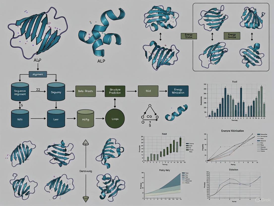
The problem of computational protein structure prediction—determining a protein's three-dimensional (3D) structure from its amino acid sequence—has been one of the most enduring challenges in computational biology and biophysics [19] [20]. Proteins, the workhorses of the cell, perform their vast array of functions through their specific 3D structures. The sequence-structure-function paradigm posits that a protein's amino acid sequence dictates its folded structure, which in turn determines its biological function [20]. For decades, scientists have relied on experimental techniques like X-ray crystallography, NMR spectroscopy, and more recently, cryo-electron microscopy (cryo-EM) to determine protein structures at atomic resolution [19] [20]. However, these methods are often time-consuming, costly, and technically demanding, creating a significant gap between the number of known protein sequences and experimentally solved structures [19] [21].
This widening sequence-structure gap has driven the development of computational methods to predict protein structure. Historically, these approaches have fallen into two main categories: template-based modeling (including homology modeling and threading) and ab initio (or de novo) methods [22] [20]. Homology modeling, which exploits evolutionary relationships between proteins, was for many years the most reliable and widely used computational approach. In parallel, ab initio methods sought to predict structure from physical principles alone, without relying on known structural templates—a computationally daunting task often considered the "holy grail" of computational structural biology [23].
This review traces the historical development and evolution of these core computational strategies, from the early dominance of homology modeling to the sophisticated ab initio methods that paved the way for today's AI revolution. We provide a technical examination of their underlying principles, methodologies, and performance, contextualizing their role in the broader landscape of protein folding research.
Homology Modeling: The Template-Based Workhorse
Principles and Historical Context
Homology modeling, also known as comparative modeling, is founded on the key observation that protein 3D structure is evolutionarily more conserved than amino acid sequence [24] [25]. Consequently, proteins with similar sequences (homologs) are very likely to possess similar 3D structures. If the structure of a homologous protein is known, it can serve as a template to model the structure of a target protein with an unknown structure [25].
The effectiveness of homology modeling is highly dependent on the degree of sequence identity between the target and template. Generally, sequence identities above 30-35% often yield models with high accuracy, potentially with root-mean-square deviation (RMSD) of 1-2 Å from experimental structures [20]. As sequence identity drops below this threshold, the accuracy decreases, requiring more sophisticated alignment and modeling techniques [21] [25].
The Stepwise Methodology of Homology Modeling
The process of building a homology model is methodical, involving several critical steps, each with its own set of tools and potential pitfalls [24] [25].
Step 1: Template Identification and Selection
The first step involves identifying potential template structures in the Protein Data Bank (PDB) that are homologous to the target sequence. This is typically done using sequence search tools like BLAST or more sensitive, iterative methods such as PSI-BLAST [21] [25]. The ideal template is chosen based on factors including sequence identity, query coverage, the resolution and quality of the template structure, and biological relevance (e.g., bound ligands, similar function) [21].
Step 2: Target-Template Alignment
Precise sequence alignment is arguably the most critical step, as errors in alignment are a major source of inaccuracies in the final model [25]. The target sequence is aligned with the template sequence(s), often using multiple sequence alignment programs like ClustalW, T-Coffee, or profile-based methods to incorporate evolutionary information [24] [25]. This alignment defines how the target sequence will be mapped onto the template's 3D coordinates.
Step 3: Model Building
The actual 3D model is constructed based on the alignment. Several strategies exist:
- Rigid-body assembly: The core regions of the target protein are built from structurally conserved regions of the template [25].
- Segment matching: Short segments from known structures are assembled based on sequence similarity and geometric constraints [25].
- Spatial restraint: The model is built by satisfying spatial restraints derived from the template structure, including bond lengths, angles, and dihedral angles. MODELLER is a widely used software that employs this method [19] [25].
Step 4: Loop Modeling
Regions where the target and template sequences are not well-aligned, often corresponding to insertions or deletions, form loops. These are structurally variable and must be modeled separately [25]. Two primary approaches are used:
- Database search: Searching for fragments from known structures that fit the flanking regions and have a matching sequence [25].
- Conformational search (ab initio loop modeling): Using physical energy functions or statistical potentials to generate and score many possible loop conformations from scratch [25]. Tools like FREAD and ModLoop are commonly used [24].
Step 5: Side-Chain Modeling
The conformations of amino acid side chains (rotamers) are predicted onto the modeled backbone. This is typically done using rotamer libraries, which are collections of preferred side-chain conformations derived from high-resolution structures [25]. Programs like SCWRL efficiently search these libraries to find the most energetically favorable side-chain packing [21] [25].
Step 6: Model Optimization and Validation
The initial model often contains steric clashes and strained geometries. Energy minimization and sometimes molecular dynamics simulations are used to relax the model into a more stable, low-energy conformation [25]. Finally, the model's quality is assessed using validation tools like PROCHECK, WHATIF, and PROSA, which evaluate stereochemistry, physical plausibility, and knowledge-based statistical potentials to identify potential errors [25].
The following workflow diagram summarizes the entire homology modeling process.
Applications and Limitations
Homology modeling has been extensively applied in drug discovery for virtual screening and ligand docking, enzyme engineering, and understanding disease-related mutations [19] [25]. Its primary strength is its reliability when a good template is available.
However, its limitations are significant. Model accuracy is wholly dependent on template selection and alignment quality. It struggles with low-homology targets and cannot predict novel folds not present in the PDB. Furthermore, it provides a static snapshot and often fails to capture protein dynamics, intrinsically disordered regions, and the structures of large protein complexes [19].
The Ab Initio Folding Challenge
Conceptual Foundation
In contrast to template-based methods, ab initio (from the beginning) or de novo protein structure prediction aims to predict the 3D structure of a protein using only its amino acid sequence and fundamental physical principles, without relying on a homologous template [22] [23]. The goal is to find the native structure as the global minimum in a complex energy landscape—a conceptual funnel where the native state resides at the bottom [21].
This approach is motivated by three factors:
- The existence of orphan proteins with no detectable homology to proteins of known structure [22].
- The desire to understand the fundamental physical forces driving protein folding [22].
- The fact that highly similar sequences can sometimes adopt different folds, making template-based methods inherently unreliable in some cases [22].
Key Methodological Strategies
Ab initio folding is a computationally intensive problem due to the vast conformational space that must be searched. Several strategies have been developed to make this problem tractable.
Fragment Assembly
A dominant strategy in modern ab initio methods is fragment assembly, pioneered by tools like Rosetta and QUARK [23] [20]. This method involves:
- Fragment Library Generation: For each short segment (typically 3-9 residues) of the target sequence, a large library of candidate structures is extracted from the PDB. These fragments are selected based on sequence similarity and predicted secondary structure compatibility [23] [26].
- Monte Carlo Assembly: The protein is folded in silico by repeatedly replacing segments of a growing model with alternative fragments from the library. Each replacement is accepted or rejected based on a scoring function through a Monte Carlo simulated annealing process [23] [20]. The scoring function typically includes terms for steric clashes, solvation energy, hydrogen bonding, and van der Waals interactions.
Simplified Protein Representations and Energy Functions
To reduce computational cost, many ab initio algorithms use simplified protein representations. Instead of modeling all atoms, they may use a Cα-trace representation or unified residue models like CABS or UNRES, where side chains are represented by a single point [22]. These coarse-grained models are paired with simplified, knowledge-based or physics-based energy functions to guide the search towards native-like structures [22].
The following diagram illustrates the core ab initio folding cycle used in systems like Rosetta.
Performance and Challenges
The performance of ab initio methods has been systematically benchmarked in competitions like the Critical Assessment of protein Structure Prediction (CASP). A 2007 review of 18 ab initio algorithms reported average normalized RMSD scores ranging from 11.17 to 3.48, with I-TASSER identified as the best-performing algorithm at the time based on a combined measure of RMSD and CPU time [22].
The primary challenge for ab initio methods is their immense computational cost, which limits their application to small proteins (typically <150 amino acids) [20]. Accuracy, while impressive for some targets, generally lags behind high-quality homology models. Furthermore, the success of the fragment assembly approach is still implicitly dependent on the existence of suitable fragments in the PDB, making it less effective for truly novel folds.
Table 1: Historical Performance Comparison of Selected Ab Initio Methods
| Method / Tool | Core Principle | Reported Performance | Key Strengths | Key Limitations |
|---|---|---|---|---|
| I-TASSER [22] [27] | Threading, fragment assembly, & iterative refinement | Top performer in early CASP; Normalized RMSD ~3.48 [22] | Full-length modeling; Active site prediction | Slow; Complex pipeline |
| Rosetta [23] [20] | Fragment assembly & Monte Carlo sampling | Excellent for proteins <100 residues [20] | Provides folding insight; Models complexes | High computational demand |
| QUARK [27] [20] | Contact-guided fragment assembly | Excellent for small proteins [20] | Uses deep learning for contact prediction | Not suited for large proteins |
The experimental implementation of these computational methods relies on a curated set of software tools, databases, and computational resources. The following table details key components of the historical computational structural biologist's toolkit.
Table 2: Key Research Reagent Solutions for Computational Structure Prediction
| Resource Name | Type | Primary Function | Relevance to Method |
|---|---|---|---|
| Protein Data Bank (PDB) [24] [21] | Database | Repository of experimentally determined 3D structures of proteins and nucleic acids. | Homology Modeling: Source of template structures. Ab Initio: Source of fragments for libraries. |
| BLAST / PSI-BLAST [24] [21] | Software Tool | Finds regions of local similarity between biological sequences to identify homologous templates. | Homology Modeling: Core tool for template identification and selection. |
| MODELLER [19] [25] | Software Tool | Builds protein 3D models by satisfaction of spatial restraints derived from a template structure. | Homology Modeling: Primary engine for model building from alignment. |
| SCWRL [21] [25] | Software Tool | Predicts side-chain conformations (rotamers) on a fixed protein backbone using a rotamer library. | Homology Modeling: Critical for the side-chain modeling step after backbone construction. |
| Rosetta [23] [26] | Software Suite | Uses fragment assembly, Monte Carlo sampling, and a sophisticated scoring function for ab initio structure prediction and protein design. | Ab Initio: A comprehensive platform for de novo structure prediction. |
| PROCHECK [25] | Software Tool | Validates the stereochemical quality of a protein structure, analyzing Ramachandran plots and other geometric parameters. | Both Methods: Essential for the final step of model validation and quality assessment. |
The historical journey from homology modeling to ab initio methods represents a concerted scientific effort to solve one of biology's most fundamental problems. Homology modeling established itself as the practical and reliable workhorse for researchers who needed a structural model for a protein with a recognizable relative in the PDB. Its stepwise methodology became a standard part of the structural bioinformatics curriculum. Meanwhile, ab initio methods like Rosetta tackled the more formidable challenge of predicting structures from scratch, driven by physical principles and statistical potentials. While computationally expensive and limited to smaller proteins, these methods provided invaluable insights into the protein folding process and offered a solution for orphan proteins without templates.
The evolution of these computational strategies, their strengths, and their limitations set the stage for the current revolution driven by deep learning. The critical need to overcome the challenges of template bias, high computational costs, and the inability to model complex assemblies efficiently fueled the development of a new generation of AI-based predictors. Tools like AlphaFold2 represent a paradigm shift, but they are built upon the foundational knowledge, conceptual frameworks, and vast structural data accumulated through decades of work in homology modeling and ab initio prediction. Understanding these historical approaches is therefore essential for appreciating the current state of the art and for guiding future innovations in computational structural biology.
The energy landscape theory represents a fundamental shift in our understanding of how proteins navigate the complex process of folding from linear polypeptide chains into functional three-dimensional structures. This theoretical framework addresses one of the most significant challenges in molecular biology: the Levinthal's Paradox, which highlights the impossibility of proteins randomly searching all possible conformations to find their native state within biologically relevant timescales [28]. Instead of conceptualizing folding as a single pathway, the energy landscape theory introduces the concept of a folding funnel, where a protein progressively moves toward its native state through a multiplicity of routes [28] [29].
At its core, the folding funnel hypothesis posits that a protein's native state corresponds to its global free energy minimum under physiological conditions [28]. The landscape is characterized by a funnel-like shape where the depth represents the energetic stabilization of the native state, while the width represents the conformational entropy of the system [28]. This conceptual framework has revolutionized the field by providing both qualitative and quantitative insights into protein folding kinetics and thermodynamics, enabling researchers to understand how proteins can fold rapidly and reliably despite the astronomical number of possible conformations [28].
The Conceptual Framework of Folding Funnels
Fundamental Principles and Theoretical Foundation
The folding funnel hypothesis, introduced by Ken A. Dill in 1987, provides a statistical mechanical approach to protein folding by considering the energetics of protein conformation across a multidimensional landscape [28]. In this representation, the y-axis corresponds to the internal free energy of a protein, encompassing contributions from hydrogen bonds, ion-pairs, torsion angle energies, hydrophobic interactions, and solvation free energies [28]. The multiple x-axes represent the vast conformational space available to the polypeptide chain, with geometrically similar structures positioned closer together in the landscape [28].
The theory is closely related to the hydrophobic collapse hypothesis, which identifies the sequestration of hydrophobic amino acid side chains into the protein interior as a major driving force for folding [28]. This process allows water molecules to maximize their entropy, thereby lowering the overall free energy of the system. Additional stabilization comes from favorable energetic contacts within the protein structure, including the isolation of electrically charged side chains on the solvent-accessible surface and the neutralization of salt bridges within the protein core [28]. The molten globule state, predicted as an ensemble of folding intermediates, represents a stage where hydrophobic collapse has occurred but many native contacts have yet to form [28].
Ruggedness and Frustration in Energy Landscapes
Real-world energy landscapes are rarely smooth, ideal funnels. Instead, they typically exhibit varying degrees of ruggedness, characterized by non-native local minima where partially folded proteins can become transiently trapped [28]. This ruggedness creates kinetic traps—energy barriers that can slow the folding process as proteins must navigate around these obstacles or occasionally overcome them to continue progressing toward the native state [28].
The concept of frustration provides a quantitative framework for understanding landscape ruggedness. Drawing analogies from spin glass physics in theoretical physics, frustration measures the competition among conflicting energy contributions within a protein structure [28]. In minimally frustrated systems, the native state exhibits optimal energetic complementarity with minimal internal conflicts. The ratio between the folding transition temperature (Tf) and the glass transition temperature (Tg) serves as an indicator of folding efficiency, with higher Tf/Tg ratios correlating with faster folding rates and fewer folding intermediates [28]. This quantitative relationship helps explain why natural selection has favored protein sequences that evolve toward minimal frustration, enabling rapid and reliable folding under physiological conditions [28].
Quantitative Parameters and Folding Kinetics
The relationship between protein structural features and folding kinetics has been quantitatively investigated through systematic analyses of folding data. The Protein Folding Database (PFD) has been instrumental in enabling these bioinformatic approaches by collecting annotated structural, methodological, kinetic, and thermodynamic data for numerous proteins [30].
Table 1: Quantitative Parameters Governing Protein Folding Rates
| Parameter | Structural Interpretation | Impact on Folding Rate |
|---|---|---|
| Contact Order [30] | Average sequence separation between contacting residues in the native structure | Higher contact order correlates with slower folding |
| Long-Range Order [30] | Proportion of contacts between residues distant in sequence | Inverse correlation with folding rate |
| Relative Contact Order [30] | Contact order normalized by protein chain length | Better predictor than absolute contact order |
| Stability (ΔG) [30] | Free energy difference between native and unfolded states | Can override topological constraints in some protein families |
| Transition Temperature (Tf/Tg ratio) [28] | Ratio of folding transition temperature to glass transition temperature | Higher ratios indicate faster folding with fewer intermediates |
Research has demonstrated that topological constraints fundamentally influence folding rates, with proteins exhibiting low contact order (e.g., α-helical bundles) typically folding faster than those with high contact order (e.g., β-sandwiches) [30]. However, studies on specific protein families like immunoglobulins and cytochrome c have revealed that stability can sometimes be a more significant determinant of folding rate than topology alone [30]. This nuanced understanding highlights the complex interplay between multiple factors in determining folding kinetics.
Computational Methodologies and Experimental Protocols
Advancements in Computational Prediction Methods
Recent advances in computational methods have revolutionized our ability to study protein folding mechanisms. These approaches can be broadly categorized into several methodological frameworks:
Simulation of Inverse Folding Pathways involves computational reconstruction of folding processes starting from the native state and moving backward to unfolded states, providing insights into possible folding routes [31]. Machine Learning for Early Folding Residues leverages artificial intelligence algorithms to identify residues that initiate the folding process, with models trained on experimental folding data [31]. Conformational Sampling explores the energy landscape through techniques like molecular dynamics simulations, generating ensembles of possible conformations to map folding pathways [31]. Template-Based Intermediate Prediction utilizes known protein structures as templates to predict potential folding intermediates, particularly for proteins with homologous folds [31].
The integration of AI technology has been particularly transformative, with systems like AlphaFold enabling remarkable advancements in predicting protein folding and interactions [31] [32]. These computational approaches have created new paradigms for studying protein folding mechanisms that complement traditional experimental methods.
The FragFold Protocol: Predicting Functional Protein Fragments
A recently developed computational method called FragFold demonstrates the power of combining AI with protein folding research. This protocol leverages AlphaFold to predict protein fragments that can bind to or inhibit full-length proteins [32]. The methodology involves several key steps:
- Computational Fragmentation: The target protein is computationally divided into short amino acid sequences representing potential functional fragments [32].
- Multiple Sequence Alignment (MSA) Optimization: Unlike standard AlphaFold implementation that calculates MSAs for every prediction, FragFold pre-calculates the MSA for the full-length protein once, then uses this result to guide predictions for each fragment, significantly improving computational efficiency [32].
- Binding Prediction: The algorithm models how each fragment would bind to relevant interaction partners, generating predicted structural models for these interactions [32].
- Experimental Validation: Predictions are tested using high-throughput experimental measurements in living cells, where millions of cells each produce one type of protein fragment to verify binding and inhibitory function [32].
- Deep Mutational Scanning: Experimentally examining thousands of mutated fragments within cells identifies key amino acids responsible for inhibition, sometimes revealing fragments with greater potency than their natural, full-length sequences [32].
This methodology has proven highly effective, with researchers confirming that more than half of FragFold's predictions for binding or inhibition were accurate, even for proteins without previous structural data on their interaction mechanisms [32].
Figure 1: The FragFold computational workflow for predicting functional protein fragments that can bind to or inhibit target proteins.
Table 2: Essential Research Resources for Protein Folding Investigations
| Resource | Function/Application | Access Information |
|---|---|---|
| Protein Folding Database (PFD) [30] | Central repository for structural, kinetic, and thermodynamic folding data | Freely available at http://pfd.med.monash.edu.au |
| AlphaFold [32] | AI system for protein structure prediction and interaction mapping | Available via public servers or local installation |
| FragFold [32] | Computational method for predicting inhibitory protein fragments | Methodology described in PNAS publication |
| ProTherm [30] | Thermodynamic database for proteins and mutants | Referenced in PFD and specialized literature |
| SCOP Database [30] | Structural classification of proteins for functional annotation | Integrated with PFD for structural analysis |
Structural Models of Folding Energy Landscapes
Classical Funnel Models and Their Variations
The folding funnel concept encompasses several distinct models that describe different topological features of protein energy landscapes:
The Ideal Smooth Funnel represents a perfectly optimized landscape where the protein consistently moves toward lower free energy without significant barriers, with increasing interchain contacts correlating with decreasing degrees of freedom until the native state is achieved [28]. In contrast, the Rugged Funnel incorporates kinetic traps and energy barriers that can temporarily impede folding progress, requiring proteins to occasionally break favorable but non-native contacts before continuing toward the native state [28]. The Moat Landscape describes a scenario where certain proteins must navigate through obligatory kinetic traps as essential steps in their folding pathway, exemplified by hen egg white lysozyme where different populations fold through distinct mechanisms [28]. The Champagne Glass Landscape features significant free energy barriers resulting from conformational entropy, particularly relevant for polar residues connecting hydrophobic clusters [28].
Figure 2: Comparative diagrams of major protein energy landscape models showing distinct topological features.
The Foldon Volcano-Shaped Funnel Model
A significant development in energy landscape theory is the Foldon Funnel Model, which proposes a volcano-shaped energy landscape rather than a simple funnel [28]. This model introduces several innovative concepts that challenge conventional folding paradigms. The outer region of the landscape is characterized by unstable secondary structures that actually increase in free energy as they form, creating an uphill slope contrary to traditional funnel models [28]. These initially unstable secondary structures become progressively stabilized by developing tertiary interactions, yet continue to increase in free energy until the final folding steps [28]. The highest free energy point occurs just before the final transition to the native state, creating a volcano-like profile with the peak at the penultimate step [28]. Despite this unusual landscape topology, the model maintains a fundamental division between native versus non-native kinetic states, consistent with the classical two-state folding behavior observed in many proteins [28].
This model aligns with experimental evidence showing that most protein secondary structures are unstable in isolation and explains the high cooperativity observed in protein folding transitions, where all steps prior to reaching the native state exist in a pre-equilibrium condition [28].
Biological Implications and Research Applications
Resolving Fundamental Paradoxes in Protein Folding
The energy landscape theory provides elegant solutions to long-standing puzzles in protein folding. The framework effectively resolves Levinthal's Paradox by demonstrating that proteins do not randomly search all possible conformations but instead follow biased stochastic paths down a funneled energy landscape [28]. This multi-dimensional search process dramatically reduces the conformational space that must be sampled, enabling biologically relevant folding timescales [28]. Similarly, the theory addresses the Blind Watchmaker's Paradox by showing how natural selection has optimized the energy landscapes of biological proteins through evolutionary pressure, favoring sequences with minimal frustration that fold reliably and efficiently [28].
The energy landscape perspective also explains the remarkable robustness of protein folding to minor sequence variations. While mutations may block specific folding routes, alternative pathways often remain available, allowing the protein to still achieve its correct native structure through different kinetic trajectories [28]. This redundancy in folding pathways provides a buffer against potentially deleterious mutations and contributes to the evolutionary stability of protein structures.
Applications in Disease Mechanisms and Therapeutic Development
Understanding protein energy landscapes has profound implications for human health and disease treatment. The framework provides mechanistic insights into protein misfolding diseases, including neurodegenerative disorders like Alzheimer's and Parkinson's disease, where proteins populate alternative stable states or kinetic traps instead of their functional native structures [31]. The ruggedness of energy landscapes explains how proteins can become trapped in misfolded conformations that nucleate harmful aggregates [28].
The application of folding landscape principles enables rational drug design strategies targeting protein folding processes. Small molecules or protein fragments can be designed to stabilize native states, destabilize pathogenic aggregates, or redirect folding trajectories toward functional conformations [32]. Tools like FragFold demonstrate how computational approaches based on folding principles can generate genetically encodable inhibitors against virtually any protein target, opening new avenues for therapeutic intervention [32]. These approaches have been successfully applied to essential cellular proteins like FtsZ (involved in cell division) and the LptF-LptG complex (involved in outer membrane biogenesis), demonstrating the broad applicability of these methods [32].
Future Perspectives and Challenges
Despite significant advances, numerous challenges remain in fully characterizing and utilizing protein energy landscapes. A major frontier involves moving from qualitative descriptions to quantitative predictions of folding pathways and rates for arbitrary protein sequences [31] [30]. This requires improved integration of physical principles with machine learning approaches to develop models with greater predictive power across diverse protein families.
The relationship between energy landscapes and biological function represents another critical research direction. Understanding how evolutionary pressure has shaped energy landscapes to optimize not just folding efficiency but also functional dynamics, allostery, and ligand binding remains an active area of investigation [30]. The integration of folding data with functional annotations through resources like the Gene Ontology database will facilitate these analyses [30].
From a technical perspective, future progress will depend on enhanced data visualization and exchange methodologies. As folding datasets grow increasingly complex and multidimensional, developing intuitive graphical representations of energy landscapes and standardizing data formats using extensible markup language (XML) will be essential for collaborative research and data mining [30]. These infrastructure developments will support the continuing integration of energy landscape theory with structural biology, biophysics, and therapeutic design, further solidifying its role as a foundational framework for understanding and manipulating protein structure and function.
AI Revolution in Structure Prediction: Methods, Mechanisms, and Real-World Applications
The prediction of a protein's three-dimensional structure from its amino acid sequence represents one of the most fundamental challenges in computational biology, a problem that remained unsolved for over five decades until recent breakthroughs in deep learning. Proteins are the essential biological machines that drive virtually every cellular process, from catalyzing metabolic reactions to facilitating cellular communication. Their function is intrinsically determined by their complex three-dimensional structure, which emerges through a folding process whereby a linear chain of amino acids collapses into a specific, energetically stable conformation. For decades, determining these structures required painstaking experimental methods such as X-ray crystallography, nuclear magnetic resonance (NMR) spectroscopy, and cryo-electron microscopy (cryo-EM)—processes that could take years of effort and substantial resources for a single protein [33] [34].
The computational protein folding problem is framed by two foundational concepts. Anfinsen's thermodynamic hypothesis posits that a protein's native structure corresponds to its minimum free energy state under physiological conditions. Conversely, Levinthal's paradox highlights the astronomical number of possible conformations a protein could theoretically adopt, making it impossible to find this native state through random search [34]. Traditional computational approaches struggled to balance accurate energy functions with efficient sampling of conformational space. Template-based modeling (TBM) relied on homology to known structures, while template-free modeling (TFM) and ab initio methods attempted predictions without templates but with limited accuracy, especially for proteins without close evolutionary relatives [34]. This landscape changed dramatically with the introduction of deep learning approaches, culminating in AlphaFold2's architectural innovations.
AlphaFold2's Architectural Breakthrough
In late 2020, DeepMind's AlphaFold2 achieved unprecedented accuracy in the CASP14 (Critical Assessment of protein Structure Prediction) competition, predicting protein structures with atomic-level accuracy rivaling experimental methods [33] [35]. This breakthrough was widely recognized as a solution to the 50-year-old protein folding problem and was honored with the 2024 Nobel Prize in Chemistry [33] [36]. Unlike previous computational methods that relied heavily on physical energy functions and complex sampling procedures, AlphaFold2 introduced a completely new deep learning architecture that could learn the complex mapping from amino acid sequence to 3D structure.
Core Architectural Components
AlphaFold2's architecture employs a novel transformer-based neural network that integrates multiple components in an end-to-end differentiable system. The model's exceptional performance stems from its ability to jointly reason about sequence relationships, geometric constraints, and spatial dependencies. At its core, AlphaFold2 utilizes an Evoformer module—a novel neural network block that jointly processes sequence and structural information [36]. The Evoformer operates on multiple sequence alignments (MSAs) and pairwise representations, enabling the system to learn evolutionary constraints and residue-residue interactions simultaneously. This is followed by a structure module that iteratively refines the atomic coordinates, directly generating the 3D structure rather than predicting intermediate features like distance maps [37].
Table: Core Components of AlphaFold2 Architecture
| Component | Function | Innovation |
|---|---|---|
| Evoformer | Processes multiple sequence alignments (MSAs) and pairwise representations | Enables co-evolutionary analysis and residue interaction modeling simultaneously |
| Structure Module | Generates atomic coordinates directly | Uses iterative refinement to build accurate 3D structures end-to-end |
| Attention Mechanisms | Captures long-range dependencies in sequences and structures | Allows the model to focus on relevant residues regardless of sequence distance |
| End-to-End Differentiability | Enables gradient flow through entire architecture | Permits joint optimization of all components for final structural accuracy |
A key innovation was AlphaFold2's use of attention mechanisms, particularly self-attention and cross-attention, which allow the model to capture long-range interactions between amino acids that may be distant in the sequence but close in the final folded structure. Unlike the first AlphaFold, which used convolutional neural networks, AlphaFold2's transformer architecture proved dramatically more effective at modeling these complex relationships [36]. The entire system is trained end-to-end, meaning all components are optimized jointly toward the final objective of accurate structure prediction, rather than having separately trained submodules.
Input Representation and Feature Engineering
AlphaFold2's input representations crucially embed evolutionary information that guides the folding process. The system takes as primary input multiple sequence alignments (MSAs) of homologous proteins, which provide information about evolutionary constraints and co-evolutionary patterns. These MSAs are complemented by template structures when available, though the system demonstrates remarkable accuracy even without templates. The model transforms these inputs into embedded representations that capture both sequential relationships and potential structural contacts [34].
The quality of these input features is paramount. MSAs are constructed by searching large sequence databases such as UniRef and BFD for homologs of the target protein. Co-evolutionary signals extracted from these alignments help identify residue pairs that maintain physical proximity through evolution, providing strong constraints for the folding process. This evolutionary data is processed through a series of embedding layers that transform the discrete sequence information into continuous vector representations suitable for deep learning processing [37].
Quantitative Performance and Impact
AlphaFold2's architectural innovations translated directly to unprecedented quantitative performance metrics. At CASP14, the system achieved a median Global Distance Test (GDT) score of 92.4 out of 100 for the most challenging protein domains, meaning its predictions were nearly indistinguishable from experimentally determined structures [33]. This represented a substantial improvement over other methods and previous versions of AlphaFold.
Table: AlphaFold2 Performance Metrics and Scientific Impact
| Metric Category | Specific Measurement | Significance |
|---|---|---|
| Prediction Accuracy | Median GDT of 92.4 at CASP14 | Atomic-level accuracy comparable to experimental methods |
| Database Scale | Predictions for >200 million proteins [33] | Coverage of nearly all known proteins |
| Research Adoption | >3 million researchers across 190 countries [33] | Widespread global utilization |
| Scientific Output | >35,000 citing papers; >200,000 methodology papers [33] | Acceleration of biological discovery |
| Experimental Enhancement | 40% increase in novel experimental structure submissions [33] | Improvement in quality and efficiency of experimental work |
The release of the AlphaFold Protein Database in partnership with EMBL-EBI marked a tipping point in accessibility, providing researchers worldwide with free access to structure predictions for virtually all known proteins [33] [35]. This database has grown to encompass over 240 million predicted structures, dramatically expanding the structural universe available to researchers. An independent analysis by the Innovation Growth Lab found that researchers using AlphaFold2 submitted 40% more novel experimental protein structures to the Protein Data Bank, and these structures were more likely to explore uncharted areas of structural space [33]. Furthermore, research incorporating AlphaFold2 was twice as likely to be cited in clinical articles and significantly more likely to be cited by patents, indicating its strong translational impact [33].
Methodological Workflow and Experimental Protocols
AlphaFold2 Experimental Pipeline
The standard workflow for protein structure prediction using AlphaFold2 involves several methodical steps, from sequence preparation to structure refinement. The following diagram illustrates this end-to-end process:
Step 1: Sequence Preprocessing and Multiple Sequence Alignment Generation The prediction process begins with the input of the target protein's amino acid sequence. The first critical step involves generating a comprehensive multiple sequence alignment (MSA) by searching large genomic databases (such as UniRef, BFD, or MGnify) for evolutionary relatives. This is typically accomplished using tools like HHblits or Jackhmmer with multiple iterations to maximize sensitivity. Simultaneously, template structures are identified from the Protein Data Bank using search tools like HHSearch, though AlphaFold2 can operate effectively without templates [34].
Step 2: Input Feature Construction and Embedding The MSAs and any identified templates are processed into structured input features. These include:
- MSA representations: One-hot encodings of the aligned sequences
- Evolutionary coupling information: Pairwise statistics derived from the MSA
- Template features: Structural information from homologous proteins
- Sequence-based features: Amino acid properties, predicted secondary structure, and solvent accessibility
These diverse features are embedded into continuous vector representations that serve as inputs to the neural network [37].
Step 3: Evoformer Processing and Information Integration The embedded features are processed through the Evoformer stack, which alternates between updating the MSA representation and the pairwise residue representation. This module uses attention mechanisms to identify long-range dependencies and co-evolutionary patterns. The MSA representation helps inform the pairwise potentials, while the evolving pairwise representation constrains the MSA updates. This iterative process allows the model to reason about both sequence relationships and spatial constraints simultaneously [36].
Step 4: Structure Module and 3D Coordinate Generation The refined pairwise representation from the Evoformer is passed to the structure module, which operates in an iterative refinement manner. Unlike earlier approaches that predicted distance maps or contact maps, AlphaFold2's structure module directly predicts atomic coordinates through a series of invariant point attention layers. The module represents the protein backbone as rigid bodies and progressively refines their positions and orientations through multiple cycles, eventually producing the full atomic structure (excluding side chains initially) [37].
Step 5: Side Chain Prediction and Confidence Estimation Once the backbone structure is established, side chain atoms are placed using a rotamer library with chi-angle predictions. Crucially, AlphaFold2 provides per-residue confidence estimates through predicted Local Distance Difference Test (pLDDT) scores, which indicate the reliability of different regions of the predicted structure. Low pLDDT scores often correspond to flexible or disordered regions, providing valuable guidance for experimental validation [33].
Research Reagent Solutions for Experimental Validation
Table: Essential Research Reagents and Tools for AlphaFold2 Workflow
| Reagent/Tool | Function | Application in AlphaFold2 Pipeline |
|---|---|---|
| Multiple Sequence Alignment Tools (HHblits, Jackhmmer) | Identification of homologous sequences | Generates evolutionary constraints for folding |
| Protein Databases (UniProt, PDB, Pfam) | Source of sequence and structural information | Provides training data and template information |
| Structure Visualization Software (PyMOL, ChimeraX) | 3D structure analysis and visualization | Enables interpretation of predicted models |
| Molecular Dynamics Packages (GROMACS, AMBER) | Simulation of protein dynamics | Refines and validates predicted structures |
| Cryo-EM/X-ray Crystallography | Experimental structure determination | Ground truth validation of predictions |
Advanced Technical Extensions
AlphaFold-Multimer for Complex Prediction
Following AlphaFold2's success with single-chain proteins, DeepMind developed AlphaFold-Multimer to predict structures of protein complexes containing multiple chains. This extension required modifications to the input representations to handle multiple sequences and their interactions simultaneously. The system learned to distinguish between intra-chain and inter-chain contacts, enabling accurate prediction of protein-protein interfaces [36]. This capability has proven invaluable for studying signaling pathways, enzyme complexes, and other multi-molecular assemblies critical to cellular function.
AlphaFold3 and Beyond
The recent development of AlphaFold3 represents a further expansion of capabilities, predicting not just proteins but also the structures of DNA, RNA, ligands, and their complexes. This unified model offers an unprecedented view of cellular machinery at the molecular level, with profound implications for drug discovery and structural biology. AlphaFold3 can model how potential drug molecules (ligands) bind to their target proteins, potentially accelerating the drug design process [33]. DeepMind has also developed specialized models inspired by AlphaFold's architecture, including AlphaMissense for predicting pathogenic genetic mutations and AlphaProteo for designing novel protein binders targeting disease-associated molecules [33].
Visualization of Architectural Components
The Evoformer's attention mechanisms represent one of AlphaFold2's most significant innovations. The following diagram illustrates the information flow within this critical component:
Limitations and Future Directions
Despite its revolutionary impact, AlphaFold2 has several important limitations. The model struggles with predicting intrinsically disordered regions that lack a fixed structure, which comprise approximately 30-40% of the human proteome [38]. It also has limitations in modeling conformational dynamics and proteins that exist in multiple states, as it primarily predicts a single, thermodynamically stable conformation [17]. Accuracy can decrease for orphan proteins with few evolutionary relatives, as the model relies heavily on co-evolutionary signals from MSAs [34]. Additionally, while AlphaFold-Multimer can predict complexes, it may not accurately capture transient protein-protein interactions or allosteric regulation mechanisms [36].
Future developments are addressing these limitations through several avenues. Ensemble methods like FiveFold combine predictions from multiple algorithms (AlphaFold2, RoseTTAFold, OmegaFold, ESMFold, and EMBER3D) to better capture conformational diversity [38]. Fine-tuning approaches are adapting the models to specific protein families or structural classes. Integration with molecular dynamics allows for refining predictions and studying folding pathways. Hybrid approaches that combine deep learning with physical energy functions are improving accuracy for challenging targets. As John Jumper of DeepMind notes, the next frontier involves "fus[ing] the deep but narrow power of AlphaFold with the broad sweep of LLMs" to enable more sophisticated scientific reasoning [36].
AlphaFold2's architectural innovations have fundamentally transformed the landscape of computational structural biology. By leveraging transformer-based architectures, sophisticated attention mechanisms, and end-to-end differentiable learning, the system solved a half-century grand challenge in science. Its impact extends far beyond academic interest, accelerating drug discovery, enabling personalized medicine approaches, and democratizing access to structural information for researchers worldwide. While challenges remain in modeling protein dynamics, disorder, and complex assemblies, AlphaFold2 has established a new paradigm for how artificial intelligence can advance scientific discovery, serving as a template for future breakthroughs at the intersection of AI and biology.
Evolutionary information derived from protein sequences is a cornerstone of modern computational biology, providing critical insights into protein structure, function, and interactions. Multiple Sequence Alignments (MSAs) and the detection of co-evolving residues represent powerful methodologies for extracting this information. MSAs enable the identification of conserved regions and evolutionary patterns across homologous sequences, while co-evolution analysis detects pairs of residues that evolve in a correlated manner, often indicating structural or functional constraints. Within computational protein folding research, these methods have transitioned from specialized tools to essential components powering the latest breakthroughs, including deep learning systems like AlphaFold2. This technical guide provides an in-depth examination of the fundamental principles, methodological approaches, and practical applications of MSAs and co-evolution analysis, framing them within the context of advanced protein structure prediction and function annotation for drug discovery and protein engineering.
Theoretical Foundations and Biological Significance
Multiple Sequence Alignments: Capturing Evolutionary Constraints
Multiple Sequence Alignments (MSAs) serve as the fundamental data structure for comparative sequence analysis, enabling the identification of evolutionarily conserved residues and regions under selective constraint. The construction of high-quality MSAs involves aligning sequences from homologous proteins to identify positions that have been conserved throughout evolution, suggesting critical structural or functional roles. The biological significance of MSAs stems from the observation that protein three-dimensional structure is more conserved than amino acid sequence over evolutionary timescales [39]. This conservation enables the transfer of structural and functional information from proteins with known characteristics to their uncharacterized homologs.
Benchmark resources like BAliBASE provide manually refined, reference alignments based on 3D structural superpositions, which are crucial for evaluating and improving MSA algorithms [40]. The latest versions of these benchmarks have significantly expanded their coverage; BAliBASE 3.0 increased from 1444 to 6255 sequences and now covers most of the protein fold space, providing more challenging test cases that represent real-world alignment problems [40]. This expansion addresses the growing need for robust benchmarks as MSA applications extend to more complex protein families and entire proteomes.
Co-evolution: From Correlation to Structural Insight
Co-evolution refers to the coordinated changes that occur between residues within a protein or between interacting proteins to maintain functional interactions through evolution. The underlying principle is that mutations at one position may require compensatory mutations at another position to preserve structural stability or functional capability. Co-evolutionary analysis has revealed that co-evolving residues are frequently found in close spatial proximity in the protein three-dimensional structure [41], making them powerful predictors of residue-residue contacts.
The detection of co-evolution has become particularly valuable for identifying functional residues that influence binding affinity, catalytic activity, or substrate specificity [41]. Specifically, Specificity-Determining Positions (SDPs) represent differentially conserved residues within particular subfamilies that can fine-tune protein activity, including binding affinity, catalytic efficiency, and environmental tolerance [41]. Unlike fully conserved catalytic residues, SDPs often control the co-adaptation of proteins to their native cellular environments and can identify residues responsible for functional divergence after gene duplication events.
Table 1: Key Concepts in Co-evolution Analysis
| Concept | Description | Biological Significance |
|---|---|---|
| Specificity-Determining Positions (SDPs) | Differentially conserved residues within protein subfamilies | Control functional specificity, substrate recognition, and cellular adaptation |
| Compensatory Mutations | Mutations at one position that offset the functional impact of mutations at another position | Maintain protein stability and function despite sequence changes |
| Direct Coupling Analysis (DCA) | Global statistical method that considers all residue pairs simultaneously | Eliminates transitivity problem in contact prediction; identifies direct residue contacts |
| Evolutionary Trace (ET) | Ranks residues by evolutionary importance based on conservation patterns | Identifies functional sites distinct from active sites, including allosteric regions |
Methodological Approaches
MSA Construction and Quality Assessment
The construction of high-quality MSAs requires careful consideration of sequence selection, alignment algorithms, and quality metrics. Key steps include:
Homolog Identification: Using tools like BLAST, HHblits, or JackHMMER to collect homologous sequences from databases such as UniRef, with careful filtering to include evolutionarily related sequences while excluding fragments and poorly characterized sequences.
Alignment Generation: Employing alignment algorithms such as Clustal Omega, MAFFT, or MUSCLE that balance accuracy with computational efficiency, particularly for large protein families.
Quality Filtering: Removing poorly aligned regions, sequences with excessive gaps, or non-homologous sequences that may introduce noise into evolutionary analyses.
Depth Optimization: Balancing the need for sufficient sequences to detect evolutionary signals with the risk of introducing phylogenetic biases or paralogous sequences. Recent approaches like clade-wise alignment integration have demonstrated that dividing large MSAs into smaller, taxonomically coherent groups can improve alignment quality and co-evolutionary signal detection [39].
Advanced strategies for MSA construction include the clade-wise integration approach, which constructs multiple distinct alignments under distinct clades in the tree of life rather than a single large alignment for each protein [39]. Co-evolutionary signals are searched separately within these clades and subsequently integrated using machine learning techniques, markedly improving overall prediction performance concomitant with better alignment quality [39].
Co-evolution Detection Methods
Computational methods for detecting co-evolving residues fall into two primary categories: local methods and global methods.
Local methods such as Mutual Information (MI) analyze each residue pair independently, calculating the statistical dependence between positions. While computationally efficient, these methods suffer from the transitivity problem, where they cannot distinguish direct correlations from indirect correlations mediated through chains of interacting residues [39].
Global methods, including Direct Coupling Analysis (DCA) and PSICOV, model all residue pairs simultaneously using global statistical models. DCA applies a maximum entropy approach to infer direct couplings between residues, effectively eliminating transitive effects and providing more accurate contact predictions [39]. The fundamental DCA equation models the probability of a sequence ( \mathbf{s} ) as:
[
P(\mathbf{s}) = \frac{1}{Z} \exp\left( \sum{i
where ( J{ij} ) represents direct coupling parameters between positions ( i ) and ( j ), ( hi ) represents local fields, and ( Z ) is the partition function. The parameters are typically inferred using mean-field approximation or pseudo-likelihood maximization to handle computational complexity.
Table 2: Computational Methods for Co-evolution Detection
| Method | Type | Key Algorithm | Advantages | Limitations |
|---|---|---|---|---|
| Mutual Information (MI) | Local | Information theory | Fast computation; simple implementation | Cannot distinguish direct from indirect correlations |
| Direct Coupling Analysis (DCA) | Global | Maximum entropy model | Eliminates transitivity; high accuracy for contact prediction | Computationally intensive for large families |
| PSICOV | Global | Sparse inverse covariance estimation | Handles limited data; reduces false positives | Requires large MSAs for best performance |
| Evolutionary Trace (ET) | Phylogenetic | Conservation ranking across tree branches | Identifies functional regions; maps surface patches | Less effective for detecting pairwise contacts |
Workflow: From Sequences to Structural Constraints
The following diagram illustrates a comprehensive workflow for extracting structural constraints from evolutionary information:
Diagram 1: Workflow for extracting structural constraints from evolutionary information.
Applications in Protein Structure Prediction and Beyond
Revolutionizing Protein Structure Prediction with Co-evolutionary Signals
The integration of co-evolutionary information, particularly through DCA, has dramatically advanced the field of protein structure prediction. Early template-free modeling approaches used predicted contacts from DCA as spatial restraints in molecular dynamics simulations to fold proteins ab initio. This approach demonstrated that evolutionary couplings alone could guide accurate structure determination for many protein families.
The revolutionary success of AlphaFold2 represents the culmination of this paradigm, with co-evolutionary information from MSAs serving as a fundamental input to its deep learning architecture [42]. AlphaFold2 processes MSAs through its Evoformer module, which jointly embeds sequence and structural information while detecting patterns of co-evolution to infer spatial relationships between residues [42]. The system learns to interpret coordinated changes across sequences as indicators of physical proximity in the folded structure, enabling atomic-level accuracy predictions even for proteins without close structural homologs.
Recent methods continue to leverage evolutionary information in innovative ways. For example, CF-random predicts alternative protein conformations by randomly subsampling input MSAs at depths too shallow for robust coevolutionary inference (as few as 3 sequences) [43]. This approach directs the AlphaFold2 network to predict structures from sparse sequence information, enabling the sampling of alternative conformations for fold-switching proteins that remodel their secondary structures in response to cellular stimuli [43].
Predicting Protein-Protein Interactions
Inter-protein co-evolution analysis extends the same principles used for intra-protein contact prediction to identify interacting protein pairs and characterize their binding interfaces. The construction of paired MSAs is critical for this application, requiring careful identification of orthologs to ensure proper pairing across species [39].
Challenges in PPI prediction include differential gene loss, gene duplications, and horizontal gene transfers that complicate orthology assignment. Clade-wise integration strategies have shown promise in addressing these challenges by building multiple distinct alignments under different taxonomic clades rather than single comprehensive alignments [39]. This divide-and-conquer approach improves alignment quality and reduces phylogenetic biases, enhancing PPI detection performance.
AlphaFold-Multimer and similar approaches have demonstrated remarkable accuracy in predicting protein-protein complexes when provided with high-quality paired MSAs [39]. This has led to proposed discover-and-refine workflows where faster coevolution-based methods pre-screen entire proteomes for potential interactions, submitting only promising candidates to more computationally intensive AI-based structure prediction [39].
Functional Annotation and Fitness Prediction
Evolutionary information provides powerful constraints for predicting the functional impact of mutations. The Evolutionary Trace (ET) method exemplifies this approach by ranking residues according to their relative evolutionary importance, enabling the identification of functional sites beyond canonical active positions [41].
Applications of ET include:
- Identification of allosteric sites in G protein-coupled receptor kinases [41]
- Determination of specificity residues in bioamine receptors that control ligand affinity and efficacy [41]
- Discovery of novel functional surfaces in bacterial RecA protein distinct from its canonical activities [41]
Recent approaches like EvoIF integrate multiple evolutionary signals for fitness prediction, combining within-family profiles from retrieved homologs with cross-family structural-evolutionary constraints distilled from inverse folding models [44]. This framework interprets natural evolution as implicit reward maximization and masked language modeling as inverse reinforcement learning, where extant sequences constitute expert demonstrations of high-fitness variants [44].
Advanced Computational Techniques
Integrating Evolutionary Information with Deep Learning
Modern protein structure prediction systems have developed sophisticated architectures for processing evolutionary information. The Evoformer module in AlphaFold2 represents a landmark innovation, employing attention mechanisms to detect patterns in MSAs and extract co-evolutionary signals [42]. This module processes both the MSA representation and a pair representation that encodes relationships between residues, allowing it to identify coupled mutations while considering the broader sequence context.
Protein language models (pLMs) like ESM provide an alternative approach by learning evolutionary constraints from millions of sequences through self-supervised training [44]. These models capture statistical patterns of natural sequence variation that reflect structural and functional constraints, enabling zero-shot fitness prediction without explicit MSA construction for each query protein.
The EvoIF framework exemplifies next-generation integration, combining sequence-based evolutionary profiles from homologous sequences with structure-based evolutionary profiles from inverse folding models [44]. This approach addresses the complementary strengths of each information source: within-family signals from MSAs provide specific conservation patterns, while cross-family structural constraints capture general physicochemical principles of fold stability.
Conformational Ensembles and Alternative States
While static structures provide valuable insights, proteins are dynamic systems that sample multiple conformational states. Traditional co-evolution analysis often captures only the dominant conformation, but advanced sampling techniques can reveal alternative states. The CF-random method achieves this by using very shallow MSAs (as few as 3 sequences) that provide insufficient information for robust co-evolutionary inference, forcing the network to explore alternative structural interpretations [43].
This approach has successfully predicted both conformations of fold-switching proteins like human XCL1, which adopts distinct structures with different hydrogen bonding networks and hydrophobic cores [43]. Similarly, CF-random has captured the alternative conformations of TRAP1-N, a mitochondrial heat shock protein domain that assumes different structures in its apo and nucleotide-bound forms [43].
The following diagram illustrates the CF-random workflow for predicting alternative conformations:
Diagram 2: CF-random workflow for predicting alternative conformations.
Experimental Protocols
Protocol 1: MSA Construction for Co-evolution Analysis
Objective: Generate a high-quality MSA suitable for co-evolution analysis and contact prediction.
Materials:
- Query protein sequence(s) in FASTA format
- Access to sequence databases (UniRef90, NR)
- Computational tools: HHblits, JackHMMER, or MMseqs2 for homolog collection; MAFFT or Clustal Omega for alignment
Procedure:
- Homolog Collection:
- Use HHblits with 3 iterations against Uniclust30 database
- Apply E-value threshold of 0.001 for inclusion
- Remove sequences with >90% identity using CD-HIT to reduce redundancy
Alignment Generation:
- Align collected homologs using MAFFT with L-INS-i algorithm for accuracy
- For larger families (>5000 sequences), use MAFFT FFT-NS-2 for faster alignment
Quality Control:
- Remove columns with >50% gaps using trimAl
- Filter sequences with >30% gaps using in-house scripts
- For paired MSAs (PPI prediction), ensure proper orthology pairing using synteny information or reciprocal best hits
Validation:
- Check alignment quality using reference benchmarks like BAliBASE [40]
- Verify expected conserved motifs are properly aligned
- Estimate effective MSA depth using Meff calculation to account for phylogenetic biases
Protocol 2: Residue-Residue Contact Prediction using DCA
Objective: Predict residue-residue contacts from MSA using Direct Coupling Analysis.
Materials:
- High-quality MSA in FASTA or A3M format
- DCA implementation (plmDCA, GREMLIN, or EVcouplings)
- Computational resources (method is computationally intensive for large proteins)
Procedure:
- MSA Preprocessing:
- Convert MSA to binary representation (20 amino acids + gap)
- Remove sequences with unusual length or composition
- For proteins >200 residues, consider splitting into domains
DCA Execution:
- Run plmDCA with default parameters for initial analysis
- Use pseudo-likelihood maximization for parameter inference
- Apply L2 regularization (λ=0.2) to prevent overfitting
Contact Extraction:
- Rank residue pairs by direct coupling scores (Frobenius norm of Jij)
- Apply Average Product Correction (APC) to remove phylogenetic biases
- Select top L/5 predictions (L = protein length) as final contact set
Validation:
- Compare predictions to known structures if available
- Calculate precision of top predictions using PDB structures as reference
- For proteins without structures, validate using known functional motifs
Protocol 3: Evolutionary Trace for Functional Site Identification
Objective: Identify functionally important residues using Evolutionary Trace analysis.
Materials:
- MSA of protein family
- Phylogenetic tree construction software (FastTree, RAxML)
- Evolutionary Trace implementation (ET-server or custom scripts)
Procedure:
- Phylogenetic Tree Construction:
- Build phylogenetic tree from MSA using FastTree with JTT+CAT model
- Divide tree into branches based on evolutionary divergence
Conservation Analysis:
- Calculate position-specific conservation scores for each branch
- Rank residues by evolutionary importance (earlier divergence = higher rank)
Functional Site Prediction:
- Map top-ranked residues to protein structure if available
- Identify spatial clusters of high-ranked residues using 4.5Å cutoff
- Compare predicted sites to known functional annotations
Experimental Validation:
- Select predicted functional residues for site-directed mutagenesis
- assay functional consequences (catalytic activity, binding affinity, etc.)
- Compare experimental results to computational predictions
The Scientist's Toolkit
Table 3: Essential Resources for Evolutionary Analysis
| Resource | Type | Function | Access |
|---|---|---|---|
| BAliBASE [40] | Benchmark database | Reference alignments for method evaluation | http://www-bio3d-igbmc.u-strasbg.fr/balibase |
| HH-suite | Software suite | Homolog detection & MSA generation | https://github.com/soedinglab/hh-suite |
| MAFFT | Alignment algorithm | Multiple sequence alignment | https://mafft.cbrc.jp/alignment/software/ |
| plmDCA | Software package | Direct Coupling Analysis | https://github.com/pagnani/plmDCA |
| EVcouplings | Framework | Co-evolution analysis pipeline | https://evcouplings.org/ |
| ET-server | Web server | Evolutionary Trace analysis | http://mammoth.bcm.tmc.edu/ET/ |
| ColabFold [43] | Software | Efficient AlphaFold2 implementation with MSA generation | https://github.com/sokrypton/ColabFold |
| CF-random [43] | Method | Alternative conformation prediction | Custom implementation |
Future Directions and Challenges
The field of evolutionary information leverage continues to evolve rapidly, with several emerging challenges and opportunities. Current limitations include the dependency on sufficient homologous sequences for robust co-evolutionary analysis, with performance degrading for protein families with few homologs [39]. Additionally, deep learning models like AlphaFold, while revolutionary, may not fully capture the physical principles underlying protein dynamics and ligand interactions [45].
Recent studies questioning whether deep learning models for co-folding truly learn the physics of protein-ligand interactions have revealed notable discrepancies when models are subjected to biologically plausible perturbations [45]. For example, binding site mutagenesis challenges show that co-folding models sometimes maintain ligand placement even after removing critical interacting residues, indicating potential overfitting to statistical correlations rather than learning underlying physical principles [45].
Future methodologies will likely integrate physical constraints more explicitly with evolutionary information, develop better approaches for modeling conformational heterogeneity, and extend robust predictions to proteins with minimal evolutionary information. The combination of evolutionary principles with physics-based simulations and experimental data will provide more comprehensive understanding of protein structure, function, and dynamics, further advancing drug discovery and protein engineering applications.
The prediction of three-dimensional protein structures from amino acid sequences represents one of the most significant challenges in computational biology. For decades, this field progressed incrementally until recent advances in deep learning catalyzed a revolutionary leap in accuracy and capability. While AlphaFold2 has garnered substantial attention, several other powerful algorithms have emerged that offer complementary strengths and capabilities. Among these, RoseTTAFold, ESMFold, and trRosetta have established themselves as foundational tools in the modern computational structural biology toolkit [16] [46].
These three methods exemplify distinct architectural philosophies in deep learning-based structure prediction. RoseTTAFold employs a three-track neural network that simultaneously reasons about protein sequence, distance constraints, and atomic coordinates. ESMFold leverages massive protein language models trained on millions of diverse sequences to predict structures directly from single sequences. trRosetta pioneered a two-step approach that first predicts inter-residue geometries then converts these into full atomic models [16] [47] [46]. Understanding their complementary strengths, limitations, and optimal application domains is crucial for researchers engaged in protein engineering, drug discovery, and functional annotation.
This technical guide provides an in-depth examination of these three complementary approaches, detailing their underlying architectures, performance characteristics, and practical implementation protocols. By framing this analysis within the broader context of computational protein folding methodologies, we aim to equip researchers with the knowledge necessary to select and utilize the most appropriate tool for their specific research challenges.
Core Architectural Principles
RoseTTAFold: Three-Track Integrated Network
RoseTTAFold implements a sophisticated three-track neural network architecture that simultaneously processes information at three levels of representation: (1) the 1D sequence track analyzes amino acid patterns and evolutionary information, (2) the 2D distance track reasons about pairwise residue interactions, and (3) the 3D spatial track models atomic coordinates [48] [46]. These tracks are connected through carefully designed attention mechanisms that allow information to flow bidirectionally between representations, enabling the network to leverage sequence patterns to inform distance constraints and geometric arrangements.
A key innovation in RoseTTAFold is its iterative refinement process, where information flows cyclically between the tracks, allowing the model to progressively improve its predictions. Starting from initial sequence features, the network generates coarse distance maps and geometric constraints, which then inform more precise atomic coordinates, which in turn refine the understanding of sequence conservation patterns. This iterative process continues until convergence, resulting in a self-consistent structural model [48].
The RoseTTAFold architecture has proven exceptionally adaptable, serving as the foundation for more advanced applications like ProteinGenerator (PG), which performs diffusion in sequence space to enable functional protein design. PG begins with a noised sequence representation and iteratively denoises it while guided by desired sequence and structural attributes, allowing designers to specify constraints like thermostability, rare amino acid enrichment, or specific structural motifs [48].
ESMFold: Language Model-Driven Prediction
ESMFold represents a paradigm shift in protein structure prediction by leveraging protein language models (pLMs) trained through self-supervision on hundreds of millions of protein sequences from diverse organisms [16] [47]. Unlike methods that rely on explicit evolutionary information from multiple sequence alignments (MSAs), ESMFold's language model internalizes evolutionary constraints and structural principles through its training objective, which involves predicting masked amino acids in sequences.
The architectural backbone of ESMFold is a transformer model with 650 million parameters, which generates contextualized residue representations that implicitly encode structural information. These representations are then passed to a structure module that directly predicts 3D coordinates, bypassing the need for intermediate geometric representations like distance maps [47]. This end-to-end approach allows ESMFold to achieve remarkable prediction speeds—often completing structure predictions within seconds for typical proteins.
A significant advantage of ESMFold's language model approach is its ability to make accurate predictions from single sequences without requiring time-consuming homology search steps. This capability makes it particularly valuable for high-throughput applications, orphan sequences with few homologs, and metagenomic discovery where MSAs are difficult to construct [47]. The ESM Metagenomics Atlas, containing over 600 million metagenomic protein structures, stands as a testament to ESMFold's scalability [16].
trRosetta: Two-Step Geometry Transformation
trRosetta (transform-restrained Rosetta) employs a two-step prediction pipeline that separates geometry prediction from structure realization [49] [46]. In the first stage, a deep neural network predicts inter-residue geometries, including distance distributions and orientation angles (ω, θ, and φ) between residue pairs. These predictions are formulated as probability distributions discretized into bins, providing rich constraints for the subsequent structure modeling stage.
The second stage converts these predicted geometric constraints into a knowledge-based potential that guides structure assembly within the Rosetta framework [49]. The network-predicted distributions are transformed into restraint energies that are minimized during the structure realization process, effectively guiding the conformational search toward models that satisfy the predicted constraints.
trRosetta's modular architecture offers practical advantages, particularly in computational efficiency and flexibility. The separation of geometry prediction from structure realization allows each component to be optimized independently and enables researchers to utilize the geometric constraints for other applications beyond full structure prediction [46]. Additionally, this approach requires fewer computational resources than end-to-end methods, making it more accessible to research groups without specialized hardware [46].
Table 1: Comparative Overview of Core Architectural Features
| Feature | RoseTTAFold | ESMFold | trRosetta |
|---|---|---|---|
| Prediction Approach | Three-track end-to-end network | Language model-driven transformation | Two-step geometry to structure |
| Evolutionary Information | MSA-derived features | Internalized in language model | MSA-derived co-evolution |
| Key Innovation | Iterative information flow between tracks | Single-sequence prediction capability | Distance/orientation probability prediction |
| Structure Representation | Atomic coordinates | Atomic coordinates | Restraint-based folding |
| External Dependencies | Rosetta (for some applications) | Standalone | Rosetta framework |
Performance Metrics and Comparative Analysis
Accuracy Benchmarks
Evaluating protein structure prediction methods requires multiple complementary metrics that capture different aspects of structural accuracy. The Template Modeling Score (TM-score) measures global fold similarity, with values above 0.5 indicating generally correct topology and values above 0.8 indicating high accuracy. The Global Distance Test (GDT) quantifies the percentage of residues positioned within specific distance cutoffs from the experimental structure, with GDT_TS providing a more reliable assessment of global accuracy than RMSD for larger proteins [16].
The pLDDT (predicted Local Distance Difference Test) score provided by AlphaFold-derived methods (including ESMFold) assesses local structure quality on a per-residue basis, with scores above 90 indicating high confidence, 70-90 indicating good confidence, and scores below 50 suggesting low reliability [16]. The Predicted Aligned Error (PAE) measures confidence in the relative positioning of different protein regions, with lower values indicating higher confidence in domain orientations [16].
In comparative assessments on standard benchmarks like CAMEO and CASP15, ESMFold generally demonstrates superior accuracy among single-sequence methods, with average TM-scores of approximately 0.80-0.85 on diverse test sets, approaching the accuracy of MSA-based methods for many targets [47]. Both RoseTTAFold and trRosetta deliver strong performance, with accuracy highly dependent on the availability of evolutionary information and structural templates. For targets with rich evolutionary information, these methods can achieve accuracy comparable to state-of-the-art approaches [46].
Computational Efficiency
Computational requirements represent a critical practical consideration when selecting protein structure prediction tools. ESMFold offers the fastest inference times, typically predicting structures in seconds to minutes depending on protein length, making it suitable for high-throughput applications [47]. This speed advantage comes from its single-sequence processing and optimized transformer architecture.
RoseTTAFold requires more substantial computational resources, particularly when generating MSAs, with prediction times ranging from minutes to hours per target. However, its accuracy generally justifies these requirements for critical applications [48]. trRosetta occupies an intermediate position, with the geometry prediction step being relatively fast and the structure realization phase consuming most of the computational time, typically totaling 30 minutes to several hours for a medium-sized protein [49].
Recent innovations like SPIRED have emerged to address efficiency constraints, achieving approximately 5-fold acceleration in inference speed and at least 10-fold reduction in training cost compared to established methods while maintaining competitive accuracy [47]. Such developments highlight the ongoing optimization of the computational protein structure prediction landscape.
Table 2: Performance and Resource Requirements Comparison
| Metric | RoseTTAFold | ESMFold | trRosetta |
|---|---|---|---|
| Typical TM-score | 0.75-0.85 (MSA-dependent) | 0.80-0.85 | 0.70-0.80 (template-dependent) |
| Prediction Speed | Minutes to hours | Seconds to minutes | 30 minutes to several hours |
| Key Strength | High accuracy with MSAs | Single-sequence speed | Robust restraint prediction |
| Limitation | MSA generation bottleneck | Lower accuracy on some orphans | Template dependency |
| Ideal Use Case | Critical high-accuracy predictions | High-throughput screening | Intermediate resource settings |
Experimental Protocols and Implementation
RoseTTAFold Implementation Protocol
Input Preparation: Begin with the target amino acid sequence in FASTA format. For optimal performance, generate multiple sequence alignments using tools like HHblits or MMseqs2 against standard sequence databases (UniClust30, BFD) [48].
Structure Prediction Execution:
- Submit the sequence and MSA to the RoseTTAFold web server or standalone package.
- The three-track network processes inputs through approximately 20-40 iterations of information exchange.
- The final output includes atomic coordinates in PDB format, per-residue confidence estimates, and potential quality metrics.
Functional Design Extension (ProteinGenerator):
- Define desired sequence and structural attributes (e.g., thermostability, specific motifs).
- Initialize with a noised sequence representation and black-hole initialized structure.
- Perform iterative denoising guided by constraint functions, with sequence logits updated at each step to steer toward desired properties [48].
- Filter generated designs by predicted confidence metrics (pLDDT > 90, RMSD to design < 2Å).
Validation: Experimentally characterize designs through size-exclusion chromatography for solubility/monomericity, circular dichroism for secondary structure, and thermal melts for stability assessment [48].
ESMFold Implementation Protocol
Input Preparation: The target amino acid sequence in FASTA format is the sole requirement—no MSA generation is needed, significantly streamlining the preparation phase [47].
Structure Prediction Execution:
- Submit the sequence to the ESMFold web server or local installation.
- The protein language model processes the sequence through its transformer layers to generate residue-wise representations.
- The folding head converts these representations into 3D atomic coordinates.
- The complete process typically requires 1-10 seconds per 100 residues on appropriate GPU hardware.
High-Throughput Applications:
- For large-scale predictions (e.g., entire proteomes or metagenomic catalogs), utilize batch processing capabilities.
- Implement quality filters based on pLDDT scores to identify high-confidence predictions.
- For the ESM Metagenomics Atlas, access precomputed predictions rather than generating new ones [16].
Integration with Fitness Prediction (SPIRED-Framework):
- Integrate ESMFold or SPIRED as the structural feature extractor within an end-to-end neural network.
- Train jointly on deep mutational scanning data to predict fitness effects from sequence alone.
- Fine-tune for specific prediction tasks like stability changes (ΔΔG) or melting temperature shifts (ΔTm) [47].
trRosetta Implementation Protocol
Input Preparation: Prepare the target amino acid sequence and, for enhanced accuracy, generate MSAs using standard tools. Optionally, identify homologous templates through structure database searches [49].
Two-Stage Prediction Execution:
- Geometry Prediction: The deep neural network predicts distance and orientation distributions for residue pairs, outputting probability distributions for different distance and angle bins.
- Structure Realization: Convert predicted distributions into restraint energies:
E_restraint = -log(P(d)) + constantwhere P(d) represents the predicted probability for a given distance bin [49]. - Minimize the restraint energies alongside Rosetta's knowledge-based force field using gradient-based optimization methods.
- Generate multiple models (typically 5-10) and select the best based on agreement with predicted restraints and Rosetta energy scores.
Advanced Applications:
- For functional motif grafting, use protocols like Rosetta FunFolDes that couple folding with design to stabilize inserted functional motifs [50].
- Implement region-specific constraints to maintain structural integrity in grafted regions.
- Validate designs through binding affinity assays (for inhibitors) or immunological assays (for vaccine candidates) [50].
The Scientist's Toolkit: Essential Research Reagents
Table 3: Key Research Reagent Solutions for Protein Structure Prediction
| Reagent/Resource | Function | Implementation Example |
|---|---|---|
| Multiple Sequence Alignment Tools (HHblits, MMseqs2) | Identify evolutionary related sequences for co-evolution analysis | Input generation for RoseTTAFold, trRosetta [16] |
| Protein Data Bank (PDB) | Repository of experimentally determined structures | Training data, template source, validation reference [34] |
| Rosetta Software Suite | Macromolecular modeling platform | Structure realization in trRosetta, functional design [50] [49] |
| ESM Metagenomics Atlas | Database of 600+ million metagenomic structures | Resource for mining novel structures without computation [16] |
| AlphaFold DB | Repository of 200+ million predicted structures | Comparison resource, template avoidance [16] |
| CAMEO Server | Continuous automated model evaluation | Independent accuracy assessment [16] |
Integrated Workflow Visualization
The following diagram illustrates how these three complementary approaches integrate into a comprehensive protein structure prediction and design workflow:
RoseTTAFold, ESMFold, and trRosetta represent complementary pillars in the modern protein structure prediction ecosystem, each with distinct strengths and optimal application domains. RoseTTAFold delivers high accuracy through its sophisticated three-track architecture and enables advanced functional design through extensions like ProteinGenerator. ESMFold offers unprecedented speed from single sequences, enabling high-throughput applications and metagenomic exploration. trRosetta provides a robust, efficient two-step approach that balances accuracy with computational accessibility.
Forward-looking researchers should view these tools not as competitors but as complementary components in a comprehensive structural biology toolkit. The emerging trend of end-to-end frameworks like SPIRED-Fitness, which integrate structure prediction with functional analysis, points toward a future where structural insights directly drive protein engineering and design. As these methods continue to evolve, their integration with experimental validation and specialized applications will further expand their impact across biochemistry, drug discovery, and synthetic biology.
Selecting the appropriate method requires careful consideration of sequence characteristics, available resources, and research objectives. For critical applications requiring maximum accuracy with evolutionary information, RoseTTAFold excels. For high-throughput screening or orphan sequences, ESMFold provides unmatched efficiency. For balanced performance in resource-constrained environments, trRosetta remains a robust choice. By understanding these complementary approaches, researchers can strategically leverage the protein structure prediction ecosystem to advance their scientific objectives.
Protein-protein interactions (PPIs) are fundamental regulators of nearly all cellular functions, influencing processes such as signal transduction, cell cycle regulation, and transcriptional control [51]. Over 80% of proteins operate within molecular complexes rather than in isolation, making the knowledge of how these complexes form crucial for understanding both physiological and pathological cellular states [52]. The map of these molecular interactions, known as the interactome, is essential for deciphering cellular functions and has significant implications for identifying therapeutic targets and advancing drug discovery [52] [53].
The cellular environment presents a major challenge for accurate PPI prediction. The cytoplasm is a crowded milieu, with macromolecules occupying up to 40% of the cytoplasmic volume at concentrations between 100 and 450 g/L [52]. This molecular crowding significantly impacts protein behavior, including structural stability, diffusion rates, and binding kinetics—factors often overlooked in traditional in vitro experiments and computational studies conducted in diluted solutions [52]. The high viscosity and dense packing of the cellular interior mean that conditions under which PPIs are typically measured in vitro can differ substantially from their native environment [52].
Computational biology has undergone a revolutionary transformation with the inclusion of deep learning and artificial intelligence, dramatically enhancing our capacity to predict PPIs with unprecedented accuracy [51] [54]. These advancements are particularly crucial for investigating interactions with no precedence in nature, known as de novo interactions, which open broad applications in biotechnology ranging from drug discovery using molecular glues to novel protein engineering [54]. This technical guide explores the core methodologies, experimental protocols, and emerging trends in computational PPI prediction, framed within the context of a broader thesis on computational protein folding methods.
Computational Foundations of PPI Prediction
The Shift from Traditional Methods to Deep Learning
Before the rise of deep learning, PPI prediction relied predominantly on experimental methods such as yeast two-hybrid screening, co-immunoprecipitation, and mass spectrometry, complemented by computational approaches based on sequence similarity and structural alignment [51]. While effective, these techniques were often time-consuming, resource-intensive, and limited in their ability to scale to large, complex biological systems [51].
Modern deep learning methods have transformed this landscape through their powerful capabilities for high-dimensional data processing and automatic feature extraction [51]. Unlike conventional machine learning algorithms such as support vector machines and random forests, which rely on manually engineered features, deep learning models can autonomously extract semantic sequence context information from protein sequence and residue data, capturing nonlinear relationships that were previously intractable [51].
Core Deep Learning Architectures for PPI Prediction
Several neural network architectures have demonstrated remarkable success in PPI prediction:
Graph Neural Networks (GNNs): GNNs and their variants excel at capturing the topological information within PPI networks by representing proteins as nodes and interactions as edges in a graph [53]. Through message-passing mechanisms, GNNs aggregate information from neighboring nodes to generate representations that reveal complex interaction patterns and spatial dependencies [51]. Key variants include:
- Graph Convolutional Networks (GCNs): Employ convolutional operations to aggregate information from neighboring nodes [51].
- Graph Attention Networks (GATs): Introduce attention mechanisms that adaptively weight the importance of neighboring nodes [51].
- GraphSAGE: Designed for large-scale graph processing through neighbor sampling and feature aggregation [51].
- Graph Autoencoders (GAE): Utilize encoder-decoder frameworks to learn compact node embeddings for reconstruction or prediction tasks [51].
Convolutional Neural Networks (CNNs): Effective for processing spatial and structural data, particularly in analyzing protein contact maps and molecular surfaces [51] [54].
Transformers and Attention Mechanisms: These architectures capture long-range dependencies and global contextual information within protein sequences and structures, with attention-free variants (AFT) also showing promise [51] [53].
Addressing Hierarchical Information with Hyperbolic Geometry
A significant advancement in PPI prediction is the recognition that protein networks exhibit natural hierarchical organization, ranging from molecular complexes to functional modules and cellular pathways [53]. Traditional Euclidean-based models struggle to represent this hierarchical structure efficiently.
Recent approaches like HI-PPI (Hyperbolic graph convolutional network and Interaction-specific learning for PPI prediction) incorporate hyperbolic geometry to better capture these hierarchical relationships [53]. In hyperbolic space, the level of hierarchy is intuitively represented by the distance from the origin, enabling more biologically meaningful embeddings that reflect the central-peripheral structure of PPI networks and identify hub proteins [53].
Table 1: Key Deep Learning Architectures for PPI Prediction
| Architecture | Key Variants | Strengths | Representative Models |
|---|---|---|---|
| Graph Neural Networks | GCN, GAT, GraphSAGE, GAE | Captures topological information and neighborhood structures | GNN-PPI, AFTGAN, HIGH-PPI, HI-PPI [51] [53] |
| Convolutional Networks | 1D, 2D, 3D CNNs | Processes spatial patterns in sequences and structures | PIPR [53] |
| Transformer-based Models | Standard Transformer, AFT | Captures long-range dependencies and global context | AFTGAN [53] |
| Multi-modal Frameworks | Heterogeneous GNNs | Integrates sequence, structure, and network data | MAPE-PPI, HI-PPI [53] |
Key Methodologies and Experimental Protocols
Feature Extraction and Representation Learning
Effective PPI prediction requires comprehensive feature extraction from multiple biological data sources:
Sequence-Based Features: Amino acid sequences are processed using pre-trained language models like ESM and ProtBERT, which capture evolutionary information and physicochemical properties [51]. These representations encode semantic sequence context that correlates with interaction potential.
Structure-Based Features: Three-dimensional protein structures, either experimentally determined or predicted by AlphaFold2, are used to construct contact maps based on the physical coordinates of residues [55] [53]. Structural features are typically encoded using graph representations or 3D convolutional networks, capturing spatial constraints that determine binding compatibility.
Network-Based Features: Topological information from existing PPI networks, including node degree, betweenness centrality, and community structure, provides context for interaction prediction [53]. Methods like HI-PPI explicitly model the hierarchical organization of these networks in hyperbolic space [53].
The HI-PPI Framework: A Case Study in Modern PPI Prediction
The HI-PPI framework represents the cutting edge in PPI prediction methodology, integrating multiple advanced concepts into a unified architecture [53]:
Feature Extraction Stage:
- Structural data is processed through a pre-trained heterogeneous graph encoder and masked codebook to generate encoded structural features [53].
- Sequence data is represented based on physicochemical properties [53].
- Structural and sequence feature vectors are concatenated to form initial protein representations [53].
Hyperbolic Graph Convolutional Network:
Interaction-Specific Learning:
Workflow Visualization: HI-PPI Architecture
Benchmark Evaluation and Performance Metrics
Rigorous evaluation of PPI prediction methods employs standard benchmark datasets and multiple performance metrics:
Commonly Used Datasets:
- SHS27K and SHS148K: Homo sapiens subsets derived from STRING database, containing 1,690 proteins with 12,517 PPIs and 5,189 proteins with 44,488 PPIs respectively [53].
- Training and test sets are constructed using Breadth-First Search (BFS) and Depth-First Search (DFS) strategies to evaluate model generalization [53].
Evaluation Metrics:
- Micro-F1 Score: Comprehensive measure of precision and recall.
- AUPR (Area Under Precision-Recall Curve): Particularly important for imbalanced datasets.
- AUC (Area Under ROC Curve): Measures overall classification performance.
- Accuracy: Proportion of correct predictions.
Table 2: Performance Comparison of PPI Prediction Methods on SHS27K and SHS148K Datasets
| Method | SHS27K (DFS) Micro-F1 | SHS148K (DFS) Micro-F1 | Key Features |
|---|---|---|---|
| HI-PPI | 0.7746 | 0.7418 | Hyperbolic GCN, interaction-specific learning [53] |
| MAPE-PPI | 0.7234 | 0.7112 | Heterogeneous GNN, multi-modal data integration [53] |
| BaPPI | 0.7536 | 0.6910 | Sequence-structure integration [53] |
| HIGH-PPI | 0.7382 | 0.7025 | Dual-view graph learning [53] |
| AFTGAN | 0.7219 | 0.6853 | Attention-free transformer with GAN [53] |
| PIPR | 0.7047 | 0.6639 | CNN-based, sequence-only [53] |
Recent benchmark evaluations demonstrate that HI-PPI achieves state-of-the-art performance, improving Micro-F1 scores by 2.62%-7.09% over the second-best method across different datasets and evaluation schemes [53]. The improvements are statistically significant (p-values < 0.05) and particularly pronounced on larger datasets with more unseen proteins, highlighting the scalability of hierarchical and interaction-specific approaches [53].
Advanced Considerations in PPI Prediction
Molecular Crowding and Cellular Environment
Traditional PPI prediction methods often overlook a critical factor: the crowded cellular environment [52]. The cytoplasm is characterized by high concentrations of macromolecules (100-450 g/L) that occupy up to 40% of the cytoplasmic volume, creating conditions that significantly impact protein behavior [52]. This molecular crowding affects:
- Structural Stability: Proteins exhibit different folding dynamics under crowded conditions.
- Diffusion Processes: Molecular movement occurs in high-viscosity environments, altering encounter rates.
- Binding Kinetics: Both functional and non-functional interactions are modulated by excluded volume effects.
Computational approaches that incorporate crowding effects include lattice simulations, hydrodynamic interaction models, and molecular dynamics simulations of realistic cytoplasmic environments [52]. These methods have revealed that crowding can significantly alter association pathways and consequently influence protein folding and binding [52].
De Novo PPI Prediction
A frontier in PPI prediction focuses on de novo interactions—those with no precedence in nature [54]. While methods based on AlphaFold2 excel at predicting endogenous interactions with an evolutionary trace, their performance drops significantly on de novo interactions [54]. Novel algorithms specifically designed for this challenge include:
- Protein-Protein Co-folding: Approaches that simulate the simultaneous folding and binding of protein pairs.
- Graph-Based Atomistic Models: Represent proteins at atomic resolution while capturing global interaction patterns.
- Molecular Surface Learning: Methods that learn from protein surface characteristics to predict interactions not found in nature, including those induced by small molecules like molecular glues [54].
These capabilities open broad applications in biotechnology, particularly for drug discovery using molecular glues that rewire cellular function and for protein engineering [54].
Visualization and Interpretation Tools
Effective visualization is crucial for interpreting complex PPI networks and their hierarchical organization:
3DProIN: A computational tool that visualizes PPI networks in both 2D and 3D views, integrating tertiary structure information with network topology [56]. It allows researchers to edit node properties, analyze interaction patterns, and export visualizations for publication.
Hierarchical Network Analysis: Methods like HI-PPI provide explicit interpretability of the hierarchical organization within PPI networks through hyperbolic embeddings, where the distance from the origin naturally reflects the hierarchical level of proteins [53].
Workflow Visualization: PPI Prediction and Validation Pipeline
Table 3: Key Research Reagent Solutions for PPI Studies
| Resource Category | Specific Examples | Function and Application |
|---|---|---|
| PPI Databases | STRING, BioGRID, IntAct, MINT, DIP [51] | Provide known and predicted PPIs for training and validation |
| Structure Databases | Protein Data Bank (PDB), AlphaFold DB [55] | Source of 3D structural data for feature extraction |
| Annotation Resources | Gene Ontology (GO), KEGG Pathways [51] | Functional annotation for result interpretation |
| Computational Tools | 3DProIN, Cytoscape, Medusa [56] | Visualization and analysis of PPI networks |
| Benchmark Datasets | SHS27K, SHS148K [53] | Standardized datasets for method evaluation and comparison |
| Deep Learning Frameworks | HI-PPI, MAPE-PPI, HIGH-PPI [53] | Specialized algorithms for PPI prediction |
The field of protein-protein interaction prediction has evolved dramatically from simple sequence-based methods to sophisticated frameworks that integrate structural information, hierarchical network topology, and interaction-specific learning. Modern approaches like HI-PPI demonstrate that incorporating biological principles such as hierarchical organization and pairwise interaction patterns significantly enhances prediction accuracy and robustness [53].
Looking forward, several challenges and opportunities remain. Effectively modeling the crowded cellular environment represents a crucial frontier for improving the physiological relevance of predictions [52]. Advancing de novo interaction prediction will unlock new possibilities in therapeutic development, particularly for molecular glue drugs and engineered protein systems [54]. Furthermore, developing more interpretable models that provide biological insights beyond mere prediction will be essential for building trust and facilitating scientific discovery.
As these computational methods continue to mature, they will increasingly serve as indispensable tools for researchers, scientists, and drug development professionals working to unravel the complexity of cellular systems and develop novel therapeutic interventions. The integration of AI-driven prediction with experimental validation creates a powerful feedback loop that promises to accelerate our understanding of the fundamental mechanisms of life and disease.
Computational protein folding methods have revolutionized molecular biology by enabling researchers to predict the three-dimensional structures of proteins from their amino acid sequences. This capability is critical for understanding biological function, elucidating disease mechanisms, and accelerating drug discovery. The field has progressed from theoretical models to highly accurate artificial intelligence systems, with AlphaFold representing a landmark achievement [35]. These tools are particularly valuable for studying complex diseases where protein misfolding plays a central role, including neurodegenerative disorders, and for tackling emerging global threats such as antibiotic resistance.
This technical guide examines applications of these methods through two case studies: investigating protein misfolding in neurodegenerative diseases and combating antibiotic resistance through protein structure analysis. We present quantitative data, experimental protocols, and visualization tools to provide researchers with practical resources for implementing these approaches in their work.
Core Computational Methods and Tools
Key Protein Structure Prediction Platforms
Table 1: Key Computational Protein Folding Platforms
| Platform/Method | Developer | Primary Approach | Key Applications | Accessibility |
|---|---|---|---|---|
| AlphaFold2 | Google DeepMind | Deep learning transformer architecture trained on PDB structures and genetic correlations | High-accuracy protein structure prediction, database generation | Free for researchers via EBI database or code download |
| AlphaFold3 | Google DeepMind | Extended AI model predicting protein-ligand and protein-protein interactions | Drug discovery, complex structure prediction | Free for academic use; restricted commercial access |
| cDNA Display Proteolysis | N/A | High-throughput experimental stability measurement via protease susceptibility | Large-scale mutational scanning, folding stability profiling | Protocol requires specialized cDNA display setup |
| Rosetta | University of Washington | Physics-based modeling and protein design | Protein engineering, de novo protein design | Academic license available |
Essential Research Reagent Solutions
Table 2: Key Research Reagents and Materials for Protein Folding Studies
| Reagent/Material | Function/Application | Technical Considerations |
|---|---|---|
| cDNA Display Library | Links protein to encoding cDNA for high-throughput screening | Enables analysis of >900,000 protein variants in single experiment [6] |
| Trypsin and Chymotrypsin | Proteases with complementary cleavage specificities for stability assays | Trypsin (basic residues); Chymotrypsin (aromatic residues); used to determine folding stability [6] |
| PA Tag | N-terminal peptide tag for pull-down assays in cDNA display | Facilitates purification of intact protein-cDNA complexes after proteolysis [6] |
| Synthetic DNA Oligonucleotide Pools | Encodes protein variant libraries for high-throughput studies | Enables testing of all single amino acid substitutions, deletions, and insertions [6] |
| AlphaFold Database | Repository of pre-computed protein structure predictions | Contains >240 million predictions; covers most known proteins [35] [57] |
Case Study 1: Protein Misfolding in Neurodegenerative Diseases
Molecular Mechanisms of Protein Misfolding
Neurodegenerative diseases including Alzheimer's disease (AD), Parkinson's disease (PD), and Huntington's disease (HD) share a common pathological feature: the misfolding and aggregation of specific proteins [58]. These proteins, which include amyloid-β and tau in AD, α-synuclein in PD, and huntingtin in HD, undergo structural transitions from their native states to β-sheet-rich conformations that assemble into toxic oligomers and ultimately form insoluble fibrils [59].
The misfolding process begins when proteins partially unfold, exposing hydrophobic regions that are normally buried. This enables abnormal intermolecular interactions that lead to oligomerization [59]. Under normal physiological conditions, cellular quality control mechanisms—including molecular chaperones, the ubiquitin-proteasome system, and autophagy pathways—prevent accumulation of misfolded proteins [58]. However, with aging, genetic mutations, or cellular stress, these protective systems can be overwhelmed, leading to "proteostatic collapse" [58].
A key feature of many neurodegenerative disease proteins is their "prion-like" behavior, whereby misfolded aggregates can template the conversion of normally folded proteins into pathological forms [58]. This enables the spread of pathology between neurons and across brain regions. Misfolded protein oligomers exert toxicity through multiple mechanisms including synaptic disruption, mitochondrial dysfunction, impairment of intracellular transport, and induction of neuroinflammation [58] [59].
Diagram 1: Protein Misfolding Pathway in Neurodegeneration
Application of Computational Methods
AlphaFold has transformed research into neurodegenerative diseases by providing high-confidence structural models of proteins implicated in these disorders [35]. Previously, determining structures of amyloidogenic proteins was challenging due to their propensity to aggregate and their structural heterogeneity. Computational approaches have helped researchers:
- Identify mutation effects: Predict how disease-associated mutations alter protein structure and stability
- Map interaction interfaces: Identify regions involved in pathogenic aggregation
- Guide therapeutic design: Enable structure-based drug design targeting specific protein conformations
For example, AlphaFold predictions have revealed structural features of proteins like Tmem81, which stabilizes a complex of sperm proteins that interact with Bouncer—a finding that emerged from neurodegenerative disease research on protein aggregation mechanisms [35].
Large-scale stability measurements using methods like cDNA display proteolysis enable comprehensive analysis of how mutations affect folding stability [6]. This approach has been used to quantify thermodynamic stability for hundreds of thousands of protein variants, providing datasets that illuminate stability determinants in both natural and designed proteins.
Experimental Protocol: cDNA Display Proteolysis for Stability Measurement
Purpose: Measure thermodynamic folding stability for hundreds of thousands of protein domains in parallel to identify destabilizing mutations associated with disease.
Workflow:
- Library Construction: Synthesize DNA oligonucleotide pools encoding protein variants (up to 900,000 sequences)
- cDNA Display: Transcribe and translate library using cell-free cDNA display to create protein-cDNA complexes
- Proteolysis: Incubate complexes with varying concentrations of trypsin or chymotrypsin
- Pull-down: Capture intact (protease-resistant) proteins via N-terminal PA tag
- Sequencing: Quantify surviving sequences via deep sequencing to determine cleavage rates
- Stability Calculation: Infer ΔG (folding free energy) using Bayesian model of cleavage kinetics
Key Parameters:
- Protease concentrations span 100-fold range to determine K50 (protease concentration at half-maximal cleavage rate)
- Two proteases with different specificities control for sequence-specific effects
- Universal kmax (maximum cleavage rate) assumed for all sequences
- K50,U (unfolded state susceptibility) inferred from position-specific scoring matrix
Diagram 2: cDNA Display Proteolysis Workflow
Case Study 2: Antibiotic Resistance Research
Protein Folding Applications in Antimicrobial Discovery
While the provided search results focus more extensively on neurodegenerative diseases, computational protein folding methods have equally transformative applications in combating antibiotic resistance. These approaches help researchers understand resistance mechanisms and develop new antimicrobial agents.
Antibiotic resistance often involves mutations in bacterial enzymes that either modify the antibiotic target or directly inactivate the drug. Computational methods can predict how these mutations affect protein structure and function, guiding the design of next-generation antibiotics that circumvent resistance mechanisms.
Experimental Protocol: Structure-Based Antibiotic Design
Purpose: Identify new antibiotic candidates or optimize existing compounds through structural analysis of drug-target interactions.
Workflow:
- Target Identification: Select essential bacterial protein as drug target
- Structure Determination: Obtain experimental structure (X-ray crystallography, cryo-EM) or generate computational model (AlphaFold2/3)
- Binding Site Analysis: Identify key residues in active site or functional domains
- Virtual Screening: computationally screen compound libraries for binding affinity
- Hit Validation: Test top candidates in biochemical and antimicrobial assays
- Optimization: Iteratively refine lead compounds using structure-activity relationship data
Key Considerations:
- AlphaFold Multimer can predict protein-protein interactions relevant to resistance mechanisms
- For β-lactam resistance, structures of β-lactamase variants reveal mutation effects
- For MRSA, structural models of PBP2a (penicillin-binding protein) guide drug design
Diagram 3: Combatting Antibiotic Resistance
Data Analysis and Interpretation
Quantitative Stability Measurements
Table 3: Stability Measurement Metrics from High-Throughput Experiments
| Metric | Definition | Interpretation | Typical Range |
|---|---|---|---|
| ΔG (kcal/mol) | Free energy of folding | Negative values favor folded state; more negative indicates greater stability | -2 to -15 kcal/mol |
| K50 (nM) | Protease concentration at half-maximal cleavage rate | Higher values indicate greater protease resistance | 10-1000 nM |
| K50,F (nM) | Protease susceptibility of folded state | Reflects cleavage in constant regions (PA tag) | Constant for all sequences |
| K50,U (nM) | Protease susceptibility of unfolded state | Sequence-dependent; based on cleavage site frequency | Varies by sequence |
Large-scale stability studies have generated datasets of unprecedented size, with one study reporting 776,298 high-quality folding stability measurements covering all single amino acid variants and selected double mutants of 331 natural and 148 de novo designed protein domains [6]. This data enables researchers to quantify how individual residues contribute to folding stability and identify thermodynamic couplings between protein sites.
Validation and Quality Control
For computational structure predictions, confidence metrics are essential for determining reliability. AlphaFold provides per-residue confidence scores (pLDDT) that indicate prediction quality [57]. High-confidence scores (>90) generally indicate reliable backbone predictions, while low-confidence regions (<70) often correspond to intrinsically disordered segments.
Experimental validation remains crucial, particularly for therapeutic applications. Cross-validation between computational predictions and experimental data (e.g., from cDNA display proteolysis or traditional biophysics) strengthens conclusions about protein stability and function.
Computational protein folding methods have fundamentally changed research in both neurodegenerative diseases and antibiotic resistance. These tools enable researchers to move from sequence to structure to function with unprecedented speed and accuracy. Future developments will likely focus on several key areas:
- Predicting conformational dynamics: Current static models will evolve to capture protein flexibility and folding pathways
- Complex assembly prediction: Improved modeling of multi-protein complexes and protein-ligand interactions
- Integration with multi-omics data: Combining structural predictions with transcriptomic, proteomic, and metabolic data
- Cellular-scale modeling: Placing protein structures in broader cellular context to understand network effects
The impact of these technologies continues to grow, with AlphaFold alone having been cited in nearly 40,000 journal articles and used by over 3.3 million researchers worldwide [35] [57]. As computational methods become more sophisticated and integrated with experimental approaches, they will play an increasingly central role in addressing challenging biomedical problems from neurodegenerative diseases to antimicrobial resistance.
Navigating Limitations and Optimizing Predictions for Challenging Scenarios
The advent of deep learning-based protein structure prediction tools, most notably AlphaFold2 (AF2), has revolutionized structural biology by enabling accurate three-dimensional modeling of proteins from their amino acid sequences alone [60]. However, the mere availability of a predicted structure is insufficient for scientific application; researchers must be able to evaluate its reliability. Within the context of computational protein folding methods, confidence metrics serve as crucial indicators of model quality, guiding interpretation and subsequent experimental design. This technical guide provides an in-depth examination of the primary confidence measures provided by AlphaFold—pLDDT and PAE—and details methodologies for their interpretation within research and drug development workflows. Proper understanding of these metrics prevents misinterpretation of predicted models and ensures that scientific conclusions are drawn from reliable structural regions [61] [60].
Core Confidence Metrics in AlphaFold
AlphaFold provides several complementary confidence metrics that assess different aspects of prediction quality. The most critical for evaluating monomeric structures are the per-residue pLDDT score and the pairwise PAE matrix.
pLDDT: Local Confidence Metric
The predicted local distance difference test (pLDDT) is a per-residue measure of local confidence scaled from 0 to 100, with higher scores indicating higher confidence in the local structure [62] [63]. pLDDT estimates how well the prediction would agree with an experimental structure based on the local distance difference test Cα (lDDT-Cα), which assesses the correctness of local distances without relying on structural superposition [62].
Table 1: Interpretation of pLDDT Scores
| pLDDT Range | Confidence Band | Structural Interpretation |
|---|---|---|
| 90 - 100 | Very high | High accuracy; both backbone and side chains typically predicted well [62]. |
| 70 - 90 | Confident | Generally correct backbone prediction with possible side chain errors [62]. |
| 50 - 70 | Low | Low confidence; potentially disordered or poorly predicted [62] [64]. |
| 0 - 50 | Very low | Very low confidence; likely disordered or unstructured regions [62] [64]. |
The pLDDT score can vary significantly along a protein chain, indicating regions where AlphaFold is confident in the structure versus areas that may be intrinsically disordered or lack sufficient evolutionary information for confident prediction [62]. Notably, low pLDDT scores (<50) are a reasonably strong predictor of intrinsic disorder, suggesting such regions are either unstructured under physiological conditions or only structured as part of a complex [65].
PAE: Global Confidence Metric
The predicted aligned error (PAE) is a pairwise residue measure that assesses confidence in the relative positioning of different parts of the structure [61]. PAE is defined as the expected positional error (in Ångströms) at residue X when the predicted and true structures are aligned on residue Y [61] [66]. Unlike pLDDT, which evaluates local accuracy, PAE specifically measures how confident AlphaFold is in the relative position and orientation of domains or secondary structure elements.
Table 2: Interpretation of PAE Values
| PAE Value (Å) | Confidence Level | Structural Interpretation |
|---|---|---|
| < 5 | High | Confident relative placement of domains [60]. |
| 5 - 10 | Medium | Moderate confidence in relative positioning. |
| > 10 | Low | Low confidence; relative positions may be essentially random [61]. |
A PAE plot is visualized as a 2D heatmap where both axes represent residue numbers, and the color at any coordinate (x,y) indicates the expected error in the position of residue x when the structure is aligned on residue y [61]. The plot always features a dark diagonal where residues are aligned against themselves, which is non-informative and can be ignored. The biologically relevant information is contained in the off-diagonal regions, which reveal inter-domain confidence [61].
Integrated Interpretation of pLDDT and PAE
For a comprehensive assessment, pLDDT and PAE must be interpreted together:
- High pLDDT, Low PAE: Indicates a high-quality model with confident local structures and domain arrangements.
- High pLDDT, High PAE: Suggests well-predected individual domains but uncertain relative orientation. This is common in multi-domain proteins with flexible linkers [61] [60].
- Low pLDDT, High PAE: Indicates regions of disorder with undefined local structure and uncertain positioning relative to other domains.
- Variable pLDDT, Localized PAE patterns: May indicate specific regions of flexibility or uncertainty that require careful interpretation.
Ignoring PAE can lead to serious misinterpretations of domain packing and relative positioning. One documented example is the mediator of DNA damage checkpoint protein 1 (AlphaFold ID: AF-Q14676-F1), where two domains appear close together in the predicted structure, but the PAE indicates that their relative positioning is essentially random [61].
Advanced Metrics and System-Specific Considerations
Additional Confidence Metrics
For complex structures, AlphaFold provides additional metrics:
- pTMS (predicted TM-score): A global metric estimating the overall quality of the predicted fold, with scores above 0.5 suggesting a correct overall fold [64] [67].
- iPTM (interface pTM): Specifically evaluates the accuracy of interfaces in multimeric complexes, with scores >0.8 indicating high-confidence interfaces [64] [67].
- Ranking confidence: A composite score used to rank multiple models, combining pLDDT for monomers or weighted PTM and iPTM for multimers [64].
Special Cases and Limitations
Confidence metric interpretation requires special consideration in certain scenarios:
- Intrinsically Disordered Regions (IDRs): Most IDRs show low pLDDT (<50), but AlphaFold may occasionally predict high-confidence structures for IDRs that undergo binding-induced folding, potentially representing their bound conformations [62].
- Antibody-Antigen Complexes: These often challenge AF2 due to limited evolutionary information across interfaces, resulting in lower confidence scores despite correct overall folds [68].
- Conformational Flexibility: Proteins undergoing large conformational changes may show high PAE between domains, reflecting genuine biological flexibility rather than prediction failure [68].
- Multimeric Complexes: iPTM is more informative than pTM for evaluating interface accuracy in complexes [67].
Experimental Protocols for Metric Analysis
Accessing Confidence Metrics from AlphaFold Output
AlphaFold outputs confidence metrics in several formats:
- PDB Files: pLDDT scores are stored in the B-factor column of output PDB files [65].
- Pickle Files: Full pLDDT arrays and PAE matrices are stored in resultmodel*.pkl files [65].
- JSON Files: ranking_debug.json contains model ranking information based on confidence scores [65].
The following workflow illustrates the process for extracting and interpreting these metrics:
Visualization and Analysis Protocol
Protocol for Visualizing Confidence Metrics:
Extract pLDDT from PDB Files:
- The B-factor column contains pLDDT scores (0-100)
- Color-code structures by pLDDT using molecular visualization software (e.g., PyMOL, ChimeraX)
- Generate per-residue plots to identify low-confidence regions [65]
Plot PAE Matrix from Pickle Files:
Integrate with MSA Information:
- Correlate low pLDDT regions with sparse MSA coverage
- Use MSA depth to contextualize prediction confidence [69]
Validation with Experimental Data
Protocol for Integrating Experimental Data:
SAXS Validation:
- Compute theoretical scattering profile from AF2 model
- Compare with experimental SAXS data
- Use ensemble optimization for flexible regions with low pLDDT [60]
NMR Validation:
Cryo-EM and Crystal Structures:
- Compute RMSD between AF2 predictions and experimental structures
- Identify systematic errors in high-pLDDT regions
- Use experimental data to validate domain orientations predicted by PAE [60]
The Scientist's Toolkit
Table 3: Essential Research Reagents and Computational Tools
| Tool/Resource | Function | Application Context |
|---|---|---|
| AlphaFold Protein Structure Database | Repository of pre-computed AF2 models | Rapid access to predicted structures for known sequences [61]. |
| ColabFold | Cloud-based AF2 implementation with accelerated MSA | Rapid modeling without local installation [60]. |
| PyMOL/ChimeraX | Molecular visualization software | 3D structure visualization with pLDDT coloring [61] [65]. |
| BioPython | Python library for biological computation | Programmatic extraction and analysis of confidence metrics [65]. |
| AMBER Force Field | Molecular dynamics energy minimization | Structure relaxation and refinement of AF2 models [64] [65]. |
| IUPred2 | Intrinsic disorder prediction | Independent validation of low pLDDT regions [69]. |
| HADDOCK | Integrative modeling platform | Combining AF2 models with experimental data [60]. |
The confidence metrics provided by AlphaFold—particularly pLDDT and PAE—are essential tools for evaluating the reliability of predicted protein structures in research and drug development. pLDDT offers per-residue local confidence estimates, while PAE assesses the relative positioning of structural domains. Their integrated interpretation allows researchers to identify well-predicted regions suitable for further analysis while flagging uncertain areas requiring experimental validation or cautious interpretation. As computational protein structure prediction becomes increasingly integrated with experimental structural biology, proper understanding and application of these confidence metrics will be crucial for generating biologically meaningful insights and advancing therapeutic development. Researchers should treat these metrics not as absolute measures of ground truth, but as guides for generating testable hypotheses about protein structure and function [60].
The paradigm of protein science is undergoing a fundamental shift from a focus on static structures to the recognition that functional dynamics and structural heterogeneity are central to protein function. This transition is particularly critical for understanding intrinsically disordered proteins (IDPs) and flexible protein regions, which constitute a substantial portion of proteomes but resist characterization by conventional structural biology methods. This technical review examines contemporary computational strategies for capturing dynamic conformational ensembles, focusing on the integration of molecular simulations, experimental data, and artificial intelligence. We provide a comprehensive analysis of methodologies, benchmarking data, and protocol specifications to guide researchers in selecting appropriate tools for investigating protein dynamics in structural biology and drug discovery contexts.
Proteins are inherently dynamic molecules whose functions are governed by transitions between multiple conformational states rather than single, static structures [70]. This dynamism is particularly pronounced in intrinsically disordered proteins (IDPs) and intrinsically disordered regions (IDRs), which lack stable tertiary structures under physiological conditions yet play critical roles in cellular signaling, transcriptional regulation, and molecular recognition [71] [72]. Approximately 70% of human proteins contain at least one stretch of 30 or more amino acids lacking stable structure, with about 5% being fully disordered [71].
The conformational heterogeneity of IDPs presents unique challenges for structural characterization and functional annotation. Traditional structural biology methods, including X-ray crystallography and cryo-EM, struggle to capture the ensemble nature of these systems [73] [70]. Likewise, conventional computational approaches for protein structure prediction have historically focused on well-folded domains, leaving disordered regions as "dark matter" in the structural proteome [74]. This review examines recent advances in overcoming these challenges through integrative approaches that combine molecular simulations, experimental data, and machine learning to determine accurate conformational ensembles of flexible proteins at atomic resolution.
Methodological Approaches
Molecular Dynamics Simulations
Molecular dynamics (MD) simulations provide atomically detailed trajectories of protein conformational changes by numerically solving equations of motion for all atoms in the system. The accuracy of these simulations depends critically on the quality of the force fields - mathematical functions and parameters describing interatomic interactions.
Table 1: Comparison of Modern Force Fields for IDP Simulations
| Force Field | Water Model | Key Features | Applicability to IDPs |
|---|---|---|---|
| a99SB-disp [73] | a99SB-disp water | Optimized for disordered proteins | Excellent agreement with NMR and SAXS data |
| CHARMM36m [73] | TIP3P water | Improved backbone torsion potentials | Good balance for folded and disordered states |
| CHARMM22* [73] | TIP3P water | Correction for helical bias | Reasonable initial agreement with experiments |
Recent methodological advances have significantly improved the accuracy of MD simulations for IDPs. The maximum entropy reweighting procedure provides a robust framework for integrating MD simulations with experimental data [73]. This approach introduces minimal perturbation to computational models while ensuring agreement with experimental observations, effectively compensating for residual force field inaccuracies.
Figure 1: Workflow for Maximum Entropy Reweighting of MD Simulations. This integrative approach combines molecular dynamics simulations with experimental data to generate accurate conformational ensembles of IDPs [73].
The protocol involves running long-timescale MD simulations (typically 30μs for systems of ~40-140 residues), predicting experimental observables from the simulation trajectory using forward models, and then reweighting the ensemble to achieve optimal agreement with experimental data while maximizing the entropy of the resulting distribution [73]. This method has demonstrated remarkable success in generating force-field independent conformational ensembles for IDPs including Aβ40, α-synuclein, and others when sufficient experimental data is available.
Artificial Intelligence and Machine Learning
Recent advances in artificial intelligence have introduced powerful alternatives to traditional simulation methods for sampling protein conformational landscapes.
BioEmu represents a breakthrough in AI-powered protein dynamics simulation [75]. This diffusion model-based generative AI system achieves a 4-5 order of magnitude speedup compared to conventional MD simulations while maintaining 1 kcal/mol accuracy in free energy predictions. The system architecture combines AlphaFold2's Evoformer module for sequence encoding with a diffusion-based denoising model that generates structural samples in 30-50 steps on a single GPU [75].
Table 2: Performance Benchmark of BioEmu on Conformational Sampling Tasks
| Sampling Task | Success Rate | Comparison to Alternatives | Key Application |
|---|---|---|---|
| Domain motions | 55-90% | Surpasses AFCluster and DiG | Substrate-induced free energy shifts |
| Local unfolding | 55-80% | Outperforms AlphaFlow | Cryptic pocket identification |
| Cryptic pockets | 55-80% | Better OOD generalization | Drug target discovery |
The training protocol for BioEmu involves three stages: (1) pretraining on a processed AlphaFold database with data augmentation, (2) further training on thousands of protein MD datasets totaling over 200ms, reweighted using Markov state models, and (3) property prediction fine-tuning (PPFT) on 500,000 experimental stability measurements [75]. This comprehensive training enables the model to generate thermodynamically accurate equilibrium ensembles.
Figure 2: BioEmu Architecture for Protein Ensemble Generation. The system uses a diffusion-based approach conditioned on sequence representations to generate thermodynamically accurate conformational ensembles [75].
Other AI approaches include gradient-based optimization methods that leverage automatic differentiation to design IDPs with tailored properties [74]. This technique treats molecular dynamics simulations as differentiable functions, enabling efficient optimization of protein sequences for desired conformational behaviors without requiring extensive training datasets.
Integrative and Hybrid Methods
Integrative approaches that combine computational and experimental techniques have emerged as particularly powerful strategies for determining accurate conformational ensembles of IDPs.
The FiveFold approach utilizes protein structure fingerprint technology based on PFSC (Protein Folding Shape Code) and PFVM (Protein Folding Variation Matrix) algorithms to expose possible conformational structures for intrinsically disordered proteins [76] [77]. This method represents protein structures as strings of alphabetic characters corresponding to local folding patterns, enabling efficient comparison and generation of multiple conformations.
For IDPs with known structures, the alignment of PFSC strings can reveal folding features, while for IDPs without known structures, local folding variations in PFVM can exhibit folding possibilities directly from sequence information [77]. This approach has been successfully demonstrated for human cellular tumor antigen P53, human alpha-synuclein, and human protamine-2 [76].
Another integrative framework combines AlphaFold-predicted distance restraints with molecular dynamics simulations to generate structural ensembles [72]. This hybrid approach leverages the evolutionary information captured by AlphaFold while incorporating the physical realism of MD simulations, particularly valuable for IDRs that undergo disorder-to-order transitions upon binding.
Experimental Protocols
Maximum Entropy Reweighting Protocol
The maximum entropy reweighting procedure has been systematically validated on multiple IDP systems [73]. The following protocol details the implementation:
System Preparation
- Obtain amino acid sequence of target IDP
- Generate initial extended structure using modeling software
- Solvate in appropriate water box with ion concentration matching experimental conditions
MD Simulation Parameters
- Use three different state-of-the-art force fields (a99SB-disp, CHARMM36m, CHARMM22*)
- Run 30μs simulations per force field using enhanced sampling if necessary
- Save frames every 1ns (resulting in 29,976 structures per ensemble)
- Employ temperature and pressure controls matching experimental conditions
Experimental Data Collection
- Acquire NMR chemical shifts (backbone and sidechain)
- Obtain NMR spin relaxation data (R1, R2, NOE)
- Collect SAXS data covering appropriate q-range
- Measure paramagnetic relaxation enhancement (PRE) if available
Reweighting Procedure
- Calculate experimental observables from each MD frame using forward models
- Define objective function quantifying agreement with all experimental data
- Optimize conformational weights to maximize entropy while minimizing disagreement with experiments
- Set Kish ratio threshold to 0.10 (retaining ~3000 structures in final ensemble)
- Validate results through cross-validation and comparison with unused experimental data
This protocol typically requires 2-4 weeks of computation time on high-performance computing clusters, followed by 1-2 days for reweighting and analysis.
AI-Based Ensemble Generation with BioEmu
For researchers seeking to implement BioEmu for protein ensemble generation [75]:
Input Preparation
- Format protein sequence in FASTA format
- Specify desired number of structures in ensemble (typically 1000-5000)
- Set computational budget (typically 1-24 hours on single GPU)
Model Configuration
- Load pretrained BioEmu weights
- Configure diffusion parameters (number of steps, noise schedules)
- Set property prediction heads if specific thermodynamic properties are targeted
Sampling Execution
- Run generation process (30-50 denoising steps per structure)
- Generate requested number of conformations
- Output structures in PDB format with associated probabilities
Validation and Analysis
- Calculate ensemble-averaged experimental observables for validation
- Compare with available experimental data
- Identify key conformational states and transitions
This protocol dramatically reduces the computational time from months on supercomputing resources to hours on a single GPU, making large-scale dynamics studies feasible for typical research groups.
The Scientist's Toolkit
Table 3: Essential Research Reagents and Computational Tools for IDP Studies
| Tool/Reagent | Type | Function | Example Applications |
|---|---|---|---|
| GROMACS [70] | MD Software | Molecular dynamics simulation | Simulating IDP conformational dynamics |
| AMBER [70] | MD Software | Molecular dynamics with enhanced sampling | Force field development and validation |
| BioEmu [75] | AI Platform | Equilibrium ensemble generation | Cryptic pocket detection, drug binding studies |
| FiveFold [77] | Structure Prediction | Multi-conformation IDP structure prediction | Exposing flexible conformations for P53, α-synuclein |
| CALVADOS [71] | Coarse-grained Model | Efficient IDP ensemble sampling | Proteome-wide disorder analysis and design |
| ATLAS Database [70] | MD Database | Access to pre-computed simulation trajectories | Reference data for specific protein families |
| GPCRmd [70] | Specialized Database | GPCR dynamics and conformational states | Membrane protein dysfunction studies |
| PDBFlex [70] | Flexibility Database | Protein flexibility from PDB structures | Comparing conformational diversity |
Challenges and Limitations
Despite significant advances, substantial challenges remain in computational prediction of dynamic conformations for flexible regions and IDPs.
Intrinsic Disorder Complexity
IDPs sample heterogeneous ensembles rather than unique structures, making their characterization fundamentally different from folded proteins [76] [77]. This heterogeneity means that experimental data represent ensemble averages, with many possible conformational distributions potentially satisfying the same constraints [73]. The sparseness of experimental data relative to the dimensionality of conformational space further complicates determining unique ensembles.
Force Field Dependencies
While recent force fields have improved dramatically for IDPs, significant discrepancies remain between different state-of-the-art models [73]. For some IDPs, unbiased MD simulations with different force fields sample distinct regions of conformational space, and reweighting may not fully resolve these differences when initial agreement with experiments is poor [73]. This highlights the need for continued force field development and validation against expanded experimental datasets.
Data Scarcity for Machine Learning
AI methods like BioEmu show remarkable performance but depend heavily on the quality and diversity of training data [75]. The limited availability of thermodynamic data with associated probabilities for conformational states presents a particular challenge [75]. Furthermore, current models primarily target single-chain proteins, with generalization to larger complexes (≥500 residues) requiring further optimization [75].
Future Directions
The field of protein dynamics prediction is rapidly evolving, with several promising research directions emerging:
Multi-scale modeling approaches that combine atomistic detail with coarse-grained representations will enable the study of larger systems and longer timescales [71]. Methods like CALVADOS already demonstrate the potential of residue-level models for proteome-wide studies [71].
Enhanced integration of experimental data through advanced forward models and maximum entropy frameworks will continue to improve the accuracy of determined ensembles [73]. Developing automated pipelines for integrating diverse data types (NMR, SAXS, single-molecule fluorescence, etc.) represents a key priority.
Expansion of AI methods to handle multi-chain systems, post-translational modifications, and environmental perturbations will greatly increase their applicability to biologically relevant systems [75] [74]. The incorporation of physical constraints into generative models represents another important frontier.
Democratization of tools through user-friendly interfaces and cloud-based implementations will make advanced dynamics prediction accessible to non-specialists, potentially transforming structural biology workflows in both academic and industrial settings [75].
As these methods mature, we anticipate a future where predicting dynamic conformational ensembles becomes as routine as predicting static structures is today, fundamentally advancing our understanding of protein function and enabling new approaches to therapeutic intervention for disorders involving protein misfolding and dysfunction.
The paradigm of protein function has historically been dominated by static structures and well-defined active sites. However, the intrinsic dynamics of proteins can give rise to cryptic binding pockets—transient, often non-obvious cavities that are not present in ground-state crystal structures yet present novel therapeutic opportunities [78]. The identification of these pockets is a pivotal challenge in modern drug discovery, particularly for targets previously considered "undruggable" due to the absence of persistent binding sites. This whitepaper examines the convergence of advanced simulation techniques and artificial intelligence in detecting these hidden pockets, a subfield positioned squarely within the broader context of computational protein folding and structure-function research.
Cryptic pockets offer a unique value proposition: they provide alternative, often more selective sites for modulating protein activity. This is especially valuable for crafting isoform-selective ligands or targeting proteins where the primary active site is conserved across many related proteins, making selective inhibition difficult [78]. The transition from analyzing static structures to deriving dynamic insights is, therefore, a critical frontier in computational chemistry and drug design [78].
Core Methodologies for Cryptic Pocket Detection
Computational methods for identifying ligand binding sites have evolved significantly, broadly falling into categories of geometry-based, simulation-based, and more recently, AI-driven approaches [79] [80]. Cryptic pocket detection demands techniques that can account for protein flexibility and conformational diversity beyond what is provided by a single static structure.
Molecular Dynamics (MD) Simulations
Molecular Dynamics simulations model the physical movements of atoms and molecules over time, providing an atomic-resolution view of protein dynamics. Conventional MD, however, is often limited in its ability to sample rare events, such as the opening of a cryptic pocket, within feasible computational timeframes.
Enhanced Sampling MD, particularly the Weighted Ensemble (WE) method, overcomes this limitation. WE runs multiple parallel simulations and strategically replicates trajectories that progress toward rare conformational states, ensuring efficient exploration of a protein's energy landscape [78]. This approach forms the backbone of state-of-the-art cryptic pocket detection pipelines, such as those implemented in OpenEye's Orion platform [78].
A typical turn-key MD workflow for cryptic pocket detection involves several automated steps [78]:
- System Preparation: The target protein is solvated in a single solvent (water) or a mixed solvent (e.g., water and xenon).
- Equilibration: The solvated system is energy-minimized and equilibrated to stable thermodynamic conditions.
- Enhanced Sampling: A Weighted Ensemble MD simulation is performed to broadly explore conformational space.
- Pocket Detection: Analysis is run on the simulation trajectories to identify potential cryptic pockets.
AI-Driven Structure Prediction
The revolution in AI-based protein structure prediction, led by tools like AlphaFold2, has opened new avenues for inferring protein function and interactions [20] [81]. While AlphaFold2 is renowned for predicting static structures, its underlying architecture is being creatively repurposed.
The FragFold algorithm exemplifies this trend. It leverages AlphaFold to predict how short protein fragments can bind to a full-length target protein [32]. By computationally fragmenting a protein and modeling the interactions of these fragments, FragFold can recapitulate native interactions and identify novel binding modes, including those that may indicate the presence of or directly occupy cryptic pockets. A key innovation of FragFold is its efficiency; it pre-calculates the evolutionarily-informed Multiple Sequence Alignment (MSA) for the full-length protein once, then uses this result to guide predictions for all fragments, bypassing a major computational bottleneck [32].
Quantitative Comparison of Detection Methods
The field employs a variety of methods, each with distinct principles, advantages, and limitations. A combination of approaches is often necessary to increase the accuracy and reliability of predictions [78].
Table 1: Comparative Analysis of Cryptic Pocket Detection Methods
| Method Category | Representative Tool/Approach | Core Principle | Key Advantages | Inherent Limitations |
|---|---|---|---|---|
| Enhanced Sampling MD | OpenEye Cryptic Pocket Detection [78] | Weighted Ensemble MD to explore conformational space and identify transient pockets. | Directly models protein dynamics & solvation; Provides physical insights & temporal data. | Computationally intensive; Requires significant resources (e.g., GPU clusters). |
| AI & Machine Learning | FragFold [32] | Uses AlphaFold to predict binding modes of protein fragments to full-length targets. | High-throughput; Can leverage evolutionary information; No pre-existing structural data on interaction needed. | "Black box" nature; Predicts binding mode but not always the functional outcome (e.g., inhibition). |
| Probe-Based Analysis | Exposon Analysis; CoSolvent Binding [78] | Analyzes changes in solvent accessibility or the binding patterns of probe molecules (e.g., xenon) during simulations. | Xenon is a non-selective hydrophobic binder with fast diffusion [78]; Provides a druggability estimate. | Results can be probe-dependent; May miss pockets with specific chemical preferences. |
| Geometric & Energy-Based | LIGSITE [79], Fpocket [79] | Identifies surface cavities and pockets based on spatial geometry or interaction energy with simple probes. | Fast computation; Suitable for initial, high-throughput scanning of static structures. | Limited to pre-existing pockets in the input structure; Cannot discover truly cryptic, conformation-dependent pockets. |
Experimental Protocols and Workflows
A Standard MD-Based Detection Protocol
The following provides a detailed methodology for running a cryptic pocket detection experiment using an enhanced sampling MD approach, as implemented in commercial and academic software [78].
1. Protein System Preparation:
- Input Structure: Obtain a high-quality initial protein structure from the PDB. Remove any native ligands if the goal is to find novel pockets.
- Solvation: Solvate the protein in a simulation box. A mixed-solvent approach can be employed using:
- Single Solvent: TIP3P water model.
- Mixed Solvent: Water with probe molecules such as Xenon. Xenon is advantageous as it is a non-selective binder to hydrophobic sites and has a fast diffusion rate [78].
- Neutralization: Add ions (e.g., Na⁺, Cl⁻) to neutralize the system's charge.
2. Simulation Equilibration:
- Energy Minimization: Use steepest descent or conjugate gradient algorithms to remove steric clashes.
- Thermalization: Gradually heat the system to the target temperature (e.g., 310 K) over 100-200 ps under NVT conditions.
- Pressure Coupling: Apply a barostat (e.g., Berendsen or Parrinello-Rahman) to equilibrate density for 1 ns under NPT conditions.
3. Enhanced Production Simulation:
- Weighted Ensemble (WE) MD: Launch the WE simulation. This involves:
- Running an ensemble of parallel trajectories.
- Periodically checking the progress of trajectories based on a progress coordinate.
- "Splitting" trajectories that have advanced to under-sampled regions and "merging" those in over-sampled regions to maintain statistical efficiency.
- Duration: The total simulation time is system-dependent but typically requires sampling hundreds of microseconds to milliseconds of aggregate simulation time.
4. Pocket Detection & Analysis: Run analysis on the resulting trajectories using one or more of these methods:
- Exposon Analysis: Calculates the cooperative changes in solvent-accessible surface area (SASA) of residues to identify regions that become exposed together [78].
- CoSolvent Binding Analysis: Identifies regions where probe molecules (like xenon in a mixed-solvent simulation) consistently cluster, indicating a potential binding site [78].
- Pocket Ranking: Use a built-in ligandability prediction model to rank the identified cryptic pockets based on their potential to bind drug-like molecules, guiding the selection of conformations for further study [78].
An AI-Driven Protocol with FragFold
For identifying inhibitory protein fragments that may bind to cryptic sites, the FragFold protocol can be applied [32].
1. Target and Fragment Selection:
- Target Protein: Define the full-length target protein sequence and its known or putative interaction partners.
- Fragment Generation: Computationally fragment the target protein and/or its partners into short peptide sequences.
2. MSA Pre-calculation:
- Generate a comprehensive Multiple Sequence Alignment for the full-length target protein. This step is performed once to save computational resources.
3. Binding Prediction:
- For each protein fragment, use the pre-calculated MSA to guide AlphaFold in predicting the structure of the fragment bound to the target protein.
- The output is a set of 3D models depicting potential binding modes.
4. Experimental Validation:
- High-Throughput Screening: Clone DNA sequences encoding the predicted binding fragments into a cellular system (e.g., E. coli) where millions of cells each produce one fragment.
- Functional Assay: Measure the biological outcome, such as the inhibition of an essential function (e.g., cell division for an FtsZ target). This validates which predicted fragments are functionally active [32].
- Deep Mutational Scanning: Experimentally mutate residues in the inhibitory fragments to identify key amino acids responsible for binding and inhibition, potentially leading to optimized fragments [32].
The following workflow diagram illustrates the key steps and decision points in a combined MD and AI approach to cryptic pocket detection.
The Scientist's Toolkit: Essential Research Reagents and Solutions
Successful cryptic pocket detection relies on a suite of computational tools and resources. The following table details key components of the research toolkit.
Table 2: Essential Research Reagents & Computational Solutions for Cryptic Pocket Detection
| Tool/Resource | Type | Primary Function | Application in Cryptic Pockets |
|---|---|---|---|
| Orion Platform (OpenEye) [78] | Commercial Software Suite | Provides automated, end-to-end workflows for biomolecular simulation. | Executes Weighted Ensemble MD simulations and subsequent cryptic pocket detection analysis in a unified, cloud-native environment. |
| Weighted Ensemble (WE) Algorithm | Computational Method | An enhanced sampling technique that improves the efficiency of simulating rare molecular events. | Enables the feasible observation of cryptic pocket opening events that occur on timescales beyond standard MD. |
| Xenon Probe Molecules [78] | Molecular Probe | A non-polar, non-selective chemical probe used in mixed-solvent simulations. | Highlights hydrophobic cryptic pockets by binding transiently due to its fast diffusion and lack of specific interactions. |
| AlphaFold2 [32] [20] | AI Model | Predicts protein 3D structure from amino acid sequence with high accuracy. | Serves as the engine for tools like FragFold to predict how protein fragments might bind, revealing potential cryptic sites. |
| FragFold [32] | AI Algorithm | A computational method built on AlphaFold to predict protein fragment binders. | Systematically identifies short protein sequences that can bind to and potentially inhibit a target, suggesting novel binding sites. |
| Multiple Sequence Alignment (MSA) [81] | Bioinformatics Data | An alignment of evolutionarily related protein sequences. | Provides co-evolutionary information that is critical for accurate structure prediction in both AlphaFold and FragFold. |
| PDB (Protein Data Bank) [79] [20] | Database | A repository of experimentally determined 3D structures of proteins and nucleic acids. | The primary source for initial, high-quality protein structures to initiate MD simulations or validate predictions. |
The integration of advanced molecular simulations and sophisticated artificial intelligence is fundamentally transforming the search for cryptic binding pockets. Methods like Weighted Ensemble MD provide a physics-based, dynamic view of protein conformational landscapes, while AI tools like FragFold offer a high-throughput, data-driven approach to infer binding modes directly from sequence information. These computational advancements are critically important, as they provide a strategic path forward for targeting proteins that have eluded traditional drug discovery efforts. As both simulation and AI technologies continue to mature and become more accessible, their systematic application in the early stages of drug discovery projects will be key to unlocking a new generation of therapeutics aimed at previously intractable protein targets.
The prediction of protein multimer structures represents a frontier challenge in computational structural biology. While deep learning methods like AlphaFold2 have revolutionized monomeric protein structure prediction, accurately modeling complexes comprising multiple polypeptide chains remains significantly more difficult [82] [83]. This technical guide examines the core challenges in multimer prediction and systematically evaluates the strategies being developed to enhance the accuracy of complex assembly modeling, with particular emphasis on methodologies validated in recent blind assessments like CASP16.
The fundamental importance of multimer prediction stems from biological reality: most proteins perform their essential functions not in isolation but by assembling into specific multimeric complexes [82]. These complexes mediate critical processes including signal transduction, immune recognition, and cellular transport [84]. Accurate computational models of these assemblies therefore provide indispensable insights for understanding disease mechanisms and guiding drug discovery efforts, particularly when targeting protein-protein interactions [82] [3].
Core Challenges in Multimer Prediction
Accurately predicting the structure of protein complexes presents unique challenges that extend beyond monomeric structure prediction. Key difficulties include:
Data Limitations: Experimental structure data for complexes is significantly scarcer than for monomeric proteins. As of December 2024, the Protein Data Bank contained approximately 115,000 structures of protein multimers or complexes, compared to 254 million known amino acid sequences in UniProt [82]. This data paucity is particularly acute for certain protein classes, including transmembrane complexes, conformationally flexible proteins, and transient interaction complexes [82].
Physical Complexity: Multimer stability depends on diverse physicochemical interactions including hydrogen bonds, hydrophobic contacts, van der Waals forces, and electrostatic effects such as π-π stacking and salt bridges [82]. Accurately modeling these interactions, especially at protein-protein interfaces, remains challenging for current computational methods.
Dynamics and Flexibility: Protein complexes often undergo substantial conformational changes and adaptive adjustments upon binding [82]. Capturing this flexibility and the associated binding-induced conformational changes represents a major hurdle, particularly for complexes involving loop motions, domain rearrangements, or hinge-like movements [68].
Insufficient Co-evolutionary Signals: Many biologically important complexes, particularly antibody-antigen systems and virus-host interactions, lack clear inter-chain co-evolutionary information in their sequences [84]. This absence of evolutionary coupling signals significantly complicates interface prediction for these complexes.
Table 1: Key Differences Between Monomer and Multimer Prediction
| Aspect | Monomer Prediction | Multimer Prediction |
|---|---|---|
| Primary Focus | Single-chain folding | Subunit assembly & interface interactions |
| Data Availability | Relatively abundant | Limited (≈115,000 complex structures) |
| Key Interactions | Intra-chain contacts | Inter-chain physicochemical interactions |
| Evolutionary Signals | Intra-chain co-evolution | Inter-chain co-evolution (often weak/absent) |
| Conformational Flexibility | Generally less critical | Essential for binding-induced changes |
| Quality Assessment | Single-chain geometry | Interface quality, affinity, stability |
Advanced Strategies for Enhanced Prediction
Paired Multiple Sequence Alignment (pMSA) Construction
Innovative methods for constructing paired multiple sequence alignments have emerged as powerful approaches for capturing inter-chain interaction signals:
DeepSCFold: This pipeline employs deep learning models to predict protein-protein structural similarity (pSS-score) and interaction probability (pIA-score) directly from sequence information [84]. These predictions enable the construction of structure-aware pMSAs that can identify biologically relevant interaction patterns even in the absence of strong co-evolutionary signals [84].
MULTICOM4: This system generates diverse MSAs by leveraging both sequence and structure comparison, integrating information from multiple sources including species annotations, UniProt accession numbers, and experimentally determined complexes from the PDB [85].
Integration of Physical Sampling with Deep Learning
Combining deep learning approaches with physics-based sampling algorithms addresses limitations of purely data-driven methods:
AlphaRED (AlphaFold-initiated Replica Exchange Docking): This approach combines AlphaFold-multimer as a structural template generator with ReplicaDock 2.0, a physics-based replica exchange docking algorithm that enhances sampling of conformational changes [68]. The method repurposes AlphaFold's confidence measures (pLDDT) to estimate protein flexibility and docking accuracy, using this information to guide the sampling process [68].
AFM-Refine-G: A fine-tuned version of AlphaFold-Multimer that refines predicted structures based on physical properties without using multiple sequence alignments or templates [83]. This method demonstrates that AlphaFold-Multimer has learned a biophysical energy function independent of MSAs or templates [83].
Stoichiometry Prediction and Model Quality Assessment
Advanced pipelines incorporate dedicated components for determining complex composition and evaluating model quality:
Stoichiometry Prediction: Methods like MULTICOM4 include dedicated subsystems for predicting complex stoichiometry (subunit composition) when this information is unavailable, a critical first step in the modeling process [85].
Deep Learning-Based Quality Assessment: Integrated model quality assessment methods, such as DeepUMQA-X used in DeepSCFold, help select the most accurate from multiple predicted models, enhancing final prediction reliability [84].
Diagram 1: Multimer Prediction Workflow. Two complementary approaches for protein complex structure prediction: structure-aware processing using sequence-derived information and physics-based sampling incorporating conformational flexibility.
Quantitative Performance Assessment
Benchmarking on CASP Targets
Rigorous evaluation on standardized benchmarks provides objective comparison of method performance:
Table 2: Performance Comparison on CASP15 Multimer Targets
| Method | TM-score | DockQ Score | Key Innovation |
|---|---|---|---|
| DeepSCFold | 0.797 (TM-score) | 0.558 (DockQ) | Sequence-derived structure complementarity |
| AlphaFold-Multimer | Baseline | Baseline | Extended AlphaFold2 for multimers |
| AlphaFold3 | -10.3% vs DeepSCFold | Not specified | Expanded biomolecular scope |
| MULTICOM_human (CASP16 Phase 1) | 0.797 | 0.558 | Integration of AF2, AF3, and in-house techniques |
DeepSCFold demonstrates significant performance improvements, achieving an 11.6% and 10.3% increase in TM-score compared to AlphaFold-Multimer and AlphaFold3, respectively, on CASP15 multimer targets [84]. This enhancement stems from its ability to capture intrinsic protein-protein interaction patterns through structure-aware information rather than relying solely on sequence-level co-evolutionary signals [84].
In the recent CASP16 assessment, MULTICOM4 achieved a TM-score of 0.752 and DockQ score of 0.584 for top-ranked predictions when stoichiometry information was unavailable (Phase 0), and improved to a TM-score of 0.797 with stoichiometry information provided (Phase 1) [85].
Performance on Challenging Targets
Antibody-antigen complexes represent particularly challenging cases due to their limited co-evolutionary signals:
Table 3: Antibody-Antigen Complex Prediction Success Rates
| Method | Success Rate | Context |
|---|---|---|
| AlphaFold-Multimer | 20% | Baseline performance on antibody-antigen targets |
| AlphaRED | 43% | Physics-based sampling approach |
| DeepSCFold | +24.7% over AF-Multimer | Structure complementarity method |
| DeepSCFold | +12.4% over AF3 | Structure complementarity method |
For antibody-antigen complexes, which are particularly challenging for evolutionary-based methods, AlphaRED demonstrates a success rate of 43%, more than doubling AlphaFold-Multimer's 20% success rate [68]. Similarly, DeepSCFold enhances prediction success rates for antibody-antigen binding interfaces by 24.7% and 12.4% over AlphaFold-Multimer and AlphaFold3, respectively [84].
Experimental Protocols
DeepSCFold Protocol for Complex Structure Modeling
The DeepSCFold protocol employs the following methodology for high-accuracy complex prediction [84]:
Input Preparation: Provide amino acid sequences for all constituent chains of the target complex.
Monomeric MSA Generation: Generate individual multiple sequence alignments for each subunit using standard sequence databases (UniRef30, UniRef90, UniProt, Metaclust, BFD, MGnify, ColabFold DB).
Structural Similarity Assessment: Apply the pSS-score deep learning model to predict structural similarity between query sequences and their homologs within monomeric MSAs.
Interaction Probability Prediction: Use the pIA-score model to estimate interaction probabilities between sequence homologs from distinct subunit MSAs.
Paired MSA Construction: Systematically concatenate monomeric homologs using interaction probabilities, supplemented with multi-source biological information including species annotations and known complex structures.
Complex Structure Prediction: Execute AlphaFold-Multimer using the constructed paired MSAs series.
Model Selection and Refinement: Select the top-ranked model using DeepUMQA-X quality assessment and use it as input template for one additional AlphaFold-Multimer iteration to generate the final structure.
AlphaRED Protocol for Docking with Conformational Flexibility
The AlphaRED protocol integrates deep learning with physics-based sampling as follows [68]:
Template Generation: Generate initial complex structures using AlphaFold-multimer (v2.3.0) with ColabFold implementation.
Flexibility Analysis: Calculate residue-specific flexibility metrics from AlphaFold confidence measures (pLDDT) to identify potentially mobile regions.
Replica Exchange Setup: Configure ReplicaDock 2.0 parameters using flexibility estimates to guide backbone movement sampling.
Enhanced Sampling: Perform replica exchange docking with temperature scaling and focused backbone moves on identified mobile residues.
Ensemble Generation: Produce diverse conformational ensembles representing potential binding modes.
Model Selection: Identify optimal docked complexes using interface quality metrics and energy evaluation.
This protocol requires approximately 6-8 hours on a 24-core CPU cluster, significantly longer than DL-only methods but substantially improving performance on flexible targets [68].
The Scientist's Toolkit
Table 4: Essential Research Reagents and Computational Tools
| Tool/Resource | Type | Function | Access |
|---|---|---|---|
| AlphaFold-Multimer | Software | Deep learning-based multimer structure prediction | GitHub/Colab |
| AlphaFold3 | Software | Expanded biomolecular interaction prediction | Online server |
| ReplicaDock 2.0 | Software | Physics-based replica exchange docking | GitHub |
| DeepSCFold | Software | Structure complementarity-based complex modeling | Not specified |
| MULTICOM4 | Software | Integrated prediction system with stoichiometry detection | Not specified |
| Protein Data Bank (PDB) | Database | Experimental structures for templates/validation | https://www.rcsb.org/ |
| UniProt | Database | Protein sequences for MSA construction | https://www.uniprot.org/ |
| SAbDab | Database | Antibody-antigen complexes for challenging cases | https://opig.stats.ox.ac.uk/webapps/sabdab/ |
| CASP/CAPRI | Benchmark | Standardized assessment for method validation | https://predictioncenter.org/ |
The accurate computational prediction of protein multimer structures remains a challenging but rapidly advancing field. Current strategies that integrate deep learning with biophysical principles, leverage structure-derived complementarity information, and implement sophisticated sampling protocols demonstrate measurable improvements over earlier approaches. As reflected in recent CASP assessments, these methodologies are progressively enhancing our capability to model complex biological assemblies, with particular gains observed in challenging cases such as antibody-antigen complexes. Future progress will likely depend on continued integration of physical principles with data-driven approaches, expanded incorporation of conformational dynamics, and development of specialized methods for protein classes that currently resist accurate modeling. These advances will further strengthen the utility of computational prediction in elucidating biological mechanisms and guiding therapeutic development.
The field of structural biology is undergoing a transformative shift, moving from a paradigm where computational predictions and experimental structure determination existed as parallel, often separate, endeavors to one of deep integration. The advent of highly accurate artificial intelligence (AI)-based structure prediction tools, most notably AlphaFold2, has fundamentally altered this landscape [86] [87]. These tools are not replacing experimental methods but are instead being woven into the fabric of structural biology workflows, accelerating discovery and enabling the study of increasingly complex biological systems [86]. This integration is particularly vital for addressing challenges that remain beyond the reach of purely computational approaches, such as characterizing conformational dynamics, disordered proteins, and large molecular complexes [88]. This guide details the methodologies, protocols, and resources that define the current state of experimental integration with computational predictions, providing a technical roadmap for researchers and drug development professionals.
Core Computational Technologies
Key Prediction Algorithms and Their Roles
The computational landscape is defined by several powerful algorithms, each with distinct strengths that make them suitable for different integrative tasks.
- AlphaFold2: A deep learning system that regularly predicts protein structures with atomic accuracy even when no similar structure is known [2]. Its key innovation lies in its neural network architecture, which incorporates physical and biological knowledge about protein structure and leverages multi-sequence alignments (MSAs) [2]. It produces a per-residue confidence metric (pLDDT) and predicted aligned error (PAE) plots that are crucial for judging model reliability before experimental validation [2] [87].
- AlphaFold-Multimer: A variant specifically trained to predict structures of protein-protein complexes, facilitating the discovery and characterization of new interactions, such as in large-scale screens of the human proteome [87].
- Distance-AF: A method designed to improve AlphaFold2 predictions by incorporating user-specified distance constraints, which is particularly valuable for fitting structures into low-resolution cryo-electron microscopy (cryo-EM) density maps or satisfying Nuclear Magnetic Resonance (NMR) data [89]. In benchmark tests, it reduced the root-mean-square deviation (RMSD) to native structures by an average of 11.75 Å compared to standard AlphaFold2 models [89].
- ABACUS-T: A multimodal inverse folding model that redesigns protein sequences for a given backbone structure. It unifies atomic sidechains, ligand interactions, a pre-trained protein language model, multiple backbone conformational states, and evolutionary information from MSAs. This allows for significant stabilization of proteins (e.g., ∆Tm ≥ 10 °C) while maintaining or even improving functional activity, a task that often eludes simpler inverse folding approaches [90].
Quantitative Performance Comparison
The table below summarizes the quantitative performance of several key structure modeling tools as reported in the literature.
Table 1: Performance Metrics of Selected Computational Tools
| Tool Name | Primary Function | Key Performance Metric | Reported Result |
|---|---|---|---|
| AlphaFold2 [2] | Protein Structure Prediction | Median Backbone Accuracy (CASP14) | 0.96 Å RMSD₉₅ |
| Distance-AF [89] | Constraint-Based Model Refinement | Average RMSD on Test Set (25 targets) | 4.22 Å |
| Rosetta [89] | Constraint-Based Model Refinement | Average RMSD on Test Set (25 targets) | 6.40 Å |
| AlphaLink [89] | Constraint-Based Model Refinement | Average RMSD on Test Set (25 targets) | 14.29 Å |
| ABACUS-T [90] | Inverse Protein Design | Thermostability Enhancement | ∆Tm ≥ 10 °C |
Integrative Methodologies and Experimental Protocols
The synergy between computation and experiment is most evident in specific, reproducible workflows. The following protocols are now standard in the field.
Protocol 1: Molecular Replacement with AlphaFold2 in X-ray Crystallography
Molecular replacement (MR) is a common phasing method in X-ray crystallography that requires a search model resembling the target structure. AlphaFold2 predictions have dramatically increased MR success rates, including for targets with no obvious homologous templates [87].
Detailed Workflow:
- Prediction and Model Preparation:
- Generate a structure prediction for the target protein using AlphaFold2 (e.g., via the AlphaFold Database or local installation).
- Use software tools like
process_predicted_modelin PHENIX or similar functions in CCP4 to prepare the model. This involves converting the pLDDT confidence score into an estimated B-factor and removing low-confidence regions (typically where pLDDT < 70) to improve phasing [87].
- Model Segmentation (if necessary):
- For large or multi-domain proteins, splitting the prediction into domains can enhance MR success. Tools like Slice'n'Dice (CCP4) or ARCIMBOLDO can automatically split models based on PAE plots or spatial clustering [87].
- Phasing and Model Building:
- Use the prepared AlphaFold2 model as a search model in standard MR pipelines within PHENIX or CCP4.
- Automated tools like MRBUMP and MRPARSE can fetch predictions from the AlphaFold Database and run MR with minimal user intervention [87].
- Subsequent rounds of refinement and model building are performed against the experimental electron density map to correct any local inaccuracies in the prediction.
Protocol 2: Integrative Model Building for Cryo-EM Maps
In cryo-EM, particularly for mid-to-low resolution reconstructions (e.g., >3.5 Å) or maps with regional heterogeneity, AlphaFold2 predictions provide a robust starting point for model building [87].
Detailed Workflow:
- Prediction of Components:
- Generate AlphaFold2 or AlphaFold-Multimer models for individual subunits or domains of the large complex being studied.
- Rigid-Body Fitting:
- Fit the predicted models into the experimental cryo-EM density map using fitting software such as UCSF ChimeraX or COOT. ChimeraX offers direct integration with ColabFold for on-the-fly prediction [87].
- Iterative Refinement:
- An advanced iterative procedure involves taking the initially fitted model and providing it back to AlphaFold2 as a template to generate a refined prediction that more closely matches the experimental density.
- Alternatively, use targeted rebuilding tools that employ deep learning-based quality scores (e.g., DAQ) to identify and rebuild low-quality regions with AlphaFold2 [87].
- Validation:
- Tools like
checkMySequenceandconkit-validatecan use AlphaFold2 predictions to identify and correct register shifts in the final model by comparing predicted and experimentally derived inter-residue contacts [87].
- Tools like
Protocol 3: Incorporating Experimental Constraints with Distance-AF
For modeling conformational states or satisfying data from NMR or other spectroscopies, Distance-AF provides a method to incorporate explicit distance restraints.
Detailed Workflow:
- Constraint Specification:
- Define distance constraints based on experimental data. For cryo-EM, this could be inter-domain distances from a low-resolution map. For NMR, this could be inter-atomic distances from NOE (Nuclear Overhauser Effect) data.
- Modeling Execution:
- Run Distance-AF, providing the target sequence and the user-defined distance constraints.
- Model Validation:
- The output model is validated by checking its consistency with the original experimental data and its improvement over a standard AlphaFold2 prediction, as measured by a reduction in RMSD to a known reference structure or better fit to the experimental density [89].
The following diagram illustrates the core iterative workflow for integrating computational predictions with experimental data, showcasing the continuous refinement process used in protocols like integrative cryo-EM and constraint-based modeling.
Successful integration requires a suite of computational and experimental resources. The table below catalogs key tools and their functions in integrative structural biology.
Table 2: Key Resources for Integrative Structural Biology
| Category | Tool/Resource | Primary Function | Use in Integration |
|---|---|---|---|
| Prediction Servers & Databases | AlphaFold Database [87] | Repository of pre-computed AlphaFold2 models | Source of initial models for MR and cryo-EM fitting. |
| ColabFold [87] | Cloud-based platform for running AlphaFold2/RoseTTAFold | Rapid generation of custom predictions and complexes. | |
| Experimental Data Analysis Suites | PHENIX [87] | Software for macromolecular structure determination | Prepares AF2 models for MR and performs refinement. |
| CCP4 Suite [87] | Software for crystallographic structure determination | Tools like Slice'n'Dice split AF2 models for MR. | |
| UCSF ChimeraX / COOT [87] | Molecular visualization and model building | Fits AF2 models into cryo-EM density maps. | |
| Specialized Modeling Tools | Distance-AF [89] | AlphaFold2 with distance constraints | Improves models to match NMR/cryo-EM data. |
| ABACUS-T [90] | Inverse folding with functional constraints | Redesigns protein sequences for stability/activity. | |
| Validation Tools | checkMySequence / conkit-validate [87] | ML-based model validation | Identifies errors like register shifts using AF2 predictions. |
Advanced Applications and Future Directions
The integration of computation and experiment is enabling new scientific frontiers. Key advanced applications include:
- Deciphering Large Macromolecular Assemblies: Researchers have used AlphaFold2 predictions of individual proteins to reconstruct massive complexes like the ~120 MDa nuclear pore complex by fitting them into intermediate-resolution cryo-EM and cryo-ET maps [87]. This approach has also been successfully applied to complexes like the Commander complex and the intraflagellar train [87].
- Rational Protein Engineering: ABACUS-T represents the next step in protein design. By using inverse folding conditioned on multiple conformational states and ligand interactions, it can create highly stable (∆Tm ≥ 10 °C) and functionally active enzyme variants, often with dozens of simultaneous mutations, bypassing the need for extensive experimental screening [90].
- Identifying Unknown Subunits in Complexes: AlphaFold2 predictions have been used to identify previously unknown protein subunits within a complex. By building a partial model from cryo-EM density and performing a structural search against the AlphaFold Database, researchers identified the LucB subunit in the mycobacterial Mce1 lipid transporter, which was later validated experimentally [87].
The following diagram outlines the specific workflow for the Distance-AF protocol, demonstrating how external constraints are integrated into the structure prediction process to produce experimentally consistent models.
The integration of computational predictions with experimental structural biology is no longer a niche approach but a central methodology that accelerates and enhances research. Tools like AlphaFold2, Distance-AF, and ABACUS-T act as powerful partners to X-ray crystallography, cryo-EM, and NMR, providing high-quality starting models, enabling the solution of challenging structures, and facilitating the rational design of improved proteins. As both computational and experimental technologies continue to advance, this synergistic relationship will undoubtedly deepen, further expanding our ability to visualize and manipulate the molecular machinery of life. For researchers, mastering these integrative workflows is now essential for pushing the boundaries of structural biology and drug discovery.
Benchmarking Performance: Validation Metrics, Comparative Analysis, and Community Standards
The Critical Assessment of protein Structure Prediction (CASP) is a community-wide, blind experiment conducted every two years to objectively determine the state of the art in modeling protein three-dimensional structure from amino acid sequence [91]. Established in 1994, CASP provides an independent mechanism for assessing protein structure modeling methods by inviting research groups to predict structures for proteins whose experimental structures have been determined but not yet publicly released [92]. This blind testing paradigm ensures objective evaluation, allowing assessors to compare submitted models with experimentally determined structures without knowing the identity of the predictors [91]. The success of CASP has made it the gold standard for benchmarking progress in the field of computational biology, driving innovation for over two decades and highlighting transformative breakthroughs such as the deep learning revolution exemplified by AlphaFold2 [2].
Historical Context and Key Breakthroughs
CASP has documented the remarkable journey of protein structure prediction from its infancy to what many now consider a solved problem for single-chain proteins. The experiments have consistently highlighted areas of progress and those requiring further development.
Table 1: Key Historical Breakthroughs in CASP Experiments
| CASP Edition | Year | Key Breakthroughs and Notable Developments |
|---|---|---|
| CASP4 | 2000 | First ab initio models of reasonable accuracy for small proteins [93]. |
| CASP11 | 2014 | Accurate prediction of a large (256 residue) protein via contact prediction; substantial progress in model refinement [93] [94]. |
| CASP12 | 2016 | Accuracy of best contact predictor nearly doubled from 27% to 47%; surge in model accuracy due to advanced statistical methods [93] [91]. |
| CASP13 | 2018 | Dramatic progress in template-free modeling driven by deep learning for distance prediction; average precision of best contact prediction reached 70% [91]. |
| CASP14 | 2020 | AlphaFold2 demonstrated atomic accuracy competitive with experimental structures; the problem of single-chain protein prediction was widely considered "solved" [2]. |
| CASP15 | 2022 | Enormous progress in modeling multimolecular protein complexes; accuracy of multimeric models almost doubled [93]. |
| CASP16 | 2024 | Experiment planned for 2024, with Google DeepMind providing temporary funding after NIH grant concluded [93] [95]. |
The quantitative progress in prediction accuracy, especially for the most challenging targets, is best visualized through the historical trends in model quality. CASP14 marked an extraordinary inflection point, where for approximately two-thirds of the targets, the computational models were considered competitive with experimental structures in terms of backbone accuracy [93].
Core Methodology of CASP Experiments
The CASP Workflow and Timeline
The CASP experiment follows a rigorous, standardized workflow to ensure a fair and blind assessment. The process begins with a call for targets from the experimental structural biology community. Target providers submit protein sequences for which they expect to have an experimental structure solved but not yet publicly released before the CASP prediction season ends [92]. The organizers then release these target sequences to predictors over a defined "modeling season." Participants, who register as human predictor groups or automated servers, submit their 3D structure models for these sequences within strict deadlines. Following the submission period, independent assessors evaluate the models against the newly solved experimental structures using a battery of metrics. The findings are then disseminated through a dedicated conference and a special issue of the journal PROTEINS [92].
Figure 1: The standardized workflow and approximate timetable of a CASP experiment, illustrating the sequence from target provision to result dissemination [92].
Key Assessment Categories and Metrics
CASP assessment is divided into several categories, each focusing on a specific aspect of the structure prediction problem. This multi-faceted approach allows for a nuanced evaluation of methodological strengths and weaknesses.
- High Accuracy/Template-Based Modeling (TBM): This category assesses domains where the majority of submitted models are of high accuracy, typically because suitable structural templates are available. It evaluates how well predictors can go beyond simply copying a template to produce models that are more accurate than the best available template [92].
- Topology/Free Modeling (FM): This category evaluates predictions for targets with no detectable homologous templates, representing the most challenging frontier of structure prediction. The focus is on correctly predicting the overall fold topology [92] [91].
- Contact and Distance Prediction: This category evaluates the accuracy of predicting which amino acid residues are in spatial proximity, which is a critical intermediate step for successful ab initio folding [91].
- Refinement: This category tests the ability of methods to improve starting models, moving them closer to the experimental structure. Even small improvements are considered significant in this challenging task [93] [94].
- Assembly: This category assesses the prediction of quaternary structures, including domain-domain, subunit-subunit, and protein-protein interactions within complexes [93] [92].
- Accuracy Estimation: This evaluates the ability of predictors to estimate the reliability of their own models, which is crucial for practical applications where experimental validation is not available [92].
- Data-Assisted Modeling: This category investigates how the accuracy of models can be improved by integrating sparse experimental data, such as from NMR, chemical cross-linking, or SAXS [93] [94].
- Biological Relevance: A newer category that assesses how well the models can answer the specific biological questions that motivated the experimental structure determination [92].
Table 2: Primary Metrics for Evaluating Model Quality in CASP
| Metric | Description | Interpretation |
|---|---|---|
| GDT_TS(Global Distance Test) | Measures the percentage of Cα atoms in the predicted structure within a threshold distance (1, 2, 4, 8 Å) of the experimental structure after optimal superposition [96]. | A higher score indicates better overall structural overlap. Scores >50 typically indicate correct topology; scores >90 are considered competitive with experiment [93] [96]. |
| GDT_HA(High Accuracy) | A more stringent version of GDT_TS using tighter distance thresholds. | Assesses high-accuracy modeling capabilities, focusing on atomic-level details. |
| lDDT(local Distance Difference Test) | A superposition-free score that evaluates local distance differences of atoms within a specific cutoff. | Provides a reliable estimate of local model quality and is used in the confidence measure pLDDT [2]. |
| RMSD(Root-Mean-Square Deviation) | Measures the average distance between corresponding atoms after superposition. | Lower values indicate higher accuracy. Sensitive to local errors, making it less favorable for global assessment. |
| TM-Score(Template Modeling Score) | A metric designed to assess global fold similarity, with a scale of 0-1. | Less sensitive to local errors than RMSD. A score >0.5 indicates generally correct topology [2]. |
| ICS/F1(Interface Contact Score) | Used for complex assembly assessment, measuring the precision and recall of interface residue contacts [93]. | A higher score (closer to 100) indicates a more accurate prediction of the binding interface. |
Engaging with CASP, whether as a predictor or a researcher utilizing the results, requires familiarity with a suite of computational tools and biological resources. The table below details key components of the modern protein structure predictor's toolkit.
Table 3: Essential Research Reagents and Resources in Protein Structure Prediction
| Resource Type | Example Resources | Function and Role in Prediction |
|---|---|---|
| Sequence Databases | UniProt, TrEMBL, GenBank | Provide the raw amino acid sequences for target proteins and are used to search for homologous sequences for MSAs [34]. |
| Structure Databases | Protein Data Bank (PDB) | The single worldwide repository for experimentally determined 3D structures of proteins, essential for template-based modeling and as a training resource for AI methods [34]. |
| Multiple Sequence Alignment (MSA) Tools | HHblits, JackHMMER | Generate deep multiple sequence alignments by searching against large sequence databases. These alignments provide evolutionary constraints that are the primary input for deep learning methods like AlphaFold2 [2]. |
| Deep Learning Frameworks | AlphaFold2, RoseTTAFold, AlphaFold3 | End-to-end deep learning systems that take MSAs and/or primary sequences as input and output 3D atomic coordinates. They have revolutionized the field by achieving unprecedented accuracy [2] [45]. |
| Molecular Dynamics Packages | GROMACS, AMBER, CHARMM | Used for physics-based refinement of initial models, helping to relax steric clashes and improve local geometry, though consistent improvement remains challenging [93] [94]. |
| Assessment & Visualization Software | CASP Assessment Tools, Mol*, PyMOL | Enable the comparison of predicted models against experimental structures using standard CASP metrics and provide visualization for qualitative analysis [93] [92]. |
CASP's Impact on the Field and Future Directions
The CASP experiments have consistently catalyzed progress by objectively identifying the most promising methodologies. The assessment in CASP13 (2018) highlighted the dramatic success of deep learning-based contact and distance prediction, which for the first time enabled accurate ab initio modeling of protein structures without templates [91]. This progress was not limited to academic benchmarks; by CASP14 (2020), models were of sufficient quality to assist in solving experimental structures for several hard targets, a task that was only occasionally possible in earlier CASPs [93].
The recent release of AlphaFold3 and RoseTTAFold All-Atom represents a shift towards "co-folding" models that predict the structures of protein-ligand, protein-nucleic acid, and other complexes within a unified framework [45]. While benchmark results are impressive, studies probing their physical robustness indicate that these models can be susceptible to adversarial examples, such as binding site mutagenesis that should displace a ligand but fails to do so in the prediction [45]. This underscores that predicting the dynamic interactions within biomolecular complexes represents the next frontier, an area where CASP's rigorous blind assessment will continue to be essential.
Looking ahead, CASP is poised to focus on several key challenges:
- Physiological Relevance: Moving beyond static structures to model conformational ensembles, dynamics, and the effects of post-translational modifications [17] [45].
- Complex Assembly: Improving the prediction of large multi-protein and multi-molecular complexes, building on the progress seen in CASP15 [93].
- Integration with Sparse Data: Further developing hybrid methods that seamlessly integrate computational predictions with low-resolution experimental data [93] [94].
- Functional Interpretation: Strengthening the link between predicted structures and biological function, ensuring models can reliably answer mechanistic questions [92].
Despite the transformative success of deep learning, a combined approach integrating in silico predictions with in vitro experimental data is envisioned as the most beneficial path forward, bridging the gaps between static models and dynamic biological function [17]. As the field evolves, CASP's role as an independent, community-driven arbiter of progress remains more critical than ever.
In computational structural biology, the accurate prediction of protein three-dimensional (3D) structures is fundamental to understanding their function. The evaluation of these computational models against experimentally determined reference structures relies on robust, quantitative metrics. These validation metrics provide objective criteria to measure the similarity between a predicted model and a known native structure, guiding the development of prediction algorithms and assessing their performance in initiatives like the Critical Assessment of protein Structure Prediction (CASP). No single metric can comprehensively capture all aspects of structural quality; each offers a different perspective on global fold similarity or local atomic-level accuracy. This guide provides an in-depth technical explanation of four cornerstone metrics—RMSD, GDT_TS, lDDT, and TM-score—framed within the context of protein folding research for scientists and drug development professionals. Their combined application offers a more complete picture of model quality, which is crucial for reliable applications in functional analysis and drug design [97] [98] [99].
Metric Fundamentals and Mathematical Definitions
Root Mean Square Deviation (RMSD)
Root Mean Square Deviation (RMSD) is one of the most traditional metrics for quantifying the average distance between corresponding atoms in two superimposed protein structures. After optimal rigid-body superposition, RMSD is calculated as the square root of the average of the squared distances between these equivalent atoms (typically backbone or Cα atoms) [100] [98]. The equation for RMSD between two sets of vectors, ( v ) and ( w ), representing atomic coordinates is:
[ \mathrm{RMSD} (v, w) = \sqrt{\frac{1}{n} \sum{i=1}^{n} \|vi - wi\|^2} = \sqrt{\frac{1}{n} \sum{i=1}^{n} ((v{ix}-w{ix})^2 + (v{iy}-w{iy})^2 + (v{iz}-w{iz})^2)} ]
An RMSD of 0 indicates a perfect match. Lower RMSD values generally indicate higher structural similarity [100]. However, a significant limitation of RMSD is its high sensitivity to local outliers; a few poorly predicted regions can disproportionately increase the overall score. It also depends entirely on the quality of the global superposition, which can be problematic for multi-domain proteins with flexible regions [98] [101].
Template Modeling Score (TM-score)
The Template Modeling Score (TM-score) was developed to provide a more balanced assessment of global fold similarity, addressing some limitations of RMSD. It is a length-normalized metric that weights smaller distance errors more strongly than larger ones, making it more sensitive to the correct prediction of the global fold than local structural variations [102] [103]. The TM-score is defined as:
[ \text{TM-score} = \max\left[ \frac{1}{L{\text{target}}} \sum{i}^{L{\text{common}}} \frac{1}{1 + \left( \frac{di}{d0(L{\text{target}})} \right)^2 } \right] ]
Here, ( L{\text{target}} ) is the length of the target protein, ( L{\text{common}} ) is the number of equivalenced residues, ( di ) is the distance between the ( i )-th pair of equivalent Cα atoms after superposition, and ( d0 ) is a normalization constant that scales with protein length to make the score size-independent [102]. The TM-score ranges between (0,1], where 1 denotes a perfect match. Empirically, scores below 0.17 indicate random structural similarity, while scores above 0.5 generally suggest that two structures share the same fold in databases like SCOP/CATH [102] [103] [99].
Global Distance Test Total Score (GDT_TS)
The Global Distance Test Total Score (GDTTS) is an agreement-based measure that quantifies the percentage of model residues that can be superimposed onto the reference structure under a series of distance thresholds. It is calculated as the average of four fractions (GDTP1, GDTP2, GDTP4, GDT_P8), each representing the percentage of Cα atoms that fall within specified distance cutoffs (1Å, 2Å, 4Å, and 8Å) after optimal superposition [98] [104]:
[ \text{GDT_TS} = \frac{\text{GDT_P1} + \text{GDT_P2} + \text{GDT_P4} + \text{GDT_P8}}{4} ]
Unlike RMSD, GDTTS is less sensitive to outliers because it measures success (atoms within a cutoff) rather than averaging all errors [98] [101]. A related variant, GDTHA (High Accuracy), uses stricter cutoffs (0.5Å, 1Å, 2Å, 4Å) for evaluating high-quality models [98] [101]. GDT_TS scores are typically expressed as percentages ranging from 0 to 100, with higher scores indicating better quality.
Local Distance Difference Test (lDDT)
The Local Distance Difference Test (lDDT) is a superposition-free metric that evaluates the local accuracy of a model by comparing inter-atomic distances within a defined neighborhood to those in the reference structure [101]. This property makes it particularly robust for assessing models of proteins that may undergo domain movements [101]. The lDDT score is computed by first defining all pairs of non-bonded atoms in the reference structure that are within a specified distance cutoff (the default inclusion radius is 15Å). For each of these atom pairs, the algorithm checks if the distance in the model is preserved within four predefined tolerance thresholds (0.5Å, 1Å, 2Å, and 4Å). The final lDDT score is the average of the fractions of preserved distances across these four thresholds [101]. Because lDDT can be computed using all heavy atoms, it validates the local atomic environment, including side-chain packing and stereochemical plausibility, without the need for global structural alignment [101]. The score ranges from 0 to 1, though it is often reported as a percentage. A per-residue lDDT (pLDDT) variant is widely used to quantify local confidence in predicted models [105] [99].
Comparative Analysis and Metric Selection
The table below provides a consolidated overview of the core characteristics of these four key metrics, serving as a quick reference for their properties and typical use cases.
Table 1: Core characteristics of key protein structure validation metrics
| Metric | Core Measurement | Score Range | Ideal Value | Dependence | Primary Use Case |
|---|---|---|---|---|---|
| RMSD | Average distance between corresponding atoms [100] [98] | 0 to ∞ (Lower is better) | < 2 Å [99] | Global superposition [98] | Measuring high-accuracy, atomic-level similarity [99] |
| TM-score | Length-normalized, weighted distance similarity [102] [103] | (0, 1] (Higher is better) | > 0.5 [102] [99] | Global superposition [102] | Assessing global fold similarity, less sensitive to local errors [102] [103] |
| GDT_TS | Percentage of residues within multiple distance cutoffs [98] [104] | 0-100% (Higher is better) | > 90% [99] | Global superposition [98] | Quantifying global similarity, model ranking in CASP [98] [105] |
| lDDT | Local distance differences without superposition [101] | 0-1 or 0-100% (Higher is better) | > 80% [99] | Superposition-free [101] | Evaluating local accuracy and quality in flexible regions/domains [101] |
A second table offers practical guidance on interpreting the scores, which is crucial for assessing model quality.
Table 2: Practical interpretation of metric scores for model quality assessment
| Metric | High Quality / Similar | Medium / Caution | Low Quality / Dissimilar | Key Interpretation Insight |
|---|---|---|---|---|
| RMSD | < 2 Å [99] | 2 - 4 Å [99] | > 4 Å [99] | Highly sensitive to outliers; a global score that may not reflect local accuracy [98]. |
| TM-score | > 0.5 [102] [99] | ~0.4 - 0.5 [99] | < 0.4 [99] | < 0.17: random similarity; > 0.5: same fold. Robust to local structural variations [102]. |
| GDT_TS | > 90% [99] | 50% - 90% [99] | < 50% [99] | A high score requires a large number of residues to be positioned with high precision [98]. |
| lDDT | > 80% [99] | 50% - 80% [99] | < 50% [99] | Low scores indicate local environmental inaccuracies; robust to domain movements [101]. |
Integrated Workflow for Metric Application
The following diagram illustrates a recommended workflow for applying these metrics in tandem to gain a comprehensive understanding of a protein structure model's quality, leveraging the complementary strengths of each metric.
Experimental Protocols for Metric Implementation
Standardized Evaluation Framework
Implementing these metrics consistently requires a structured protocol. The following workflow outlines the key steps for a robust comparative analysis of protein structures, from data preparation to final interpretation. This is essential for reproducible research, especially in benchmark studies like CASP.
This table lists key software tools and resources essential for calculating these validation metrics, many of which are used in community-wide assessments.
Table 3: Essential tools and resources for protein structure validation
| Tool Name | Type / Function | Key Metrics Provided | Notes |
|---|---|---|---|
| US-Align / TM-align [97] [102] | Structure Alignment & Scoring | TM-score, RMSD | Commonly used for structure comparison and template-based modeling assessment. |
| LGA [102] | Structure Alignment Program | GDTTS, GDTHA, LCS | Used as a primary evaluation method in CASP experiments. |
| lDDT [101] | Local Quality Assessment | lDDT | Superposition-free; available as standalone tool and within servers like SWISS-MODEL. |
| RNAdvisor 2 [97] | Unified Evaluation Platform | Multiple metrics & meta-metrics | Extends evaluation to RNA structures; includes RMSD, TM-score, GDT, lDDT, and more. |
| MolProbity [98] | All-Atom Contact Analysis | Clash Score, Ramachandran | Assesses stereochemical quality and atomic clashes to complement similarity metrics. |
The integrated use of RMSD, GDTTS, TM-score, and lDDT provides a multi-faceted and robust framework for validating computational protein structure models. While RMSD offers a traditional measure of atomic-level precision, TM-score and GDTTS provide a more holistic view of global fold correctness. The superposition-free lDDT score adds a critical dimension by enabling the assessment of local accuracy, even in flexible systems. For researchers in computational folding and drug development, no single metric is sufficient; their strengths are complementary. The ongoing development of meta-metrics—which combine Z-scores or normalized values of individual metrics into a unified score—represents the cutting edge in creating more robust and automated quality assessment pipelines [97]. By applying these metrics through standardized protocols and interpreting them in the context of their specific research goals, scientists can make informed decisions on the reliability and applicability of their protein structural models.
The advent of artificial intelligence (AI) has revolutionized the field of protein structure prediction, moving it from a challenging computational problem to a practically viable tool for research and drug discovery. Among the various AI-driven approaches developed in recent years, AlphaFold2, RoseTTAFold, and ESMFold represent leading methodologies with distinct architectural philosophies and performance characteristics [17]. These tools have democratized access to high-quality protein structural information, yet each possesses unique strengths and limitations that researchers must consider for specific applications [38]. This review provides a comprehensive comparative analysis of these three prominent protein structure prediction methods, evaluating their technical architectures, accuracy metrics, computational requirements, and suitability for different biological contexts. Understanding these distinctions is crucial for structural biologists, computational researchers, and drug development professionals seeking to leverage these tools for studying protein function, interaction networks, and therapeutic development.
Methodological Frameworks and Architectural Principles
The three prediction methods employ fundamentally different approaches to the protein folding problem, with significant implications for their performance characteristics and application suitability.
AlphaFold2: MSA-Dependent Deep Learning
AlphaFold2 utilizes an advanced deep learning architecture that leverages evolutionary information through multiple sequence alignments (MSAs) to predict protein structures with remarkable accuracy [38]. Its neural network architecture integrates attention mechanisms and novel training procedures based on physical and biological knowledge of protein structure [106]. The system employs a Evoformer module that processes MSAs and pairwise representations, followed by a structure module that generates atomic coordinates [17]. This MSA-dependent approach allows AlphaFold2 to capture long-range interactions and complex fold topologies, particularly for proteins with sufficient evolutionary information in sequence databases [38].
RoseTTAFold: Integrated Three-Track Network
RoseTTAFold implements a three-track neural network that simultaneously processes sequence, distance, and coordinate information, enabling iterative information exchange between these different levels of structural representation [106]. Developed as a more computationally efficient alternative to AlphaFold2, RoseTTAFold provides a tighter connection between residue-residue distances, orientations, sequences, and atomic coordinates [106]. While also MSA-dependent, RoseTTAFold's architecture is particularly optimized for modeling protein-protein complexes through sequence information alone, making it valuable for studying interaction networks [107]. The method demonstrates remarkable capability in accurately predicting complex structures despite lower hardware requirements compared to AlphaFold2 [106].
ESMFold: Language Model-Based Prediction
ESMFold represents a paradigm shift in protein structure prediction by leveraging a protein language model trained on millions of protein sequences without explicit evolutionary information [38] [108]. This MSA-independent approach uses the ESM-2 (Evolutionary Scale Modeling) language model to extract structural insights directly from single sequences, dramatically accelerating prediction speed [108]. The method operates by first processing the protein sequence through the language model to generate residue representations, which are then passed through a structure module similar to AlphaFold2's to produce 3D coordinates [38]. This architecture allows ESMFold to perform rapid predictions for orphan sequences with limited homologous information, though with some potential trade-offs in accuracy for complex folds [38].
Table 1: Core Architectural Comparison of Protein Structure Prediction Methods
| Architectural Feature | AlphaFold2 | RoseTTAFold | ESMFold |
|---|---|---|---|
| Primary Input | Multiple Sequence Alignments (MSAs) | MSAs | Single sequence |
| Core Methodology | Evoformer + Structure module | Three-track network | Protein language model |
| Evolutionary Signals | Explicit co-evolutionary analysis | Co-evolutionary analysis | Implicit in language model |
| Hardware Requirements | High (GPU memory intensive) | Moderate | Low |
| Prediction Speed | Slow | Moderate | Very fast |
Performance Benchmarking and Accuracy Metrics
Rigorous benchmarking against experimental structures provides critical insights into the relative performance of these prediction methods across different protein classes and structural contexts.
Global Accuracy Metrics
A systematic benchmark conducted on 1,327 protein chains deposited in the PDB between July 2022 and July 2024 (ensuring no overlap with training data) revealed distinct performance patterns [109]. AlphaFold2 achieved the highest median accuracy with a TM-score of 0.96 and lowest median RMSD of 1.30 Å, confirming its position as the most accurate method overall [109]. ESMFold demonstrated strong performance with a TM-score of 0.95 and RMSD of 1.74 Å, remarkable given its single-sequence input [109]. OmegaFold was also included in this benchmark for reference, achieving a TM-score of 0.93 and RMSD of 1.98 Å [109].
Evaluation on the human reference proteome further clarified these relationships, indicating that when AlphaFold2 and ESMFold produce similar structures, AlphaFold2 models consistently receive higher quality assessment scores [108]. However, in cases where predictions diverge significantly, ESMFold models represent the best choice for approximately 49% of human proteins according to a consensus of three quality assessment tools [108]. This suggests that ESMFold captures complementary structural information that may be valuable for specific protein families.
Specialized Performance in Complex Prediction
The assessment of protein-protein complex modeling capabilities reveals more nuanced performance patterns. A comprehensive evaluation of heterodimeric complex prediction found that interface-specific scoring metrics such as ipTM (interface pTM) and model confidence provide more reliable discrimination between correct and incorrect predictions compared to global scores [110]. RoseTTAFold's specialized extension, RoseTTAFold2-PPI, demonstrates particular strength in predicting protein-protein interactions (PPIs) by using paired multiple-sequence alignments and structural information to estimate interaction likelihoods and residue-level contact probabilities [107].
For antibody modeling—a particularly challenging case due to hypervariable regions—RoseTTAFold has demonstrated capability in accurately predicting 3D structures of antibodies, with especially promising performance for the difficult-to-predict H3 loop [106]. While its overall antibody modeling accuracy may not surpass specialized tools like ABodyBuilder, RoseTTAFold exhibits better H3 loop modeling than ABodyBuilder and achieves comparable performance to SWISS-MODEL for this critical structural element [106].
Table 2: Quantitative Performance Comparison Across Protein Types
| Performance Metric | AlphaFold2 | RoseTTAFold | ESMFold |
|---|---|---|---|
| Overall TM-score | 0.96 [109] | Information Missing | 0.95 [109] |
| Overall RMSD (Å) | 1.30 [109] | Information Missing | 1.74 [109] |
| Complex Prediction | High accuracy (ipTM key metric) [110] | Optimized for PPIs [107] | Information Missing |
| Antibody Modeling | Information Missing | Accurate for H3 loop [106] | Information Missing |
| IDP Handling | Limited [38] | Information Missing | Limited [38] |
| Speed | Slowest | Moderate | Fastest |
Experimental Protocols for Method Evaluation
Standardized benchmarking protocols are essential for meaningful comparison between prediction methods. The following section outlines representative experimental methodologies cited in the literature for evaluating protein structure prediction tools.
Standard Benchmarking Protocol
A comprehensive benchmarking approach should utilize a non-redundant set of experimentally determined structures released after the training cut-off dates of all methods being evaluated to prevent data leakage [109]. The protocol should include:
Dataset Curation: Select protein chains or complexes with high-resolution experimental structures (e.g., <2.5 Å for monomeric proteins). For complexes, focus on heterodimeric interfaces rather than homodimeric ones to introduce greater diversity and more challenging evaluation conditions [110]. Appropriate filtering should ensure that biological assemblies match asymmetric units to avoid alignment artifacts during evaluation [110].
Structure Generation: Generate predictions using default parameters for each method. For ensemble methods like FiveFold, generate multiple conformations by sampling from consensus and variation data using probabilistic selection algorithms [38].
Quality Assessment: Calculate both global and local quality metrics. For monomers, use TM-score and RMSD relative to experimental structures [109]. For complexes, employ interface-specific metrics such as ipTM, ipLDDT, interface PAE (iPAE), and pDockQ2 in addition to global scores [110].
Statistical Analysis: Perform comparative analysis of scores across the dataset, identifying features (sequence properties, structural families, experimental contexts) that drive significant accuracy discrepancies between methods [109].
Specialized Assessment for Protein Complexes
Evaluating complex prediction requires additional considerations:
Paired MSA Construction: For methods relying on co-evolutionary signals (AlphaFold2, RoseTTAFold), construct deep paired multiple-sequence alignments using tools that integrate structural similarity predictions and interaction probability estimates [84].
Interface-Focused Metrics: Prioritize interface-specific scores over global metrics. The ipTM score and model confidence have demonstrated the best discrimination between correct and incorrect complex predictions [110].
CAPRI Criteria Application: Classify prediction quality using established CAPRI criteria based on DockQ scores: 'high' quality (DockQ >0.8), 'medium' quality, and 'incorrect' (DockQ <0.23) [110].
Workflow Integration and Practical Applications
Understanding the practical implementation requirements and synergistic potential of these tools enhances their utility in research pipelines.
Computational Requirements and Resource Considerations
The three methods present significantly different computational profiles. AlphaFold2 requires substantial hardware resources, including high-end GPUs with significant memory, making it less accessible for high-throughput applications [106]. RoseTTAFold offers a more favorable hardware profile with lower computational demands while maintaining competitive accuracy, particularly for complex prediction [106]. ESMFold represents the most computationally efficient option, enabling rapid predictions on less powerful hardware or for large-scale screening applications [38] [108].
This efficiency gradient directly impacts their practical application: ESMFold excels for high-throughput screening of sequence-structure relationships; RoseTTAFold balances accuracy and efficiency for interaction network mapping; while AlphaFold2 provides the highest accuracy for detailed structural analysis of individual proteins [109].
The Scientist's Toolkit: Essential Research Reagents
Table 3: Key Computational Tools for Protein Structure Prediction and Analysis
| Tool/Resource | Function | Application Context |
|---|---|---|
| HH-suite [106] | Multiple Sequence Alignment generation | Evolutionary analysis for MSA-dependent methods |
| ChimeraX [110] | Molecular visualization and analysis | Model inspection, analysis, and quality assessment |
| PICKLUSTER v.2.0 [110] | ChimeraX plug-in for complex analysis | Interactive access to scoring metrics for protein complexes |
| DockQ [110] | Quality assessment for complexes | Evaluating prediction accuracy of protein-protein interfaces |
| GMQE [106] | Global Model Quality Estimate | Template-based quality estimation for homology modeling |
| C2Qscore [110] | Weighted combined quality score | Improved model quality assessment for complexes |
Ensemble Approaches and Method Synergies
Rather than relying on a single method, emerging approaches leverage the complementary strengths of multiple prediction algorithms through ensemble strategies [38]. The FiveFold methodology, for example, integrates predictions from AlphaFold2, RoseTTAFold, OmegaFold, ESMFold, and EMBER3D to generate conformational ensembles that capture structural diversity [38]. This approach specifically addresses limitations of individual methods through several mechanisms:
MSA Dependency Reduction: Combining MSA-dependent methods (AlphaFold2, RoseTTAFold) with MSA-independent methods (ESMFold) reduces reliance on sequence alignment quality [38].
Structural Bias Compensation: Different algorithms have varying biases toward structured versus disordered regions, with ensemble approaches balancing these biases through weighted consensus [38].
Conformational Sampling Enhancement: Single methods may miss alternative conformations due to computational constraints, while ensemble sampling explores broader conformational space [38].
Visualization of Method Workflows
The following workflow diagrams illustrate the fundamental architectural differences between the three protein structure prediction methods, highlighting their unique approaches to processing sequence information and generating structural models.
Protein Structure Prediction Workflows. This diagram illustrates the fundamental architectural differences between AlphaFold2, RoseTTAFold, and ESMFold, highlighting their distinct approaches to processing sequence information and generating 3D structural models.
Method Selection Decision Framework. This decision tree provides guidance for researchers selecting the most appropriate protein structure prediction method based on their specific accuracy requirements, application focus, and computational resources.
The comparative analysis of AlphaFold2, RoseTTAFold, and ESMFold reveals a nuanced landscape where each method occupies a distinct performance niche. AlphaFold2 remains the uncontested leader in prediction accuracy for single-chain structures with sufficient evolutionary information [109]. RoseTTAFold provides a balanced solution with particular strength in modeling protein-protein interactions and complexes [107] [106]. ESMFold offers unmatched speed and efficiency for high-throughput applications and proteins with limited evolutionary context [38] [108].
Rather than viewing these tools as mutually exclusive, researchers can maximize insights by leveraging their complementary strengths through ensemble approaches [38] or strategic selection based on specific project requirements. As the field advances, addressing current limitations in modeling conformational dynamics, disordered regions, and transient interactions will further enhance the utility of these remarkable tools in structural biology and drug discovery [17].
Within the broader context of computational protein folding methods, the revolutionary accuracy of deep learning-based structure prediction tools has created a pressing need for equally reliable confidence metrics. For researchers, scientists, and drug development professionals, a predicted model is only as useful as the trust one can place in it. These metrics are crucial for determining whether a prediction can guide experimental design, inform hypothesis generation, or be trusted for in-silico drug docking studies. This guide provides an in-depth examination of the confidence scores associated with modern protein complex prediction methods, detailing their correlation with observed accuracy, protocols for their validation, and practical advice for their application in research.
Core Confidence Metrics in Protein Complex Prediction
Accurately estimating the reliability of a predicted protein structure is a critical challenge. Confidence metrics are statistical measures designed to quantify this reliability, providing users with an estimated accuracy for a model without prior knowledge of its true, experimentally-determined structure.
In protein monomer (single-chain) prediction, the primary confidence metric is the predicted Local Distance Difference Test (pLDDT). This per-residue score estimates the local confidence of the model on a scale from 0 to 100. A pLDDT score above 90 indicates high confidence, 70-90 indicates good confidence, 50-70 suggests low confidence, and below 50 signifies very low confidence, often corresponding to unstructured regions.
For protein complexes (multimers), the situation is more complex because the quality of the prediction depends not only on the accuracy of each individual chain but also on the correctness of their relative orientation and the atomic details of their binding interface. AlphaFold-Multimer, a specialized version for complexes, provides two additional key metrics derived from the Template Modelling (TM) score [111]:
- Interface predicted Template Modelling Score (ipTM): This score measures the accuracy of the predicted relative positions of the subunits forming the protein-protein complex. It is particularly focused on the quality of the binding interface. Values higher than 0.8 represent confident, high-quality predictions, while values below 0.6 suggest a likely failed prediction. The range between 0.6 and 0.8 is a "grey zone" where predictions could be correct or wrong and require further scrutiny [111].
- predicted Template Modelling Score (pTM): This is an integrated measure of how well the overall structure of the entire complex has been predicted. It is the predicted TM-score for a superposition between the prediction and the hypothetical true structure. A pTM score above 0.5 indicates that the overall predicted fold for the complex is likely similar to the true structure, whereas a score below 0.5 means the predicted structure is likely incorrect [111].
In practice, for multimers, the ipTM score is often more informative than the pTM score because the quality of the subunit positioning and the quality of the whole complex prediction are highly interdependent. If the relative positions of the subunits are correct (high ipTM), one can expect that the whole complex is also correct. However, overall confidence should always be based on a combination of all metrics—pLDDT, pTM, and ipTM [111].
Quantitative Correlation of Metrics with Accuracy
Extensive benchmarking against experimental structures has established quantitative correlations between these predicted metrics and actual model accuracy. The following tables summarize key performance data from recent state-of-the-art methods.
Table 1: Global Structure Prediction Accuracy on CASP15 Multimer Targets. TM-score improvement demonstrates enhanced performance of advanced methods.
| Prediction Method | Average TM-score | Improvement over Baseline |
|---|---|---|
| AlphaFold-Multimer (Baseline) | Benchmark Result | -- |
| DeepSCFold (2025) | Benchmark Result | +11.6% [84] |
| AlphaFold3 | Benchmark Result | +1.3% (implied) [84] |
Table 2: Local Interface Prediction Success on Antibody-Antigen Complexes (SAbDab Database). Success rate measures correct prediction of binding interfaces.
| Prediction Method | Interface Success Rate | Improvement over Baseline |
|---|---|---|
| AlphaFold-Multimer (Baseline) | Benchmark Result | -- |
| DeepSCFold (2025) | Benchmark Result | +24.7% [84] |
| AlphaFold3 | Benchmark Result | +12.4% (implied) [84] |
The data demonstrates that newer methods like DeepSCFold, which leverage sequence-derived structural complementarity, show a marked improvement in accuracy, particularly for challenging targets like antibody-antigen complexes that may lack clear co-evolutionary signals [84]. It is crucial to remember that these metrics are predictions of accuracy, not direct measurements. Instances of significant deviation between AI predictions and experimental structures have been documented, underscoring the necessity of experimental validation for critical applications [112].
Experimental Protocols for Validating Confidence Metrics
To establish the correlations described in the previous section, rigorous benchmarking experiments are essential. The following protocol outlines the standard methodology for validating the performance of a new protein complex prediction method and calibrating its confidence metrics.
Benchmark Construction
- Dataset Curation: Assemble a diverse set of protein complexes with experimentally determined, high-resolution structures (e.g., from the PDB). Common benchmarks include targets from the CASP (Critical Assessment of protein Structure Prediction) competition and specialized databases like SAbDab for antibody-antigen complexes [84].
- Temporal Separation: Ensure all experimental structures in the benchmark were released after the training data cutoff of the methods being tested. This prevents data leakage and ensures a blind assessment, mimicking real-world prediction scenarios [84].
- Complexity Stratification: Categorize targets based on difficulty, such as the presence or absence of clear co-evolutionary signals, to understand method performance across different scenarios [84].
Model Generation and Analysis
- Prediction Execution: Run the structure prediction method (e.g., DeepSCFold, AlphaFold-Multimer) on the benchmark set using only sequence information as input.
- Accuracy Calculation: For each predicted model, compute standard accuracy metrics by comparing it to the experimental ground truth:
- TM-score: Measures global fold similarity; scores >0.5 indicate a generally correct fold.
- Interface RMSD (I-RMSD): Measures the accuracy of the binding interface after superimposing the complex.
- Correlation Analysis: Perform a statistical analysis to correlate the method's predicted confidence scores (ipTM, pTM) with the observed accuracy metrics (TM-score, I-RMSD). A strong positive correlation between ipTM and I-RMSD, for example, validates the utility of ipTM as a reliable indicator of interface quality.
This workflow for validating confidence metrics involves a cyclical process of prediction, comparison, and correlation analysis, which can be visualized as follows:
The Scientist's Toolkit: Research Reagent Solutions
Success in computational protein structure prediction relies on a suite of software tools and databases. The table below details essential "research reagents" for the field.
Table 3: Essential Tools and Databases for Protein Complex Structure Prediction and Validation.
| Item Name | Type | Primary Function in Research |
|---|---|---|
| AlphaFold-Multimer | Software | Specialized version of AlphaFold2 for predicting structures of protein complexes; provides ipTM and pTM scores [111]. |
| DeepSCFold | Software | A pipeline that uses sequence-based deep learning to predict structural similarity and interaction probability for improved complex modeling [84]. |
| ColabFold | Software/Web Server | A highly accessible platform combining fast MSA generation (MMseqs2) with AlphaFold-Multimer, enabling rapid prototyping and prediction without local installation [113]. |
| Protein Data Bank (PDB) | Database | The global repository for experimentally determined 3D structures of proteins and nucleic acids; serves as the source of ground-truth data for training and benchmarking [84] [113]. |
| SAbDab | Database | The Structural Antibody Database; a curated resource of antibody structures, commonly used as a benchmark for antibody-antigen complex prediction [84]. |
| UniProt/UniRef | Database | Comprehensive databases of protein sequences and clusters; used as primary sources for constructing Multiple Sequence Alignments (MSAs), which are critical inputs for deep learning predictors [84]. |
| CASP Targets | Benchmark Dataset | A set of blind protein and complex structure prediction targets from the biennial CASP experiment; the gold standard for rigorous, independent method assessment [84] [2]. |
A Practical Workflow for Reliable Estimation
Integrating the concepts and tools described, the following diagram provides a practical workflow for researchers to reliably estimate the accuracy of a predicted protein complex using a combination of confidence metrics. This workflow emphasizes the hierarchy of metrics, from global to local assessment.
To execute this workflow:
- Start with pLDDT: First, inspect the per-residue pLDDT plot. A model with a high average pLDDT (e.g., >70) is a good candidate for further analysis of the complex.
- Evaluate Global Fold with pTM: Examine the pTM score to assess whether the overall quaternary structure of the complex is plausible (pTM > 0.5).
- Critically Assess the Interface with ipTM: The ipTM score is the most critical metric for the complex's function. Trust the binding interface prediction only if ipTM is high (>0.8). An ipTM below 0.6 indicates a likely incorrect interface, regardless of other scores [111].
- Synthesize Evidence: Always consider all metrics together. Be aware that a high pTM can be dominated by a large, well-predicted subunit, masking a wrong prediction for a smaller partner [111]. Conversely, a moderate ipTM (0.6-0.8) warrants caution and may require experimental follow-up.
Confidence metrics like ipTM and pTM are indispensable tools for translating raw protein complex predictions into actionable biological hypotheses. As the field advances with methods like DeepSCFold, these metrics continue to improve in their correlation with observed accuracy. However, they remain sophisticated estimates, not infallible guarantees. A rigorous, multi-metric approach, combined with an understanding of their empirical validation, empowers researchers to leverage the full potential of computational structure prediction while critically appraising its results.
The field of structural biology has been transformed by the advent of accurate computational protein structure prediction. Within this landscape, AlphaFold DB (AlphaFold Protein Structure Database) and ColabFold have emerged as pivotal community resources that democratize access to state-of-the-art prediction technologies. Developed through a collaboration between EMBL-EBI and Google DeepMind, AlphaFold DB provides open access to hundreds of millions of pre-computed protein structure predictions, serving as a massive repository for the research community [114]. In contrast, ColabFold operates as an accelerated, accessible platform that combines the fast homology search of MMseqs2 with the structure prediction power of AlphaFold2 or RoseTTAFold, enabling researchers to generate new predictions efficiently [115]. Together, these platforms address different but complementary needs within the scientific ecosystem: AlphaFold DB offers instant access to predicted structures for known sequences, while ColabFold provides the tools for generating novel predictions, including protein complexes and structures with customized modifications.
The significance of these resources extends across multiple domains, from basic biological research to targeted drug discovery. For researchers and drug development professionals, they provide critical insights into protein function, interaction networks, and molecular mechanisms of disease. The integration of these tools into major databases, visualization platforms, and analysis pipelines has established them as fundamental resources in modern bioinformatics and structural biology [114].
Platform Architectures and Technical Foundations
AlphaFold Database: Infrastructure and Capabilities
The AlphaFold Protein Structure Database (AFDB) has undergone significant enhancements in its 2025 release, featuring a redesigned interface and expanded structural coverage. The database aligns with the UniProt 2025_03 release, incorporating annotations directly integrated with an interactive 3D viewer and introducing dedicated domains and summary tabs [114]. This architectural improvement enhances usability, accessibility, and structural interpretation for researchers. The database's infrastructure now includes structural coverage of isoforms alongside underlying multiple sequence alignments, providing a more comprehensive view of protein structural diversity.
Data accessibility remains a core strength of AlphaFold DB, with multiple distribution channels including the website, FTP, Google Cloud, and updated APIs [114]. This multi-channel access strategy ensures that researchers can integrate AFDB data into diverse computational workflows, from simple visual inspection to large-scale bioinformatics analyses. The database's sustainability as a community resource is reinforced through these continuous improvements in data representation and access patterns.
ColabFold: Accelerated Prediction Pipeline
ColabFold's architecture employs several innovative strategies to accelerate protein structure prediction while maintaining high accuracy. The system consists of three integrated components: (1) an MMseqs2-based homology search server that builds diverse multiple sequence alignments (MSAs) and finds templates by efficiently aligning input sequences against UniRef100, PDB70, and environmental sequence sets; (2) a Python library that communicates with the search server, prepares input features for structure inference, and visualizes results; and (3) Jupyter notebooks for basic, advanced, and batch use [115].
A key innovation in ColabFold is the replacement of traditional sensitive search methods HMMer and HHblits with MMseqs2, achieving a 40-60-fold acceleration in homology search [115]. This optimization addresses what was traditionally the most time-consuming component of structure prediction pipelines. The MSA generation is further optimized through a sequence space sampling filter that ensures diversity while keeping the MSA small enough to run on computers with limited RAM, making the platform accessible even with constrained computational resources.
Table 1: Core Components of the ColabFold Architecture
| Component | Function | Advantage |
|---|---|---|
| MMseqs2 Server | Homology search against multiple databases | 40-60× faster than HMMer/HHblits |
| Python Library | Feature preparation, model inference, visualization | Unified interface for single chains and complexes |
| Jupyter Notebooks | Web-based interactive environment | No installation required, free GPU access |
ColabFold incorporates specialized environmental databases to enhance prediction quality. The system combines the Big Fantastic Database (BFD) and MGnify database into a redundancy-reduced version called BFD/MGnify, and further extends it with ColabFoldDB [115]. This enhanced database includes eukaryotic proteins, phage catalogs, and an updated version of MetaClust, addressing the underrepresentation of eukaryotic protein diversity in standard databases caused by limitations in assembly and gene calling due to complex intron and exon structures.
Performance Benchmarks and Comparative Analysis
Single-Chain Protein Structure Prediction
Comprehensive benchmarking against CASP14 targets demonstrates ColabFold's competitive performance in single-chain protein structure prediction. When evaluated on free-modeling targets, ColabFold-AlphaFold2-BFD/MGnify achieved a mean TM-score of 0.826, slightly outperforming the standard AlphaFold2 implementation (TM-score: 0.79) and significantly exceeding AlphaFold-Colab (TM-score: 0.744) [115]. Across all CASP14 targets, ColabFold's performance nearly matched the standard AlphaFold2 implementation (TM-scores of 0.887 and 0.888 respectively), indicating that its massive acceleration in processing time does not compromise accuracy.
The speed advantages of ColabFold are particularly noteworthy for research applications requiring rapid iteration. ColabFold achieves an approximately fivefold reduction in total processing time for single predictions compared to AlphaFold2 and AlphaFold-Colab when considering both MSA generation and model inference [115]. This acceleration enables researchers to predict close to 1,000 structures per day on a single GPU-equipped server, dramatically increasing the scale of feasible structural investigations.
Table 2: Prediction Accuracy (TM-score) on CASP14 Targets
| Method | Free-Modeling Targets | All CASP14 Targets |
|---|---|---|
| ColabFold-AlphaFold2-BFD/MGnify | 0.826 | 0.887 |
| ColabFold-AlphaFold2-ColabFoldDB | 0.818 | 0.886 |
| AlphaFold2 (with templates) | 0.790 | 0.888 |
| AlphaFold-Colab (no templates) | 0.744 | N/A |
| ColabFold-RoseTTAFold-BFD/MGnify | 0.620 | 0.754 |
Protein Complex Structure Prediction
ColabFold extends its capabilities to protein complex prediction through several approaches. The platform supports both the Glycine linker method (combining two sequences with a glycine linker) and the residue-index modification (increasing the model's internal parameter) for complex structure prediction [115]. For highest accuracy, ColabFold implements a pairing procedure that provides sequences in paired form to AlphaFold2, similar to approaches used in specialized complex prediction tools.
The evaluation of protein complex prediction reveals that ColabFold achieves its highest accuracy with the AlphaFold-multimer model, though some targets perform better using the residue-index mode [115]. The inclusion of the inter-chain predicted alignment error (inter-PAE) metric provided by AlphaFold2 assists researchers in ranking and evaluating predicted complexes, offering valuable insights into the confidence of interface predictions.
Recent advancements beyond ColabFold include DeepSCFold, a pipeline that uses sequence-based deep learning models to predict protein-protein structural similarity and interaction probability [84]. This approach demonstrates significant improvements in protein complex structure prediction, achieving an 11.6% and 10.3% improvement in TM-score compared to AlphaFold-Multimer and AlphaFold3 respectively on CASP15 multimer targets [84]. For antibody-antigen complexes, DeepSCFold enhances the prediction success rate for binding interfaces by 24.7% and 12.4% over AlphaFold-Multimer and AlphaFold3, indicating its particular value for immunology and therapeutic antibody development.
Side Chain Conformation Prediction
While global protein topology prediction has achieved remarkable accuracy, side chain conformation prediction presents ongoing challenges. Analysis of ColabFold's performance in predicting side chain rotamer states reveals that for χ1 dihedral angles, the prediction error is approximately 14%, increasing to about 48% for χ3 dihedral angles [116]. This accuracy gradient reflects the increasing conformational complexity and degrees of freedom in side chain torsion angles further from the protein backbone.
The performance varies significantly by residue type, with nonpolar side chains showing smaller prediction errors compared to polar residues [116]. ColabFold demonstrates a discernible bias toward the most prevalent rotamer states in the Protein Data Bank, potentially limiting its ability to capture rare side chain conformations effectively. The use of structural templates improves side chain prediction accuracy, particularly for residues in structured regions with well-conserved conformations.
Comparative analysis with AlphaFold3 indicates slightly better side chain prediction accuracy compared to ColabFold [116]. This improvement likely reflects architectural advancements in the more recent model, though both systems face fundamental challenges in capturing the full diversity of side chain conformational states, especially for flexible surface residues and in regions with limited evolutionary information.
Experimental Protocols and Methodologies
Standard Protein Structure Prediction Protocol
For standard protein structure prediction using ColabFold, researchers should follow a systematic protocol to ensure optimal results. The process begins with sequence preparation, ensuring the protein sequence is in standard FASTA format. For single-chain predictions, the sequence can be used directly, while for complexes, sequences should be provided with appropriate chain separation or using the glycine linker approach for initial screening.
The MSA generation phase utilizes the MMseqs2 server to search against UniRef100, PDB70, and the ColabFold environmental databases [115]. Users can adjust the MSA diversity parameters based on their specific needs, with more diverse MSAs generally benefiting from the sampling filter that evenly covers sequence space. For proteins with few homologs, enabling the expanded ColabFoldDB database may improve results, particularly for eukaryotic proteins [115].
During model inference, the default recycle count of 3 is typically sufficient for most applications, but for difficult targets or designed proteins without known homologs, increasing recycling iterations to 12 can yield quality improvements [115]. ColabFold exposes multiple internal AlphaFold2 parameters that advanced users can adjust, including the number of models to generate, structural templates usage, and relaxation steps. The entire process can be executed through the web-based Colab notebooks requiring no local installation, or via local installation for batch processing and high-throughput applications.
Advanced Complex Prediction Methodology
For challenging protein complex predictions, particularly those involving multiple chains or novel interactions, advanced methodologies beyond the standard protocol are recommended. The DeepSCFold pipeline represents a state-of-the-art approach that integrates structural complementarity predictions with co-evolutionary information [84]. The protocol begins with comprehensive MSA generation for individual chains from multiple sequence databases including UniRef30, UniRef90, UniProt, and specialized environmental databases.
The key innovation in DeepSCFold is the computation of two sequence-based metrics: the protein-protein structural similarity score (pSS-score) and interaction probability score (pIA-score) [84]. These metrics are predicted using deep learning models trained on known structures and interactions. The pSS-score quantifies structural similarity between input sequences and their homologs, enhancing the selection of relevant MSA sequences, while the pIA-score predicts interaction probabilities between sequences from different subunits.
The methodology continues with the construction of paired MSAs using the predicted scores combined with multi-source biological information including species annotations, UniProt accession numbers, and known complexes from the PDB [84]. These paired MSAs are then used as input to AlphaFold-Multimer for structure prediction. Finally, model selection employs specialized quality assessment methods like DeepUMQA-X, and top-ranked models can be used as templates for additional refinement iterations to generate the final output structures.
Side Chain Conformation Analysis Protocol
To assess the accuracy of side chain predictions for folded proteins, researchers can implement a systematic validation protocol. This involves predicting structures for proteins with well-determined experimental coordinates, then comparing dihedral angles between predicted and experimental structures [116]. The analysis should include calculation of χ1, χ2, χ3, and χ4 dihedral angle errors, with particular attention to the distribution of errors across different residue types and secondary structure elements.
For quantitative assessment, the protocol should include rotamer state analysis to determine whether predicted side chains fall within experimentally observed rotamer libraries. This evaluation should specifically examine the bias toward high-prevalence rotamers and the method's ability to recover rare conformations [116]. The integration of structural templates in prediction comparisons can quantify their impact on side chain accuracy, particularly for buried residues versus surface-exposed side chains.
Application of these protocols to mutational analysis represents an advanced methodology. By combining Potts sequence-based statistical energy models with ColabFold prediction, researchers can explore cooperative mutations and their structural consequences [116]. This integrated approach enables large-scale mutational scans to identify strongly cooperative mutational pairs and predict their effects on side chain rearrangements, linking sequence variation to structural and functional changes.
The Scientist's Toolkit: Essential Research Reagents
Table 3: Key Resources for Computational Protein Structure Prediction
| Resource | Type | Function | Access |
|---|---|---|---|
| AlphaFold DB | Database | Pre-computed structures for known proteins | Open access [114] |
| ColabFold | Prediction platform | Generate new structures/complexes | Open source, free [115] |
| MMseqs2 Server | Homology service | Fast MSA generation for sequences | Public server [115] |
| ColabFoldDB | Custom database | Enhanced metagenomic sequences | Downloadable [115] |
| DeepSCFold | Advanced pipeline | Protein complex structure prediction | Research use [84] |
| UniProt | Protein database | Reference sequences & annotations | Open access [84] |
| PDB70 | Template database | Structural templates for modeling | Open access [115] |
Future Perspectives and Development Trends
The landscape of accessible protein structure prediction continues to evolve rapidly, with several significant trends emerging for future development. The recent release of AlphaFold3 in 2024 represented a substantial advancement in predicting molecular complexes beyond just proteins, including ligands, nucleic acids, and modified residues [117]. However, its initial restricted access for commercial use has stimulated increased development of fully open-source alternatives such as OpenFold and Boltz-1 [117]. This trend toward open-source implementations is likely to accelerate throughout 2025, driven by the research community's need for unrestricted access to state-of-the-art prediction tools.
The RoseTTAFold All-Atom framework from David Baker's lab represents another significant direction, offering capabilities similar to AlphaFold3 but under different licensing terms that permit non-commercial use [117]. The coexistence of multiple advanced platforms with different access policies is creating a complex ecosystem where researchers must select tools based on both technical capabilities and licensing constraints, particularly for drug discovery applications.
Methodological innovations continue to address persistent challenges in protein structure prediction. The integration of Potts models with deep learning approaches demonstrates how combining evolutionary information with physical principles can enhance predictions of mutational effects and cooperative interactions [116]. Similarly, the success of structural complementarity approaches in DeepSCFold highlights the value of moving beyond purely sequence-based co-evolutionary signals to capture conserved interaction patterns [84]. These hybrid methodologies represent a promising direction for overcoming current limitations, particularly for complexes lacking clear co-evolutionary signatures such as antibody-antigen and virus-host systems.
AlphaFold DB and ColabFold have established themselves as cornerstone resources in the computational structural biology toolkit, making high-accuracy protein structure prediction accessible to researchers worldwide. While AlphaFold DB provides comprehensive coverage of predicted structures for known sequences, ColabFold enables customized predictions including novel complexes and designed proteins. Performance benchmarks demonstrate that these resources achieve accuracy comparable to specialized implementations while offering dramatic improvements in accessibility and computational efficiency.
Despite remarkable progress, challenges remain in predicting precise side chain conformations, rare structural states, and complexes with weak evolutionary signals. The emerging generation of tools, including DeepSCFold for complex prediction and integrated pipelines combining Potts models with structure prediction, address these limitations through innovative methodologies. As the field continues to evolve toward open-source implementations and hybrid approaches, researchers and drug development professionals can anticipate even more powerful and accessible resources for understanding protein structure and function.
Conclusion
Computational protein folding has transitioned from a theoretical challenge to a practical tool revolutionizing structural biology and drug discovery. The integration of deep learning with evolutionary and physical principles has enabled unprecedented prediction accuracy, as demonstrated by AlphaFold2 and related systems. However, significant frontiers remain, including modeling conformational dynamics, protein-complex interactions, and condition-dependent folding. Future directions will likely focus on integrating temporal dimensions to simulate folding pathways, improving multimer prediction reliability, and developing specialized approaches for membrane proteins and disordered regions. As these computational methods become increasingly embedded in biomedical research pipelines, they promise to accelerate therapeutic development from target identification to drug design, ultimately enabling personalized medicine approaches through rapid analysis of genetic variants and their structural consequences.
References
- 1. Anfinsen's dogma [https://en.wikipedia.org/wiki/Anfinsen%27s_dogma]
- 2. Highly accurate protein structure prediction with AlphaFold [https://www.nature.com/articles/s41586-021-03819-2]
- 3. Advances of computational protein design: Principles ... [https://www.sciencedirect.com/science/article/abs/pii/S0734975025001429]
- 4. Machine-Learning Model Reveals Protein-Folding Physics [https://link.aps.org/doi/10.1103/Physics.15.183]
- 5. On the thermodynamic hypothesis of protein folding - PMC [https://pmc.ncbi.nlm.nih.gov/articles/PMC20414/]
- 6. Mega-scale experimental analysis of protein folding ... [https://www.nature.com/articles/s41586-023-06328-6]
- 7. ' Levinthal - Wikipedia s paradox [https://en.wikipedia.org/wiki/Levinthal%27s_paradox]
- 8. Navigating the landscape of protein folding and proteostasis [https://www.nature.com/articles/s41392-025-02439-w]
- 9. Solution of Levinthal's Paradox and a Physical Theory of ... [https://pmc.ncbi.nlm.nih.gov/articles/PMC7072185/]
- 10. Levinthal's paradox - PMC [https://pmc.ncbi.nlm.nih.gov/articles/PMC48166/]
- 11. Solution of Levinthal ’ s is possible at the level of the formation... paradox [https://link.springer.com/article/10.1134/S0006350916010085]
- 12. Mapping conformational landscape in protein folding [https://www.sciencedirect.com/science/article/abs/pii/S0301462225000018]
- 13. Mapping conformational landscape in protein folding [https://pubmed.ncbi.nlm.nih.gov/39862593/]
- 14. Protein Misfolding and Aggregation in Proteinopathies [https://pmc.ncbi.nlm.nih.gov/articles/PMC9944956/]
- 15. Misfolding and aggregation in neurodegenerative diseases [https://biosignaling.biomedcentral.com/articles/10.1186/s12964-024-01791-8]
- 16. Unlocking the power of AI models: exploring protein folding ... [https://pmc.ncbi.nlm.nih.gov/articles/PMC11377126/]
- 17. AlphaFold and what is next: bridging functional, systems ... [https://pubmed.ncbi.nlm.nih.gov/39824781/]
- 18. Computational protein design: Advancing biotechnology ... [https://www.sciencedirect.com/science/article/abs/pii/S0079610725000380]
- 19. A critical address to advancements and challenges in ... [https://www.sciencedirect.com/science/article/abs/pii/S1476927125000908]
- 20. Computational Methods for Protein Structure Prediction [https://www.ijert.org/computational-methods-for-protein-structure-prediction]
- 21. Ten quick tips for homology modeling of high-resolution ... [https://pmc.ncbi.nlm.nih.gov/articles/PMC7117658/]
- 22. A comparative study of the reported performance of ab ... [https://pmc.ncbi.nlm.nih.gov/articles/PMC2405928/]
- 23. Ab Initio Protein Folding - Mukundh Murthy - Medium [https://mukundh-murthy.medium.com/ab-initio-protein-folding-be27509d134a]
- 24. Homology Modeling in the World of Proteins [https://bitesizebio.com/38005/homology-modeling-proteins/]
- 25. Homology Modeling- Definition, Steps, Diagram, Uses [https://microbenotes.com/homology-modeling/]
- 26. Protein Folding using the Broker [https://docs.rosettacommons.org/demos/latest/tutorials/advanced_denovo_structure_prediction/folding_tutorial]
- 27. Advances in Computational Intelligence-Based Methods of ... [https://pmc.ncbi.nlm.nih.gov/articles/PMC11430421/]
- 28. Folding funnel [https://en.wikipedia.org/wiki/Folding_funnel]
- 29. Funnels, pathways, and the energy landscape of protein folding [https://pubmed.ncbi.nlm.nih.gov/7784423/]
- 30. PFD: a database for the investigation of protein folding kinetics ... [https://pmc.ncbi.nlm.nih.gov/articles/PMC539970/]
- 31. Recent Advances in Protein Pathway Prediction through... Folding [https://pubmed.ncbi.nlm.nih.gov/37828669/]
- 32. AI system predicts protein fragments that can bind to or inhibit a target [https://news.mit.edu/2025/ai-system-fragfold-predicts-protein-fragments-0220]
- 33. AlphaFold: Five Years of Impact [https://deepmind.google/blog/alphafold-five-years-of-impact/]
- 34. Protein structure prediction via deep learning: an in-depth ... [https://www.frontiersin.org/journals/pharmacology/articles/10.3389/fphar.2025.1498662/full]
- 35. AlphaFold is five years old — these charts show how it ... [https://www.nature.com/articles/d41586-025-03886-9]
- 36. What's next for AlphaFold: A conversation with a Google ... [https://www.technologyreview.com/2025/11/24/1128322/whats-next-for-alphafold-a-conversation-with-a-google-deepmind-nobel-laureate/]
- 37. Deep Learning in Protein Structural Modeling and Design - PMC [https://pmc.ncbi.nlm.nih.gov/articles/PMC7733882/]
- 38. A comprehensive application of FiveFold for conformation ... [https://www.nature.com/articles/s41598-025-17022-0]
- 39. Enhancing coevolutionary signals in protein– ... [https://www.nature.com/articles/s41598-024-55655-9]
- 40. BAliBASE 3.0: latest developments of the multiple ... [https://pubmed.ncbi.nlm.nih.gov/16044462/]
- 41. The functional importance of co-evolving residues in proteins [https://pmc.ncbi.nlm.nih.gov/articles/PMC11113390/]
- 42. Protein structure prediction via deep learning: an in-depth ... [https://pmc.ncbi.nlm.nih.gov/articles/PMC12003282/]
- 43. Large-scale predictions of alternative protein ... [https://pmc.ncbi.nlm.nih.gov/articles/PMC12219334/]
- 44. Evolutionary Profiles for Protein Fitness Prediction [https://arxiv.org/html/2510.07286v1]
- 45. Investigating whether deep learning models for co-folding ... [https://www.nature.com/articles/s41467-025-63947-5]
- 46. Protein structure prediction in the deep learning era [https://www.sciencedirect.com/science/article/abs/pii/S0959440X22001749]
- 47. An end-to-end framework for the prediction of protein ... [https://www.nature.com/articles/s41467-024-51776-x]
- 48. Multistate and functional protein design using ... [https://www.nature.com/articles/s41587-024-02395-w]
- 49. The trRosetta server for fast and accurate protein structure ... [https://www.nature.com/articles/s41596-021-00628-9]
- 50. Rosetta FunFolDes – A general framework for the ... [https://pmc.ncbi.nlm.nih.gov/articles/PMC6277116/]
- 51. Recent advances in deep learning for protein-protein interaction [https://pmc.ncbi.nlm.nih.gov/articles/PMC12168265/]
- 52. Computational Approaches to Predict Protein–Protein ... [https://pmc.ncbi.nlm.nih.gov/articles/PMC11009965/]
- 53. Prediction of protein–protein interaction based ... - BMC Biology [https://bmcbiol.biomedcentral.com/articles/10.1186/s12915-025-02359-9]
- 54. Review Machine learning to predict de novo protein– ... [https://www.sciencedirect.com/science/article/pii/S0167779925001581]
- 55. AlphaFold Protein Structure Database [https://alphafold.ebi.ac.uk/]
- 56. 3DProIN: Protein - Protein Networks and Structure... Interaction [https://pmc.ncbi.nlm.nih.gov/articles/PMC4316736/]
- 57. Google DeepMind's AlphaFold shows why science may be ... [https://fortune.com/2025/11/28/google-deepmind-alphafold-science-ai-killer-app/]
- 58. Protein misfolding in neurodegenerative diseases [https://pmc.ncbi.nlm.nih.gov/articles/PMC5348787/]
- 59. Exploring Protein Misfolding and Aggregate Pathology in ... [https://www.mdpi.com/2076-3417/15/18/10285]
- 60. The power and pitfalls of AlphaFold2 for structure ... [https://pmc.ncbi.nlm.nih.gov/articles/PMC11956457/]
- 61. : A measure of global confidence in AlphaFold2 predictions PAE [https://www.ebi.ac.uk/training/online/courses/alphafold/inputs-and-outputs/evaluating-alphafolds-predicted-structures-using-confidence-scores/pae-a-measure-of-global-confidence-in-alphafold-predictions/]
- 62. pLDDT: Understanding local confidence | AlphaFold [https://www.ebi.ac.uk/training/online/courses/alphafold/inputs-and-outputs/evaluating-alphafolds-predicted-structures-using-confidence-scores/plddt-understanding-local-confidence/]
- 63. Predicted local distance difference test (pLDDT) - Fastfold | Docs [https://docs.fastfold.ai/learn-about/glossary-of-terms/predicted-local-distance-difference-test-plddt]
- 64. Confidence Metrics | google-deepmind/alphafold | DeepWiki [https://deepwiki.com/google-deepmind/alphafold/6.1-confidence-metrics]
- 65. Explained: how to plot the prediction quality metrics with AlphaFold2 [https://blog.biostrand.ai/explained-how-to-plot-the-prediction-quality-metrics-with-alphafold2]
- 66. Confidence Metrics | RC Learning Portal [https://learning.rc.virginia.edu/notes/rivanna-alphafold/alphafold/7-confidence-metrics/]
- 67. Interpreting Chai-1 (AlphaFold3) Metrics and Visualizations on... [https://neurosnap.ai/blog/post/interpreting-chai-1-alphafold3-metrics-and-visualizations-on-neurosnap/67943bec1d1c9fa86479c694]
- 68. protein-protein docking with AlphaFold, Rosetta, and... Reliable [https://elifesciences.org/reviewed-preprints/94029]
- 69. AlphaFold2 models indicate that protein sequence ... [https://www.nature.com/articles/s41598-022-14382-9]
- 70. Beyond static structures: protein dynamic conformations ... [https://pmc.ncbi.nlm.nih.gov/articles/PMC12262120/]
- 71. Computational design of intrinsically disordered proteins [https://arxiv.org/html/2509.12460v1]
- 72. Decoding disorder: Advances in predicting intrinsically ... [https://www.sciencedirect.com/science/article/pii/S157016462500030X]
- 73. Determining accurate conformational ensembles of ... [https://www.nature.com/articles/s41467-025-64098-3]
- 74. Order from disordered proteins [https://seas.harvard.edu/news/2025/10/order-disordered-proteins]
- 75. BioEmu: AI‐Powered Revolution in Scalable Protein ... [https://pmc.ncbi.nlm.nih.gov/articles/PMC12643044/]
- 76. Expose flexible conformations for intrinsically disordered ... [https://www.sciencedirect.com/science/article/pii/S2665928X25000078]
- 77. Expose flexible conformations for intrinsically disordered ... [https://pmc.ncbi.nlm.nih.gov/articles/PMC12270658/]
- 78. Biomolecular Modeling in Orion® - Cryptic Pocket Detection [https://www.eyesopen.com/cryptic-pocket]
- 79. Exploring the computational methods for protein-ligand ... [https://pmc.ncbi.nlm.nih.gov/articles/PMC7049599/]
- 80. Computational approaches to druggable site identification [https://www.sciencedirect.com/science/article/pii/S2211383525007105]
- 81. Advancing Protein Structure Prediction with a New Model [https://scisimple.com/en/articles/2025-10-31-advancing-protein-structure-prediction-with-a-new-model--a3dodze]
- 82. Multimeric protein interaction and complex prediction : Structure... [https://pmc.ncbi.nlm.nih.gov/articles/PMC12149419/]
- 83. Advancements in Protein Structure Prediction - Simple Science [https://scisimple.com/en/articles/2025-02-03-advancements-in-protein-structure-prediction--ak4o21w]
- 84. High-accuracy protein complex structure modeling based ... [https://www.nature.com/articles/s41467-025-65090-7]
- 85. Improving AlphaFold2 and 3-based protein complex structure... | CoLab [https://colab.ws/articles/10.1101%2F2025.03.06.641913]
- 86. How AI Revolutionized Protein Science, but Didn't End It [https://www.quantamagazine.org/how-ai-revolutionized-protein-science-but-didnt-end-it-20240626/]
- 87. AlphaFold two years on: Validation and impact - PMC [https://pmc.ncbi.nlm.nih.gov/articles/PMC11348012/]
- 88. Experimentally-Driven Protein Structure Modeling - PMC [https://pmc.ncbi.nlm.nih.gov/articles/PMC7214187/]
- 89. Distance-AF improves predicted protein structure models by ... [https://pubmed.ncbi.nlm.nih.gov/41028845/]
- 90. Enhancing functional proteins through multimodal inverse ... [https://www.nature.com/articles/s41467-025-65175-3]
- 91. Critical Assessment of Methods of Protein Structure ... [https://pmc.ncbi.nlm.nih.gov/articles/PMC6927249/]
- 92. Home - CASP14 [https://predictioncenter.org/casp14/]
- 93. Home - Prediction Center [https://predictioncenter.org/]
- 94. Critical Assessment of Methods of Protein Structure ... [https://pmc.ncbi.nlm.nih.gov/articles/PMC5394799/]
- 95. Google DeepMind to fund CASP, as NIH funding runs out | STAT [https://www.statnews.com/2025/07/21/casp-new-funding-from-alphafold-developer-google-deepmind-after-nih-grant-runs-out/]
- 96. Critical Assessment of protein Structure Prediction (CASP) [https://elearning.vib.be/courses/alphafold/lessons/introduction-to-alphafold/topic/critical-assessment-for-structure-prediction-casp/]
- 97. RNAdvisor 2: A unified platform for RNA 3D model quality ... [https://www.nature.com/articles/s41598-025-23326-y]
- 98. Comparative analysis of methods for evaluation of protein ... [https://pmc.ncbi.nlm.nih.gov/articles/PMC6419896/]
- 99. I have several protein structures – what to do and where ... [https://310.ai/blog/several-protein-structures]
- 100. Root mean square deviation of atomic positions [https://en.wikipedia.org/wiki/Root_mean_square_deviation_of_atomic_positions]
- 101. : a local superposition-free lDDT for comparing score ... protein [https://pmc.ncbi.nlm.nih.gov/articles/PMC3799472/]
- 102. Template modeling score - Wikipedia [https://en.wikipedia.org/wiki/Template_modeling_score]
- 103. TM-score: Quantitative assessment of similarity between ... [https://aideepmed.com/TM-score/]
- 104. RMSD [https://predictioncenter5.genomecenter.ucdavis.edu/casp15/doc/rna_help.html]
- 105. Structure Prediction : A Primer (Part 2) | by Siddhant... | Medium Protein [https://medium.com/@rsiddhant73/protein-structure-prediction-a-primer-part-2-fa23f1ec35f2]
- 106. Differential performance of RoseTTAFold in antibody modeling [https://pmc.ncbi.nlm.nih.gov/articles/PMC9487640/]
- 107. RoseTTAFold2-PPI: Predicting Protein Interactions with AI [https://rosettacommons.org/2025/10/25/rosettafold2-ppi-predicting-protein-interactions-with-ai/]
- 108. Evaluation of the structural models of the human reference ... [https://www.sciencedirect.com/science/article/pii/S2665928X25000042]
- 109. A Comparison of AlphaFold2, ESMFold, and OmegaFold [https://icml.cc/virtual/2025/51245]
- 110. Assessing scoring metrics for AlphaFold2 and AlphaFold3 ... [https://pmc.ncbi.nlm.nih.gov/articles/PMC12516916/]
- 111. Confidence scores in AlphaFold-Multimer [https://www.ebi.ac.uk/training/online/courses/alphafold/inputs-and-outputs/evaluating-alphafolds-predicted-structures-using-confidence-scores/confidence-scores-in-alphafold-multimer/]
- 112. Severe deviation in protein fold prediction by advanced AI [https://pubmed.ncbi.nlm.nih.gov/39922965/]
- 113. Emerging frontiers in protein structure prediction following ... [https://pmc.ncbi.nlm.nih.gov/articles/PMC11999738/]
- 114. AlphaFold Protein Structure Database 2025: a redesigned ... [https://pubmed.ncbi.nlm.nih.gov/41273079/]
- 115. ColabFold: making protein folding accessible to all [https://www.nature.com/articles/s41592-022-01488-1]
- 116. Predicting Side Chain Conformations in Folded Proteins by ... [https://www.sciencedirect.com/science/article/pii/S0006349525006101]
- 117. What's Hot in 2025: Protein Structure Prediction using AI [https://www.criver.com/eureka/whats-hot-2025-protein-structure-prediction-using-ai]