Decoding Evolution: Phylogenomic Insights into tRNA Diversification and Amino Acid Recruitment
This article explores the powerful synergy between phylogenomics and the study of transfer RNA (tRNA) and aminoacyl-tRNA synthetase (aaRS) evolution.
Decoding Evolution: Phylogenomic Insights into tRNA Diversification and Amino Acid Recruitment
Abstract
This article explores the powerful synergy between phylogenomics and the study of transfer RNA (tRNA) and aminoacyl-tRNA synthetase (aaRS) evolution. We trace the ancient origins of the translation apparatus, from the last universal common ancestor (LUCA) to the expansion of the genetic code's amino acid alphabet. For researchers and drug development professionals, the piece details cutting-edge computational methodologies, addresses common analytical challenges, and validates phylogenomic findings against structural and biochemical data. Finally, it highlights the direct applications of this research, from understanding pathogen evolution for antibiotic development to exploring the repurposing of ancient aaRS modules in synthetic biology and therapeutic design.
The Evolutionary Bedrock: Tracing tRNA and Aminoacyl-tRNA Synthetases to LUCA
Transfer RNAs (tRNAs) represent one of the most ancient and well-conserved biological molecules, serving as living fossils that record evolutionary history. Their exceptional sequence and structural conservation across all domains of life, coupled with their fundamental role in translation, make them powerful markers for phylogenetic analysis and studying the origin and evolution of the genetic code. This whitepaper examines the molecular basis for tRNA conservation, details experimental methodologies for tRNA analysis, and demonstrates how tRNA data can be leveraged to reconstruct deep evolutionary relationships and trace the historical recruitment of amino acids into the genetic code.
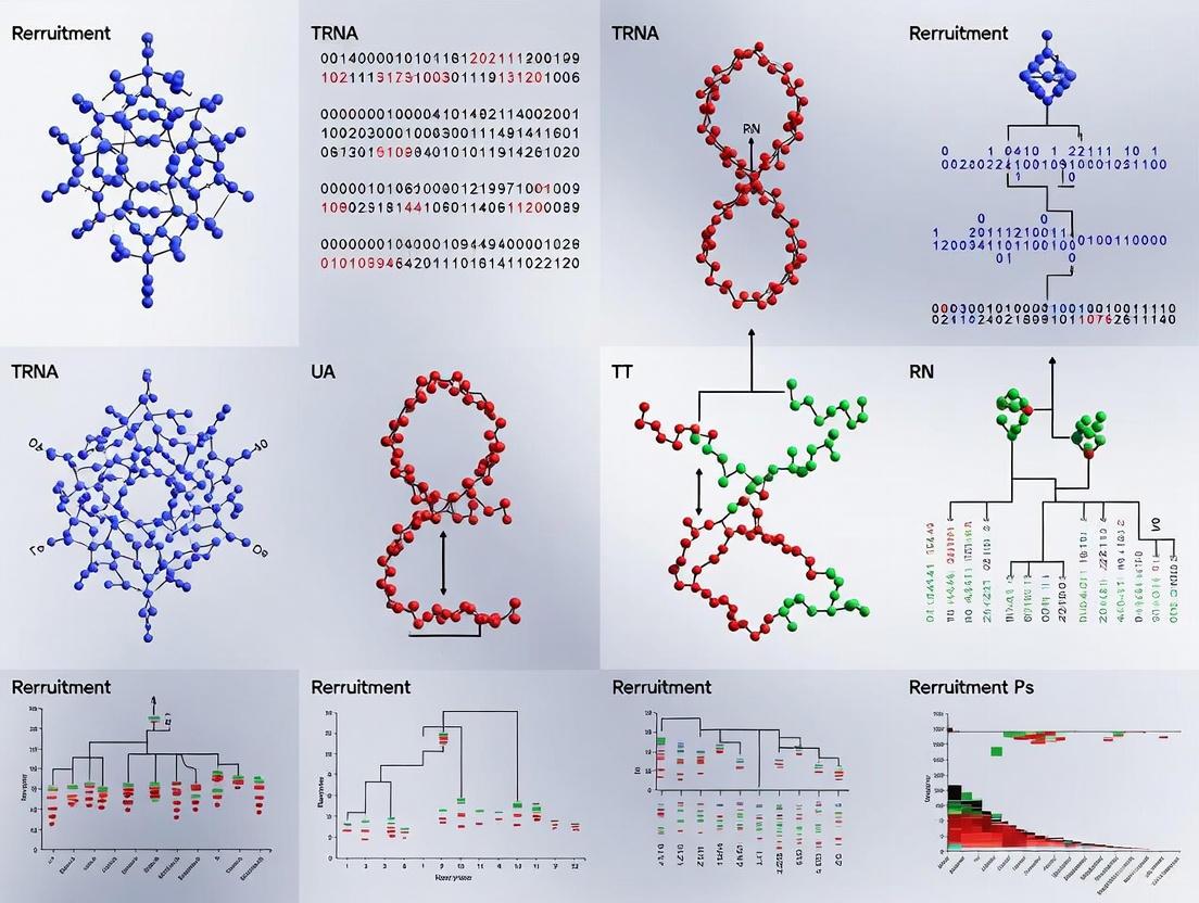
Transfer RNA molecules stand as remarkable relics in the evolutionary history of life, often termed "living fossils" due to their conservation across billions of years of evolution [1]. The tRNA scaffold preserves molecular information dating back to the origin of the translation system approximately 3 billion years ago, providing a window into early biological evolution [2] [3]. Their utility as phylogenetic markers stems from several unique properties: universal distribution across all domains of life, highly conserved secondary and tertiary structures, and functional conservation in translation despite sequence variation in specific positions.
The molecular fossil record preserved in tRNAs reveals evidence of the gradual evolution of the genetic code itself. Phylogenetic analyses suggest that amino acids were incorporated into the genetic code in a specific chronological sequence, with tyrosine, serine, and leucine representing some of the earliest amino acids (Group 1), followed by eight additional amino acids (Group 2), and finally the remaining standard amino acids (Group 3) [2]. This pattern of amino acid recruitment is preserved in the evolutionary relationships between different tRNA isoacceptors and their corresponding aminoacyl-tRNA synthetases.
Molecular Basis of tRNA Conservation
Structural Conservation
The canonical L-shaped three-dimensional structure of tRNA remains highly conserved across all domains of life [4]. This conserved architecture arises from two orthogonal helices consisting of the acceptor and anticodon domains, which fold independently to stabilize the overall structure through intramolecular interactions between the D- and T-arms [4]. Despite substantial sequence variation across tRNA genes, analysis of tRNA alignments shows that specific tRNA sequence motifs are highly conserved across multicellular eukaryotes [5].
Table 1: Conserved Structural Elements in tRNA Molecules
| Structural Element | Conservation Pattern | Functional Significance |
|---|---|---|
| Acceptor stem | 7 base pairs with specific non-Watson-Crick pairs | Amino acid attachment site |
| D-arm | Conserved GG sequence in D-loop | Tertiary stabilization |
| Anticodon stem | 5 base pairs with specific geometry | mRNA codon recognition |
| TΨC arm | GTΨC sequence highly conserved | Ribosomal binding |
| Tertiary interactions | Base triples between D/T loops | Maintain L-shaped fold |
The conservation extends throughout isoacceptors (tRNAs charging the same amino acid) and isodecoders (tRNAs with the same anticodon but different sequences), with some cases showing two sets of conserved isodecoders [5]. This structural conservation is maintained despite the potential for significant sequence variation, as the secondary structure must be preserved for proper tRNA function.
Sequence-Level Conservation
At the sequence level, tRNA genes demonstrate remarkable conservation across vast evolutionary distances. A comprehensive analysis of 50 plant species identified 28,262 tRNA genes with lengths ranging from 62-98 bp, showing strong conservation in gene length, intron length, GC content, and sequence identity [1]. tRNA gene length was found to peak at 72 bp and 82 bp across the plant species studied.
Non-Watson-Crick base pairs, particularly GoU pairs, represent important conserved elements in tRNA helical stems. Each of the four helical stems may contain one or more conserved GoU pairs, with some being amino acid-specific and potentially representing identity elements for cognate aminoacyl-tRNA synthetases [5]. The distribution of these conserved pairs reflects a balance between accommodating isotype-specific functions and those shared by all tRNAs essential for ribosomal translation.
tRNA Evolution and Phylogenetic Signal
Patterns of tRNA Gene Evolution
The evolution of tRNA genes occurs through several distinct mechanisms, with gene recruitment emerging as a common phenomenon in tRNA multigene family evolution [6]. This process involves a tRNA gene evolving horizontally from a copy of an alloacceptor tRNA gene in the same genome, typically accompanied by a single nucleotide substitution at the middle position of the anticodon. This substitution results in changes to both the tRNA's amino acid identity and the class of aminoacyl-tRNA synthetase involved in aminoacylation [6].
Tandem duplication represents another fundamental evolutionary force producing homologous tRNA clusters through localized genomic amplification. Studies have identified 578 identical tandemly duplicated tRNA gene pairs grouped into 410 clusters across plant species, with some clusters containing up to 26 tRNA genes [1]. In Arabidopsis thaliana, notable examples include a cluster of 27 tandemly duplicated tRNAPro genes and 27 consecutive tRNATyr–tRNATyr–tRNASer repeat units on chromosome 1 [1].
Table 2: Evolutionary Mechanisms in tRNA Gene Families
| Mechanism | Frequency | Evolutionary Impact |
|---|---|---|
| Gene recruitment | 11 cases in nuclear genomes of primates | Enables diversification of tRNA families |
| Tandem duplication | 578 pairs in 50 plant species | Generates homologous tRNA clusters |
| Anticodon modification | Most common recruitment mechanism | Changes tRNA aminoacylation specificity |
| Segmental duplication | Creates tRNA gene arrays | Amplifies specific tRNA isoacceptors |
Phylogenetic Information Content
The conserved nature of tRNA molecules makes them particularly valuable for resolving deep evolutionary relationships. Studies of mitochondrial DNA sequences including multiple tRNA genes (tRNAIle, tRNAGln, tRNAMet, ND2, tRNATrp, tRNAAla, tRNAAsn, tRNACys, tRNATyr) have provided well-resolved phylogenetic hypotheses with strong statistical support, demonstrating the utility of tRNA sequences for molecular systematics [7].
The pattern of diversification in tRNA molecules reveals important insights into the evolution of the genetic code. Phylogenetic analyses suggest that tRNA diversification occurred primarily through changes in the second base of the anticodon, leading to correlated changes in both the hydropathy of the anticodon and the class of aminoacyl-tRNA synthetase responsible for tRNA recognition [3]. This pattern indicates that the evolution of tRNAs and aminoacyl-tRNA synthetases occurred symmetrically, with Class I synthetases binding the acceptor stem from the minor groove side while Class II synthetases bind to the major groove side [3].
Experimental Methodologies for tRNA Analysis
tRNA Gene Identification and Annotation
The Genomic tRNA Database (GtRNAdb) represents the primary resource for genomic tRNA gene identification, containing alignments of tRNA genes based on the tRNAscan-SE prediction algorithm [5]. This covariance model-based approach classifies potential tRNA genes, assigning a bit score that measures how closely each tRNA resembles a prototypical tRNA. For phylogenetic analyses, researchers typically focus on tRNA genes with bit scores of at least 55, as scores below this threshold may indicate pseudogenes [5].
Protocol: High-Confidence tRNA Identification
- Download genome sequences from appropriate databases (e.g., Phytozome for plants)
- Annotate tRNA-coding genes using tRNAscan-SE with "-H" and "-y" parameters for eukaryotic tRNAs
- Filter for high-confidence sets using EukHighConfidenceFilter
- Calculate Minimum Fold Energy (MFE) for each tRNA gene using RNAFold
- Annotate secondary structures using visualization tools like VARNA GUI [1]
Advanced Sequencing Methods
Recent methodological advances have revolutionized tRNA analysis, particularly nanopore sequencing of intact aminoacylated tRNAs. The "aa-tRNA-seq" method uses chemical ligation to sandwich the amino acid of a charged tRNA between the body of the tRNA and an adaptor oligonucleotide, followed by high-throughput nanopore sequencing [8]. This approach enables simultaneous resolution of tRNA sequence, modification status, and aminoacylation at the single-molecule level.
Protocol: Nanopore Sequencing of Aminoacylated tRNAs
- Perform chemical ligation of aminoacyl-tRNA using HEI-catalyzed reaction at pH 5.5 for 30 minutes
- Purify ligation products via gel electrophoresis
- Enzymatically ligate 5' adapter using T4 RNA ligase 2 (RNL2)
- Prepare nanopore direct RNA sequencing library (ONT RNA004 chemistry)
- Sequence and analyze data using machine learning models to identify amino acid identities based on signal distortions [8]
Phylogenetic Analysis Workflow
Construction of phylogenetic trees from tRNA sequences requires specialized approaches to handle their conserved nature and limited length:
Protocol: tRNA Phylogenetic Reconstruction
- Compile tRNA gene sequences and perform multiple sequence alignment using tools like Multialin or ClustalO
- Identify best-fit evolutionary models using model testing software (e.g., IQ-TREE 2 model finder)
- Construct phylogenetic trees with high bootstrap replicates (≥1000)
- Calculate synonymous substitution rates (Kn/Ks) using KaKs_Calculator 3.0 with default parameters
- Perform comparative analysis of tree topologies and ancestral sequence reconstruction [1]
Figure 1: Experimental workflow for tRNA phylogenetic analysis, showing parallel paths for sequence-based and functional characterization.
The Scientist's Toolkit: Essential Research Reagents
Table 3: Key Research Reagents for tRNA Phylogenetic Analysis
| Reagent/Resource | Function | Application |
|---|---|---|
| tRNAscan-SE 2.0 | tRNA gene identification | Genomic annotation of tRNA genes |
| GtRNAdb 2.0 | Genomic tRNA database | Reference database for comparative analysis |
| MODOMICS | tRNA modification database | Catalog of posttranscriptional modifications |
| RNAFold | Secondary structure prediction | MFE calculation and structural modeling |
| Nanopore aa-tRNA-seq | Direct sequencing of charged tRNAs | Simultaneous analysis of sequence, modification, and aminoacylation |
| Flexizyme | In vitro aminoacylation | Experimental charging of synthetic tRNAs |
| HEI catalyst | Chemical ligation enhancement | Efficient adapter ligation for nanopore sequencing |
| KaKs_Calculator | Evolutionary rate calculation | Synonymous/non-synonymous substitution analysis |
Case Studies in tRNA Phylogenetics
Deep Evolutionary Relationships
Analysis of tRNA sequences has proven particularly valuable for resolving deep evolutionary relationships where standard markers provide insufficient signal. Studies of anguid lizards and related taxonomic families utilized 2001 aligned bases of mitochondrial DNA sequence including multiple tRNA genes (tRNAIle, tRNAGln, tRNAMet, tRNATrp, tRNAAla, tRNAAsn, tRNACys, tRNATyr) to generate a well-resolved phylogenetic hypothesis containing 1013 phylogenetically informative characters [7]. This analysis provided statistical support for major clades and enabled reconstruction of historical biogeographic patterns.
The evolutionary changes in mitochondrial tRNACys genes revealed distinctive patterns of D-stem reduction through successive base deletions in some lineages, contrasting with the parallel elimination of D-stems in other reptile groups through replication slippage [7]. These lineage-specific evolutionary patterns provide additional phylogenetic signal for resolving relationships.
Plant tRNA Evolution
Comprehensive analysis of 50 plant species representing eight divisions within the plant kingdom has revealed remarkable conservation of tRNA genes despite billions of years of evolutionary divergence [1]. The study identified 28,262 high-confidence tRNA-coding genes with strong conservation in gene length, intron length, GC content, and sequence identity. Notably, tRNA gene abundance showed no significant correlation with genome size (r = 0.18, p = 0.21), indicating other evolutionary forces maintain tRNA gene copy number.
Tandemly duplicated tRNA gene pairs with anticodons to proline were found to be widely distributed across 33 plant species, including both lower and higher plants, suggesting this arrangement represents an ancient evolutionary feature [1]. Different types of tandem duplication were identified, including double-, triple-, and quintuple-tRNA genes repeated varying numbers of times.
Amino Acid Recruitment Patterns
The pattern of tRNA evolution provides critical insights into the historical recruitment of amino acids into the genetic code. Phylogenetic analyses reveal that changes in the second base of the anticodon served as the primary mechanism for tRNA diversification, with these changes resulting in coordinated shifts in both the hydropathy of the anticodon and the class of aminoacyl-tRNA synthetase responsible for recognition [3].
This diversification pattern minimized binding of tRNAs from the same ancestry with aminoacyl-tRNA synthetases having similar recognition patterns, driving the co-evolution of tRNAs and their corresponding synthetases. The correlation between anticodon hydropathy and amino acid properties suggests that the genetic code evolved to maintain specific chemical relationships between codons and their encoded amino acids.
Figure 2: Evolutionary pathways in tRNA diversification, showing the relationship between molecular changes and functional consequences.
tRNA molecules serve as exceptional molecular fossils that preserve deep evolutionary signals dating back to the origin of the translation system. Their strong structural conservation, coupled with specific patterns of sequence evolution, provides powerful markers for resolving phylogenetic relationships across vast evolutionary timescales. The ongoing development of novel sequencing technologies, particularly nanopore-based methods for analyzing intact aminoacylated tRNAs, promises to further enhance our ability to extract phylogenetic information from these ancient molecules.
Future research directions include more comprehensive integration of tRNA sequence data with structural information and modification profiles, expansion of tRNA databases across underrepresented taxonomic groups, and development of more sophisticated evolutionary models that account for the unique constraints on tRNA evolution. As these methodological advances continue, tRNAs will remain indispensable tools for reconstructing the deep history of life and understanding the origin and evolution of the genetic code.
Aminoacyl-tRNA synthetases (aaRS) stand as essential molecular interpreters at the heart of genetic coding, performing the critical task of covalently linking amino acids to their cognate tRNAs with remarkable fidelity. These enzymes implement the genetic code by ensuring that the information encoded in mRNA sequences is accurately translated into corresponding protein sequences [9]. What makes this superfamily particularly remarkable is its fundamental bifurcation into two structurally and evolutionarily distinct classes—Class I and Class II—that share no significant sequence similarity or common structural fold [10] [11] [9]. This division represents one of the most ancient splits in enzyme evolution, predating the Last Universal Common Ancestor (LUCA) [9] [12]. The existence of two unrelated superfamilies performing the same essential biochemical function but employing different structural solutions has fascinated scientists for decades, prompting investigations into whether this duality emerged from an ancestral gene that coded for both classes simultaneously [10] [9]. Understanding the origin and evolution of these two superfamilies provides a unique window into the earliest stages of biological evolution and the emergence of the genetic code itself.
Structural and Functional Characteristics of Class I and Class II aaRS
The division between Class I and Class II aaRS is manifested through profound differences in their structural architectures, catalytic mechanisms, and approaches to substrate recognition. These differences extend beyond mere structural variation to encompass fundamentally different solutions to the problem of aminoacylation.
Architectural and Catalytic Differences
Table 1: Fundamental Structural and Catalytic Differences Between Class I and Class II aaRS
| Feature | Class I aaRS | Class II aaRS |
|---|---|---|
| Catalytic Fold | Rossmann dinucleotide binding fold [10] | Antiparallel β-sheet structure [10] |
| Active Site Location | Formed at interface between parallel β-strands and amino termini of two helixes [10] | Formed from antiparallel β-strands [10] |
| ATP Binding Motif | Backbone Brackets (backbone hydrogen bonds) [9] | Arginine Tweezers (pair of arginine residues) [9] |
| Approach to tRNA | Recognize tRNA acceptor stem from minor groove side [11] | Recognize tRNA acceptor stem from major groove side [11] |
| Characteristic Motifs | HIGH and KMSKS signatures [10] [11] | Motifs 1, 2, and 3 [11] |
Class I aaRS active sites assume a Rossmann dinucleotide binding fold first observed in lactate dehydrogenase and flavodoxin, while Class II active sites are constructed from antiparallel β-strands [10]. This fundamental architectural difference extends to their catalytic mechanisms, particularly in how they bind ATP. Class I enzymes utilize a "Backbone Brackets" mechanism where ATP is bound via backbone hydrogen bonds, while Class II enzymes employ "Arginine Tweezers" formed by a pair of arginine residues that create salt bridges toward the ATP molecule [9]. These different approaches to the same biochemical problem—amino acid activation—suggest independent evolutionary solutions that converged on the same functional outcome.
Amino Acid Specificity and Recognition Mechanisms
The division of labor between the two classes is non-random with respect to amino acid specificity. Class I typically handles larger and less polar amino acids, while Class II generally charges smaller and more polar amino acids [10]. This separation is remarkably consistent, with each class being responsible for exactly ten of the twenty canonical amino acids in most contemporary organisms [12]. The recognition mechanisms also differ substantially between the classes. Computational analysis of crystallographic structures has revealed that hydrogen bonds are the most prevalent interaction type in Class II aaRS (59.23% of interactions), whereas hydrophobic interactions dominate in Class I aaRS (44.60% of interactions) [9]. This difference in recognition strategy reflects the different chemical properties of their cognate amino acids and their distinct structural frameworks for constructing binding pockets.
Evolutionary Origins: The Rodin-Ohno Hypothesis and Complementary Coding
The most compelling explanation for the fundamental bifurcation of aaRS is the Rodin-Ohno hypothesis, which proposes that Class I and Class II aaRS originated from opposite strands of the same ancestral gene [10] [9] [12].
Evidence for Bi-directional Coding
The hypothesis, first proposed by Rodin and Ohno in the 1990s, emerged from observations of remarkable complementarity between conserved motifs in the two aaRS classes [10]. Multi-family sequence alignments revealed that codons for Class I signature motifs (PxxxxHIGH and KMSKS) were almost exactly anticodons for Class II Motifs 2 and 1, respectively [10]. This statistically significant, in-frame complementarity (with probabilities of 10⁻⁸ to 10⁻¹⁸ under the null hypothesis) suggests that contemporary aaRS superfamilies descended from a single ancestral gene where one strand coded for the ancestral Class I synthetase while the opposite strand coded for the ancestral Class II synthetase [10]. This arrangement represents a form of genetic economy where both strands of the ancestral gene were utilized to create functionally related but structurally distinct enzymes.
Structural Consequences of Complementary Coding
The inversion symmetry inherent in complementary coding of opposite DNA strands has recognizable consequences for protein secondary and tertiary structures [10]. The complementary relationship potentially explains the structural antipodality observed between Class I and Class II active sites—while Class I enzymes approach tRNA from the minor groove side, Class II enzymes approach from the major groove side [11]. This fundamental difference in interaction geometry may have originated from the complementary base-pairing relationships between the ancestral coding sequences. The bi-directional genetic coding of some of the oldest genes in the proteome places major limitations on the likelihood that any RNA World preceded the origins of coded proteins, suggesting instead that the genetic code arose from a peptide•RNA partnership [10].
Experimental Deconstruction and Phylogenetic Analysis
Modern experimental approaches have provided compelling support for the deep evolutionary relationships between Class I and Class II aaRS through both protein engineering and computational phylogenetics.
Urzyme and Protozyme Studies
Experimental deconstruction of contemporary aaRS has revealed parallel losses in catalytic proficiency at novel modular levels termed protozymes and Urzymes [10]. These represent progressively smaller and more ancestral forms of the enzymes that retain catalytic activity despite their simplified architectures. Structural biology of synthetase Urzymes suggests they are catalytically active molten globules, broadening the potential manifold of polypeptide catalysts accessible to primitive genetic coding [10]. This experimental approach demonstrates that even minimal versions of both Class I and Class II aaRS retain their distinct catalytic mechanisms, supporting the hypothesis that these mechanisms represent ancient and fundamental solutions to the aminoacylation problem.
Phylogenomic Reconstructions
Table 2: Key Findings from Phylogenomic Analyses of aaRS Evolution
| Study Type | Key Findings | Implications |
|---|---|---|
| Large-scale Genomic Analysis (2,500+ prokaryotic genomes) [11] | Horizontal gene transfer, gene duplication, and gene loss are more frequent than originally thought; some AARS often absent or have paralogs | Evolutionary history more complex than simple vertical inheritance; alternative pathways exist for aminoacylation |
| Bayesian Phylogenetic Analysis [12] | Identified 36 families of AARS catalytic domains; small structural modules (insertion modules) key to discriminating between amino acids | Piecewise assembly of aaRS through evolutionary time; code expansion via modular acquisition |
| tRNA Pool Analysis (UniFrac algorithm) [13] | tRNA pools cluster by organismal phylogeny despite individual tRNA horizontal transfer | Overall pattern of tRNA evolution tracks universal phylogeny |
Recent phylogenetic reconstructions of extant AARS genes, enhanced by analyzing modular acquisitions, reveal six AARS with distinct bacterial, archaeal, eukaryotic, or organellar clades, resulting in a total of 36 families of AARS catalytic domains [12]. These analyses show that small structural modules—insertion modules (IM)—that differentiate one AARS family from another played pivotal roles in discriminating between amino acid side chains, thereby expanding the genetic code and refining its precision [12]. The most probable evolutionary route for an emergent amino acid type to establish a place in the code was by recruiting older, less specific AARS, rather than adapting contemporary lineages—a process termed retrofunctionalisation [12].
Diagram: Proposed evolutionary trajectory from an ancestral bi-directional gene to modern Class I and Class II aaRS through intermediate forms including Urzymes and modular acquisitions.
Methodologies for Experimental Investigation
Phylogenetic Reconstruction Protocols
Detailed Bayesian phylogenetic analysis of aaRS evolution involves multiple carefully orchestrated steps [12]. The protocol begins with building sequence alignments using annotated AARS sequence entries from GenBank, selecting taxonomically representative samples for each family. Protein structures are predicted with AlphaFold v2.3.0 and secondary structures defined using DSSP v3.0.0. Pairwise structural alignments are generated by DeepAlign, followed by per-family multiple sequence alignments using 3DCOMB with refinement of contiguous regions lacking secondary structure using ClustalW based on primary sequence [12]. Bayesian phylogenetic inference is performed using BEAST v2.7.3 with two independent Markov chain Monte Carlo chains run for each class, assessing convergence by confirming effective sample sizes over 200 using Tracer v1.7 [12]. This comprehensive approach integrates both sequence and structural information to reconstruct evolutionary relationships.
Structural Analysis of Binding Sites
The characterization of amino acid recognition mechanisms involves computational analysis of crystallographic structures from the Protein Data Bank (PDB) [9]. Researchers typically use the Protein-Ligand Interaction Profiler (PLIP), a rule-based tool for characterizing non-covalent interaction patterns in protein-ligand complexes [9]. The analytical workflow involves identifying all available structures of aaRSs co-crystallized with their amino acid ligands, selecting each protein chain containing a catalytic aaRS domain, and systematically annotating interaction types (hydrogen bonds, hydrophobic interactions, salt bridges, π-stacking, and metal complexes) [9]. This approach allows for quantitative comparison of recognition strategies across different aaRS classes and subclasses, revealing how specificity is achieved through distinct physicochemical solutions.
tRNA Gene Identification and Analysis
The reliable identification of functional tRNA genes in genomes containing numerous tRNA-derived repetitive elements requires a multi-step bioinformatics approach [14]. The standard protocol involves initial analysis using tRNAscan-SE to identify putative tRNA genes, followed by filtering with RepeatMasker to identify and remove repetitive elements, particularly short interspersed elements (SINEs) containing tRNA-derived sequences [14]. Comparative genomics is then employed using multiple vertebrate genomes to identify highly conserved tRNA genes, typically applying a 95% sequence similarity threshold to distinguish functional genes from neutrally evolving repetitive elements [14]. This approach successfully reduces thousands of putative tRNA predictions to a refined set of likely functional genes.
Table 3: Key Research Reagents and Computational Tools for aaRS and tRNA Research
| Resource Category | Specific Tools/Reagents | Primary Function | Application Context |
|---|---|---|---|
| Structure Prediction | AlphaFold v2.3.0 [12] | Protein structure prediction | Phylogenetic analysis of aaRS catalytic domains |
| Structural Alignment | DeepAlign [12], 3DCOMB [12] | Pairwise and multiple structural alignment | Identifying conserved structural modules in aaRS |
| Phylogenetic Analysis | BEAST v2.7.3 [12], Tracer v1.7 [12] | Bayesian evolutionary analysis | Dating evolutionary events in aaRS history |
| tRNA Identification | tRNAscan-SE [14] | Genome-wide tRNA detection | Initial identification of tRNA genes in genomes |
| Interaction Analysis | Protein-Ligand Interaction Profiler (PLIP) [9] | Characterization of non-covalent interactions | Analyzing amino acid binding sites in aaRS |
| Sequence Analysis | MEME software [11] | Motif discovery | Identifying conserved motifs in aaRS classes |
| Structure Visualization | PV [12] | Molecular visualization | Display of predicted protein structures |
Implications for Genetic Code Evolution and Modern Applications
The deep evolutionary history of aaRS bifurcation has profound implications for understanding the origin and evolution of the genetic code, with direct relevance to modern biotechnology and drug development.
The division of aaRS into two classes appears to have been essential for the gradual expansion of the genetic code. The model emerging from phylogenetic studies shows "a tendency for less elaborate enzymes, with simpler catalytic domains, to activate amino acids that were not synthesised until later in the evolution of the code" [12]. This suggests that the binary choice implemented by the two aaRS classes provided a flexible framework for incorporating new amino acids as biosynthetic pathways evolved. The existence of two fundamentally different structural solutions to aminoacylation may have allowed the genetic code to cover a broader range of amino acid physicochemical properties than would have been possible with a single structural framework [9].
From a practical perspective, understanding aaRS evolution and specificity has direct applications in antibiotic development and synthetic biology. Numerous microorganisms have evolved low molecular weight toxins that target essential AARS enzymes in other microorganisms, with commercial antibiotics like mupirocin (which targets IleRS) representing prominent examples [11]. The discovery that divergent AARS paralogs confer resistance to natural AARS inhibitors has been documented for MetRS, TrpRS, IleRS and SerRS paralogs, providing both potential antibiotic targets and resistance mechanisms [11]. Furthermore, in synthetic biology, the manipulation and extension of the genetic code for incorporating unnatural amino acids relies heavily on understanding how AARS specificity is determined, making evolutionary studies of aaRS directly relevant to engineering enzymes with novel properties [11].
The bifurcation of aminoacyl-tRNA synthetases into Class I and Class II superfamilies represents one of the most fundamental and ancient divisions in biology, predating the last universal common ancestor. The Rodin-Ohno hypothesis of complementary coding from opposite strands of an ancestral gene provides a compelling explanation for this duality, with substantial support from structural studies, phylogenetic analyses, and experimental deconstruction of modern enzymes to their ancestral Urzyme forms. The piecewise assembly of aaRS through the acquisition of structural modules, particularly insertion modules that enhanced amino acid discrimination, enabled the gradual expansion and refinement of the genetic code. This evolutionary history not only illuminates the deep past of biological information processing but also provides valuable insights for contemporary applications in antibiotic development and synthetic biology, where understanding and engineering aaRS specificity remains a central challenge.
The standard 20-amino acid alphabet is a conserved feature of life, yet evidence from phylogenomics, prebiotic chemistry, and experimental evolution indicates it expanded from a smaller, primordial set. Phylogenetic analyses of transfer RNA (tRNA) and aminoacyl-tRNA synthetases (aaRS) reveal a co-evolutionary pattern where the diversification of these molecules directly correlated with the incorporation of new amino acids into the genetic code [15]. Furthermore, data from astrochemistry and simulated prebiotic environments suggest that a subset of the modern amino acids was likely available for early life, supporting the hypothesis of a reduced initial alphabet [16]. This whitepaper synthesizes phylogenetic, biochemical, and synthetic biological data to explore the evidence for a simpler genetic alphabet and the mechanisms of its expansion, providing a framework for understanding this fundamental evolutionary transition.
The universal presence of a 20-amino acid alphabet across the tree of life is a testament to its evolutionary optimization. However, this alphabet is not static; the existence of a 21st genetically encoded amino acid, selenocysteine, and a 22nd, pyrrolysine, demonstrates the potential for natural expansion [17]. The central question is not whether the alphabet could change, but what forces drove its evolution to the current standard of 20 and whether it originated from a more limited set.
The "metabolism first" hypothesis suggests that early life operated with a reduced set of amino acids, with more complex members being biosynthetically derived later [16]. This is supported by analyses of prebiotic chemistry, which show that meteorites like Murchison contain a limited number of proteinogenic amino acids (e.g., glycine, alanine, and aspartic acid), while others such as lysine, arginine, and histidine are notably absent [16] [17]. The order of amino acid entry into the genetic code, as deduced from biosynthetic pathways and genomic analyses, provides a phylogenetic roadmap for this expansion [17]. This report details the phylogenetic and experimental evidence for this model, leveraging insights from tRNA evolution and modern synthetic biology.
Phylogenomic analysis of tRNA and aaRS diversification
A monophyletic origin and anticodon-driven diversification
tRNA molecules are central to interpreting the genetic code, and their evolutionary history provides critical insights into the alphabet's expansion. A prevailing view is that tRNAs have a monophyletic origin, with all modern tRNAs descending from a universal ancestral molecule [15]. Strong evidence for this includes the high conservation of tRNA structure, specific sequence regions, and the position of introns across diverse organisms [15].
The diversification of this ancestral tRNA is characterized by changes in the second base of the anticodon. This pattern is significant because the second base is a major determinant of the amino acid's hydropathy. A change at this position typically alters the hydropathy of the anticodon, which in turn correlates with the physical-chemical properties of the corresponding amino acid [15]. This suggests an early, direct chemical relationship between anticodons and their amino acids. The driving force behind this diversification was likely the need to minimize mischarging by aaRS as the alphabet grew, ensuring that tRNAs from the same ancestral group were distinguished by aaRS with different recognition patterns [15].
Table 1: Evidence for tRNA and aaRS Co-evolution
| Evidence Type | Key Finding | Implication for Genetic Code Expansion |
|---|---|---|
| Anticodon Mutation | Changes in the second base of the anticodon alter tRNA hydropathy [15]. | Enabled the coding of amino acids with novel chemical properties. |
| aaRS Class Divergence | Class I and Class II aaRS bind the acceptor stem from opposite grooves [15]. | Symmetrical co-evolution ensured accurate tRNA recognition as the alphabet expanded. |
| Experimental Evolution | Yeast deleting a tRNA-AGG gene evolved a mutation in a tRNA-AGA gene to AGG, restoring growth [18]. | Demonstrates anticodon switching is a rapid, adaptive mechanism to meet novel translational demands. |
The role of anticodon mutations in adaptive evolution
The evolution of the tRNA pool is not merely a historical relic but an ongoing adaptive process. Experimental evolution studies in Saccharomyces cerevisiae have directly demonstrated that anticodon mutations are a key mechanism for adapting to new translational demands. When a yeast strain was engineered to lack the gene for a rare arginine tRNA (corresponding to the AGG codon), it initially grew slowly [18]. After evolving for 200 generations, the population recovered its growth rate by acquiring a point mutation in the gene for another arginine tRNA (corresponding to the AGA codon), changing its anticodon to match the deleted AGG-specific tRNA [18]. This shows that anticodon switching is a direct and efficient evolutionary solution to correct an imbalance between tRNA supply and codon demand.
A systematic genomic analysis across hundreds of species confirmed that this mechanism is not confined to the laboratory. Anticodon mutations have occurred throughout the tree of life, highlighting their general role in the evolution of the translational machinery [18].
Figure 1: Adaptive evolution via anticodon switching. A deletion of a tRNA gene creates a translational deficit, which is compensated for by an anticodon mutation in a different tRNA gene, restoring growth.
Prebiotic chemistry and the case for a reduced early alphabet
The theory of a reduced early alphabet is strongly supported by prebiotic chemistry. Analysis of carbonaceous meteorites, such as the Murchison meteorite, has revealed the presence of over 80 amino acids, but only a limited subset of the standard 20 [16]. Twelve proteinogenic amino acids have been identified in these extraterrestrial sources, including glycine, alanine, and valine, while others like arginine, lysine, and histidine have not been found [16]. This suggests that the early Earth had access to a non-random, restricted pool of amino acids of both terrestrial and extraterrestrial origin.
Laboratory experiments simulating early Earth conditions, such as Miller-Urey spark discharge experiments, further support this. These experiments produce a similar subset of amino acids, with more complex ones like cysteine and methionine only appearing under specific modified conditions [16]. The absence of certain amino acids in prebiotic simulations and meteorites, coupled with their biosynthetic complexity, indicates they were likely incorporated into the genetic code at a later stage through evolutionary innovation.
Table 2: Evidence for a Reduced Early Amino Acid Set from Prebiotic Chemistry
| Amino Acid | Detected in Murchison Meteorite | Produced in Classic Miller-Urey Experiment | Inferred Status in Early Alphabet |
|---|---|---|---|
| Glycine | Yes [16] | Yes [16] | Early |
| Alanine | Yes [16] | Yes [16] | Early |
| Valine | Yes [16] | Yes [16] | Early |
| Aspartic Acid | Yes [16] | Yes [16] | Early |
| Serine | Yes [16] | Yes (in variants) [16] | Early |
| Lysine | No [17] | No | Late |
| Arginine | No [17] | No | Late |
| Histidine | No [17] | No | Late |
| Cysteine | No (or debated) | Yes (in variants with H₂S) [16] | Late |
| Methionine | No (or debated) | Yes (in variants with H₂S) [16] | Late |
Experimental expansion of the genetic code
Modern synthetic biology approaches
Synthetic biology provides direct experimental evidence that the genetic code is expandable. Traditional methods have relied on repurposing stop codons (e.g., TAG, TGA) to encode non-canonical amino acids (ncAAs). This approach utilizes an orthogonal tRNA/aaRS pair that charges a ncAA and recognizes the stop codon. However, competition with release factors often limits incorporation efficiency to less than 5% [19].
A more efficient strategy involves repurposing rare sense codons. Because rare codons have low corresponding tRNA abundance in the cell, an introduced orthogonal tRNA faces less competition, leading to higher incorporation yields [19]. For example, in human cell lines, the TCG codon (a rare serine codon) was identified as the most effective for incorporating a ncAA with minimal disruption to cellular proteins, achieving incorporation efficiencies above 80% [19]. This method has been successfully extended to incorporate multiple different ncAAs simultaneously by repurposing several rare codons (e.g., TCG, TAG, TGA) within a single gene [19].
Detailed protocol: Incorporating non-canonical amino acids via rare codon recoding
This protocol outlines the key steps for efficient ncAA incorporation in mammalian cells, as developed by Lin et al. [19].
1. Identification of Rare Codons:
- Method: Perform RNA sequencing (RNA-seq) on the target cell line (e.g., HEK293T) to transcriptome-wide codon usage frequency.
- Output: Generate a ranked list of the least used codons. In human cells, these include TCG, TAG, TGA, and others [19].
2. Selection of Optimal Codon for Recoding:
- Method: Clone the gene for a reporter protein (e.g., enhanced green fluorescent protein, eGFP), introducing the candidate rare codon at a permissive site.
- Procedure: Transfect cells with the modified reporter construct and an orthogonal tRNA/aaRS pair that is specific for the desired ncAA and the rare codon.
- Validation: Quantify ncAA incorporation efficiency via fluorescence (for eGFP) or Western blot. Assess background incorporation by staining the cellular proteome for the traceable ncAA. The TCG codon consistently shows high efficiency and low background [19].
- Note: Incorporation efficiency is context-dependent and can vary from 5% to 99% based on the surrounding nucleotide sequence [19].
3. Multi-site Incorporation:
- Method: Introduce different rare codons (e.g., TCG, TAG, TGA) at distinct positions in the target gene.
- Procedure: Co-express multiple orthogonal tRNA/aaRS pairs, each charged with a distinct ncAA and specific to one of the repurposed rare codons.
- Output: A single polypeptide chain containing multiple, distinct ncAAs with unique chemical properties [19].
Figure 2: Workflow for incorporating non-canonical amino acids via rare codon recoding.
Research reagent solutions for genetic code expansion
Table 3: Essential Reagents for Genetic Code Expansion Experiments
| Reagent / Tool | Function | Application Example |
|---|---|---|
| Orthogonal tRNA/aaRS Pair | Charges a specific non-canonical amino acid and recognizes a designated codon (stop or rare sense codon) without cross-reacting with endogenous host systems. | An orthogonal pair from archaea or engineered in vitro is used to incorporate a photocrosslinking amino acid in response to the TCG codon in human cells [19]. |
| Reporter Gene Constructs (e.g., eGFP) | A genetically modified gene containing the target codon at a specific site; allows for rapid quantification of incorporation efficiency. | An eGFP gene with a TCG codon at a permissive site is used to screen and optimize ncAA incorporation efficiency [19]. |
| Non-Canonical Amino Acid (ncAA) | The novel chemical building block to be incorporated into the protein. Can possess unique reactivity (e.g., cross-linkers, fluorophores). | Amino acids with ketone, azide, or alkyne functional groups for bioorthogonal conjugation post-translation [19]. |
| Recoded Synonymous Genes | Alternative gene sequences (e.g., alt1l.e., alt2l.e.) that encode the same protein but use a different codon schema to explore a larger mutational landscape. | Used in directed evolution of integrases to access beneficial mutations not available in the wild-type sequence space [20]. |
Discussion and future directions
The convergent evidence from phylogenomics, prebiotic chemistry, and synthetic biology presents a compelling case for the expansion of the genetic alphabet from a reduced initial set. The co-diversification of tRNAs and aaRS, driven by anticodon changes, provided the mechanistic pathway for incorporating new amino acids with diverse chemical properties [15] [18]. The prebiotic availability of a subset of amino acids likely constrained the initial composition of the code, with more complex amino acids being added through biosynthetic pathways as life evolved [16] [17].
From a therapeutic perspective, the ability to expand the genetic code experimentally opens new frontiers in drug development. Proteins with site-specifically incorporated ncAAs can be used to create:
- Antibody-Drug Conjugates (ADCs): Enabling high-yield, homogeneous conjugation of cytotoxic drugs to antibodies directly during biosynthesis [19].
- Biologics with Enhanced Properties: Incorporating amino acids with unique chemistries (e.g., cross-linkers, stable isotopes) to improve protein stability, half-life, or functionality [19].
- Probes for Studying Protein Interactions: Incorporating photo-reactive or bio-orthogonal amino acids to map protein-protein interactions and complex cellular pathways.
Future research will continue to refine our understanding of the primordial amino acid set and optimize the tools for genetic code expansion, further blurring the line between what life uses and what chemistry allows.
The aminoacyl-tRNA synthetases (aaRS) represent a unique paradigm in molecular evolution, serving as the essential enzymes that interpret the genetic code by catalyzing the attachment of specific amino acids to their cognate tRNAs. These enzymes form two distinct, apparently unrelated superfamilies (Class I and Class II) that appear to have originated from opposite strands of the same ancestral gene [10]. This bi-directional genetic coding hypothesis, first proposed by Rodin and Ohno, suggests that the contemporary aaRS superfamilies descended from a single ancestral gene where one strand encoded the ancestral Class I synthetase while the opposite strand encoded the ancestral Class II synthetase [10]. The statistical support for this hypothesis is remarkably strong, with probabilities of 10⁻⁸ – 10⁻¹⁸ for the observed alignments under the null hypothesis [10].
The division of labor between Class I and Class II aaRS is non-random: Class I aaRS typically charge larger, less polar amino acids, while Class II aaRS generally charge smaller, more polar amino acids [10] [21]. This fundamental partition reflects deeper principles about how amino acids behave in water and in protein folding, suggesting that the aaRS were intimately involved in shaping the genetic code itself [10]. The modular architecture of aaRS, characterized by progressive levels of structural organization from compact catalytic units to complex multi-domain enzymes, provides a unique window into the earliest evolution of coded protein synthesis and challenges the traditional RNA World hypothesis [10] [22].
Theoretical Foundation: The Bi-directional Coding Hypothesis
Historical Context and Evidentiary Basis
The Rodin-Ohno hypothesis emerged from observations of striking complementarity between Class I and Class II active-site motifs. Multi-family sequence alignments revealed that codons for Class I signature sequences (PxxxxHIGH and KMSKS) were nearly exact anticodons for Class II Motifs 2 and 1, respectively [10]. This in-frame complementarity suggested an ancestral gene where both strands were functional coding sequences for the two synthetase classes [10]. Subsequent experimental work has substantially strengthened this hypothesis through several key findings:
- Phylogenetic metrics based on middle base-pairing frequencies in sense/antisense alignments provide deeper evolutionary insights than traditional multiple sequence alignments [10] [21]
- Structural inversion symmetry between Class I and II active sites reflects the inversion symmetry of complementary coding strands [10]
- tRNA coding elements record information about how amino acids behave in water, connecting the operational RNA code to protein folding properties [10]
Implications for the Origin of Genetic Coding
The bi-directional coding hypothesis has profound implications for understanding the origin of biological information systems. The aaRS represent a unique, reflexive interface between genes and gene products - they are themselves translated according to the genetic code, yet once folded, they enforce that same code by aminoacylating tRNAs [10]. This self-referential relationship suggests that the earliest coding systems likely emerged as a collaboration between ancestral peptides and RNAs rather than from an RNA-only world [10] [22]. The catalytic capabilities of relatively simple ancestral peptides challenge the necessity of sophisticated ribozymes for initiating translation, pointing instead to a Peptide•RNA World where both polymers cooperated from the earliest stages [22].
Table 1: Core Evidentiary Support for the Bi-directional Coding Hypothesis
| Evidence Type | Key Findings | Implications |
|---|---|---|
| Sequence Complementarity | Codons for Class I motifs are anticodons for Class II motifs | Common ancestral gene for both aaRS classes |
| Structural Phylogenetics | Inversion symmetry between Class I and II active sites | Opposite strand coding preserved in structural features |
| Catalytic Modularity | Parallel deconstruction reveals similar protozyme/urzyme organization | Common evolutionary trajectory for both classes |
| tRNA Recognition | Operational RNA code in acceptor stems predates anticodon code | Early aaRS-tRNA coevolution shaped the genetic code |
Hierarchical Deconstruction: Urzymes and Protozymes
Defining the Modular Architecture
Experimental deconstruction of both Class I and II aaRS has revealed a hierarchical modular architecture characterized by several distinct levels of organization:
- Full-length synthetases: Contemporary enzymes with complete complement of domains for catalysis, editing, and tRNA recognition
- Catalytic domains: Core folds retaining amino acid activation and tRNA acylation capabilities
- Urzymes: ~120-130 residue constructs containing the essential catalytic machinery [22] [23]
- Protozymes: ~46 residue segments containing ATP-binding sites [23]
This modular hierarchy is conserved across both aaRS classes, despite their extensive structural differences. Class I aaRS active sites assume a Rossmann dinucleotide binding fold with parallel β-strands, while Class II active sites are formed from antiparallel β-strands [10]. Yet both classes yield functionally analogous urzymes and protozymes when deconstructed, supporting their parallel evolutionary trajectories from simpler ancestral peptides.
Catalytic Proficiency of Minimal Constructs
Remarkably, both Class I and II urzymes retain significant catalytic capabilities despite their dramatically reduced size. Quantitative analyses reveal:
- Amino acid activation: Urzymes accelerate cognate amino acid activation by ATP ~10⁸-fold compared to uncatalyzed rates [22]
- tRNA acylation: Class I TrpRS and Class II HisRS urzymes acylate tRNA 10⁶ times faster than the uncatalyzed rate of nonribosomal peptide bond formation [22]
- Transition state stabilization: Urzymes exhibit ~60% of contemporary catalytic proficiencies despite their simplicity [22]
Table 2: Catalytic Parameters of Representative Urzymes Compared to Full-length Enzymes
| Catalyst | Reaction | kcat/Km (s⁻¹M⁻¹) | Rate Enhancement | Transition State Stabilization (kcal/mol) |
|---|---|---|---|---|
| Uncatalyzed reference | Amino acid activation | 2.70×10⁻⁸ | 1× | 10.4 |
| TrpRS Urzyme | Amino acid activation | 1.5 | 5.6×10⁷ | -0.3 |
| Full-length TrpRS | Amino acid activation | 1.8×10⁴ | 6.7×10¹¹ | -5.9 |
| Uncatalyzed reference | tRNA acylation | 8.00×10⁻⁵ | 1× | 5.6 |
| TrpRS Urzyme | tRNA acylation | 3.0×10² | 3.8×10⁶ | -3.4 |
| Full-length TrpRS | tRNA acylation | 8.9×10⁵ | 1.1×10¹⁰ | -8.2 |
The unexpected catalytic proficiency of urzymes suggests they are themselves highly evolved descendants of even simpler ancestral peptides [22]. Their catalytic properties, combined with sense/antisense coding and modular architecture, imply considerable prior protein-tRNA co-evolution before the emergence of modern aaRS [22].
Experimental Methodologies and Protocols
Urzyme Construction and Expression
The experimental pipeline for studying aaRS urzymes involves multiple stages of protein engineering and biochemical characterization:
Figure 1: Experimental workflow for constructing and characterizing aaRS urzymes
Key methodological considerations for urzyme studies include:
- Solubility challenges: Urzymes typically require expression as maltose-binding protein (MBP) fusions due to exposed hydrophobic patches and inherent instability [23]
- Active site titration: Essential for accurate kinetic parameter determination due to variable fractions of active enzyme (typically 0.35-0.7) [22]
- tRNA preparation: Cognate tRNAs must be prepared with care, as aminoacylatability often ranges from 0.2-0.55 [23]
Kinetic Characterization Assays
Multiple complementary assays are employed to authenticate urzyme catalytic activities and eliminate potential artifacts:
Amino acid activation is typically measured via the ATP-PP₁ exchange assay, which monitors the incorporation of ³²P from labeled pyrophosphate into ATP in the presence of cognate amino acid [23]. For single-turnover studies, active site titrations measuring burst sizes in the time dependence of ³²P transfer from the γ-position of ATP provide crucial validation [23].
tRNA aminoacylation is assessed using ³²P-labeled tRNA substrates, with reaction products separated by thin-layer chromatography and quantified by phosphor imaging analysis [22]. The fraction of acylated A76 base provides a direct measure of aminoacylation efficiency.
Non-canonical activities must also be considered, as urzymes may exhibit promiscuous phosphoryl-transfer reactions. For example, LeuAC catalyzes production of ADP in addition to canonical aminoacylation, suggesting conformational flexibility in ATP binding sites [23].
Research Reagents and Experimental Tools
Table 3: Essential Research Reagents for aaRS Urzyme Studies
| Reagent/Category | Specific Examples | Function/Application | Technical Considerations |
|---|---|---|---|
| Expression Systems | MBP fusion vectors, TEV protease sites | Enhance urzyme solubility and purification | TEV cleavage often essential for full activity [23] |
| Activity Assays | ATP-PP₁ exchange, active site titration, tRNA aminoacylation | Quantify catalytic parameters | Multiple complementary assays required for validation [22] [23] |
| Site-directed Mutagenesis | Active site signature motifs (HIGH, KMSKS) | Establish catalytic mechanisms | Conservative mutations often retain partial function [23] |
| tRNA Preparation | In vitro transcription, 3'-end labeling | Generate substrates for aminoacylation | Aminoacylatability typically 20-55% [22] [23] |
| Structural Analysis | X-ray crystallography, NMR | Determine urzyme structures | Urzymes may represent "molten globule" states [21] |
Structural Biology of Ancestral Catalytic Modules
Structural studies of aaRS urzymes reveal they likely represent catalytically active molten globules - compact but conformationally dynamic states that broaden the potential manifold of polypeptide catalysts accessible to primitive genetic coding [21]. This structural plasticity has important implications for early evolution:
- Conformational diversity may have enabled primitive peptides to catalyze multiple reactions with modest specificity
- Modular assembly of discrete structural elements progressively enhanced specificity and efficiency
- tRNA coevolution shaped the structural refinement of urzymes toward contemporary enzymes
The LeuAC urzyme derived from Pyrococcus horikoshii leucyl-tRNA synthetase exemplifies the structural organization of these minimal catalysts. Despite containing only the A (protozyme) and C (KMSKS domain) modules and lacking the B (CP1 insertion) and D (anticodon-binding) domains, LeuAC authentically catalyzes both amino acid activation and tRNALeu aminoacylation [23]. Mutation of the three active-site lysine residues to alanine causes significant but modest reduction in both activities, confirming the role of these residues in catalysis while suggesting additional stabilizing interactions [23].
Implications for the Evolution of the Genetic Code
The modular evolution of aaRS provides compelling insights into how the genetic code might have emerged through progressive stages of refinement. Phylogenomic analysis of dipeptide sequences across 1,561 proteomes supports an evolutionary chronology where an early operational RNA code in the acceptor arm of tRNA preceded implementation of the standard genetic code in the anticodon loop [24]. This timeline reveals:
- Early emerging dipeptides containing Leu, Ser, and Tyr, followed by those containing Val, Ile, Met, Lys, Pro, and Ala [24]
- Synchronous appearance of dipeptide-antidipeptide sequences, supporting ancestral duality of bidirectional coding [24]
- Late development of protein thermostability, suggesting origins in mild Archaean environments [24]
The aaRS urzymes represent crucial experimental models for understanding how peptide•RNA partnerships could have established the first coding systems without requiring pre-existing sophisticated ribozymes [22]. Their catalytic proficiency demonstrates that relatively simple peptides could have catalyzed key reactions in translation, while their modular architecture provides a plausible pathway for progressive evolutionary refinement.
The experimental deconstruction of aaRS into urzymes and protozymes has established a new paradigm for understanding the modular evolution of these essential enzymes and their role in origin of genetic coding. The striking parallel between Class I and II aaRS, extending from their bi-directional genetic coding to their hierarchical modular organization, provides compelling evidence for their descent from a common ancestral gene. The catalytic capabilities of urzymes demonstrate that relatively simple peptides could have catalyzed critical steps in translation, challenging the requirement for an RNA World preceding the emergence of coded protein synthesis.
Future research directions in this field include:
- Expanding the urzyme repertoire to include representatives from additional aaRS families
- Structural characterization of urzyme•tRNA complexes to understand primitive recognition mechanisms
- In vitro evolution experiments to explore potential evolutionary pathways from urzymes to contemporary enzymes
- Integration with origins of life models that incorporate peptide•RNA partnerships from the earliest stages
The study of aaRS modular evolution continues to provide profound insights into one of biology's most fundamental processes, revealing how molecular complexity can emerge through the progressive assembly and refinement of simple functional modules.
Transfer RNA (tRNA) pools, comprising the complete set of tRNA genes in a genome, serve as evolutionary records that extend beyond their canonical role in translation. This technical guide explores the premise that tRNA complements function as genomic signatures for phylogenomic analysis. We synthesize evidence from studies on organisms spanning yeast, plants, and mammals, demonstrating how quantitative features of tRNA pools—including gene copy number, sequence conservation, anticodon distribution, and genomic organization—provide a robust framework for inferring evolutionary relationships. The integration of these features with mechanistic insights into tRNA gene regulation and function offers a powerful approach for reconstructing organismal phylogeny and understanding the evolutionary recruitment of amino acids.
The nuclear genome of an organism encodes a full complement of tRNA genes, collectively known as its tRNA pool. Historically studied for its role in determining translation efficiency and fidelity, the tRNA pool is increasingly recognized as a rich source of phylogenetic information. The fundamental hypothesis is that the characteristics of these pools are not random but are shaped by evolutionary pressures, leaving distinct signatures that can be traced across lineages.
The architecture of tRNA pools is defined by several quantifiable parameters: the absolute number of tRNA genes, their sequence identity, their genomic organization into clusters or singleton genes, and the distribution of isoacceptors (tRNAs with different anticodons carrying the same amino acid) and isodecoders (tRNAs with the same anticodon but different body sequences) [25] [26]. The conservation of these features, driven by functional constraints on translation and beyond, makes them excellent markers for deep evolutionary studies. Furthermore, the evolution of tRNA genes through mechanisms such as tandem duplication provides a record of genomic events that can be used to delineate phylogenetic relationships [27].
Architectural Features of tRNA Pools and Their Quantitative Analysis
tRNA Gene Copy Number and Conservation
The total number of tRNA genes varies significantly between species, but within phylogenetic groups, patterns of expansion and contraction emerge.
Table 1: Variation in tRNA Gene Abundance Across Species
| Species Group | Representative Species | Total tRNA Genes | Notes | Primary Source |
|---|---|---|---|---|
| Angiospermae | Camelina sativa | 1,451 | High gene count observed | [27] |
| Angiospermae | Gossypium hirsutum | >1,000 | High gene count observed | [27] |
| Bryophyta | Ceratodon purpureus | >1,000 | High gene count observed | [27] |
| S. cerevisiae | - | 275 | Systematic deletion library created | [25] |
| Rhodophyta | Porphyra umbilicalis | 56 | Among the lowest abundances found | [27] |
A comprehensive study of 50 plant species identified 28,262 high-confidence tRNA genes, revealing that tRNA gene abundance shows a weak, non-significant positive correlation with genome size (r=0.18) [27]. This indicates that tRNA gene number is not a simple function of genome size but is likely under specific selective pressures. The length of these tRNA genes is highly conserved, ranging from 62 to 98 base pairs, with peaks at 72 bp and 82 bp [27].
Sequence Identity and Structural Conservation
Sequence analysis reveals a high degree of conservation in tRNA genes. In plants, the sequence identity of tRNA genes, particularly in the acceptor stem and anticodon loop, is notably high, supporting the concept of tRNAs as "living fossils" [27]. This strong sequence conservation is a critical prerequisite for using tRNA pools in phylogeny, as it ensures that similarities are due to common ancestry rather than convergent evolution.
The secondary and tertiary structures of tRNAs are universally conserved, governed by the need to interact with the ribosome and aminoacyl-tRNA synthetases (aaRS) [28]. This functional constraint on structure creates a framework within which sequence-level evolutionary changes can be reliably interpreted.
Genomic Organization: Tandem Duplications and Clusters
The arrangement of tRNA genes within the genome provides a distinct layer of phylogenetic information. Tandem duplication of tRNA genes is a fundamental evolutionary force, producing homologous tRNA clusters through localized genomic amplification [27].
Table 2: Examples of tRNA Gene Clusters in Plant Genomes
| Species | Chromosome | tRNA Gene Cluster Composition | Number of Repeats |
|---|---|---|---|
| Arabidopsis thaliana | Chromosome 1 | tRNA-Pro | 27 genes |
| Arabidopsis thaliana | Chromosome 1 | tRNATyr–tRNATyr–tRNASer | 27 repeat units |
| Zea mays | Chromosome 2 | tRNA-Ile | 28 genes |
A systematic analysis identified 578 identical tandemly duplicated tRNA gene pairs, grouped into 410 clusters, in the 50 plant species studied. These clusters included various duplication types, such as double-, triple-, and quintuple-tRNA genes, which were repeated varying numbers of times [27]. Notably, tandemly located tRNA gene pairs with anticodons for proline were widely spread across 33 plant species, from lower to higher plants, suggesting an ancient and conserved duplication event [27]. The presence, absence, or specific pattern of such clusters can serve as a phylogenetic marker.
Experimental Methodologies for tRNA Pool Analysis
A robust phylogenetic analysis based on tRNA pools requires accurate gene identification and quantification. Below are detailed protocols for key methodologies.
Protocol: Identification and Annotation of tRNA Genes
Objective: To comprehensively identify and annotate tRNA genes from a sequenced genome. Reagents:
- Genome Sequence File: FASTA format.
- tRNAscan-SE Software: Version 2.0.12 or higher. This is the primary tool for computational tRNA detection [27].
- EukHighConfidenceFilter: For generating a high-confidence set of tRNA predictions in eukaryotes [27].
- Computing Environment: Unix/Linux server or high-performance computing cluster.
Procedure:
- Data Preparation: Download the nuclear genome sequence of the target organism in FASTA format.
- Software Execution: Run tRNAscan-SE using the command:
tRNAscan-SE -H -y [genome.fasta]. The-Hflag suppresses high-score secondary structure hits, and-yinvokes the algorithm for eukaryotic tRNAs. - Result Filtration: Process the raw output through
EukHighConfidenceFilterto remove low-confidence predictions. - Data Extraction: From the filtered output, extract the following data for each tRNA gene: genomic coordinates, anticodon, isotype (amino acid), intron coordinates, and sequence.
- Secondary Structure Validation (Optional): Calculate the minimum free energy (MFE) of predicted tRNA genes using
RNAFoldfrom the ViennaRNA package to assess structural plausibility [27].
Protocol: Quantitative Profiling of Mature tRNA Transcripts
Objective: To quantify the abundance of mature, functionally available tRNA transcripts using mim-tRNAseq. Reagents:
- Cell or Tissue Sample: From the organism of interest.
- mim-tRNAseq Library Kit: For library preparation, leveraging modification-induced misincorporation for accurate quantification [26].
- High-Throughput Sequencer: e.g., Illumina platforms.
- Bioinformatic Pipeline: For processing mim-tRNAseq data, including aligners and deconvolution algorithms specific to tRNA [26].
Procedure:
- RNA Isolation: Extract total RNA, preserving small RNAs.
- Library Preparation: Construct sequencing libraries using the mim-tRNAseq protocol, which involves adapter ligation and reverse transcription that is sensitive to tRNA modifications.
- High-Throughput Sequencing: Sequence the libraries to sufficient depth (typically millions of reads).
- Bioinformatic Analysis:
- Pre-processing: Trim adapter sequences.
- Alignment: Map reads to a curated reference of nuclear and mitochondrial tRNA genes.
- Deconvolution: Use the misincorporation patterns to distinguish between highly similar isodecoders.
- Quantification: Generate counts of reads mapping to each unique tRNA transcript.
- Differential Expression Analysis: Use tools like DESeq2 to compare tRNA transcript levels between different species, tissues, or conditions [26].
Protocol: Analyzing tRNA Gene Evolution and Phylogeny
Objective: To infer phylogenetic relationships based on tRNA gene features. Reagents:
- Annotated tRNA Genes: From multiple species (Output from Protocol 3.1).
- Sequence Alignment Tool: e.g., ClustalOmega or MAFFT.
- Phylogenetic Software: e.g., IQ-TREE 2 for maximum likelihood trees [27].
- KaKs_Calculator 3.0: For calculating non-synonymous (Kn) and synonymous (Ks) substitution rates [27].
Procedure:
- Feature Matrix Construction: Create a data matrix for phylogenetic analysis. Features can include:
- Presence/absence of specific anticodon families.
- Copy number of each isoacceptor family.
- Sequence of a specific, highly conserved tRNA (e.g., tRNA-His).
- Sequence Alignment: For sequence-based phylogenies, perform multiple sequence alignments of orthologous tRNA genes from different species.
- Model Selection: Use ModelFinder in IQ-TREE 2 to identify the best substitution model for the aligned sequences [27].
- Tree Construction: Build a phylogenetic tree using maximum likelihood method in IQ-TREE 2 with 1000 bootstrap replicates to assess branch support [27].
- Evolutionary Rate Analysis: Calculate Kn/Ks ratios for tRNA gene pairs to identify genes under positive or purifying selection [27].
Visualization of tRNA Pool Analysis and Evolution
The following diagrams illustrate the core workflows and evolutionary concepts described in this guide.
Workflow for Phylogenetic Analysis of tRNA Pools
Figure 1: A workflow for reconstructing phylogeny from tRNA pools, from genome sequence to phylogenetic tree.
Evolution of tRNA Pools via Tandem Duplication
Figure 2: A model of tRNA pool evolution, where tandem duplication events create gene clusters that diverge over time, becoming phylogenetic markers.
Table 3: Key Research Reagents and Computational Tools for tRNA Pool Analysis
| Item Name | Type | Primary Function in Analysis | Example/Reference |
|---|---|---|---|
| tRNAscan-SE | Software | Automated identification and annotation of tRNA genes in genomic sequences. | [27] |
| mim-tRNAseq | Wet-lab / Bioinformatic Protocol | High-accuracy quantification of mature tRNA abundance by leveraging modification-induced misincorporation. | [26] |
| EukHighConfidenceFilter | Software Filter | Generates a high-confidence set of eukaryotic tRNA predictions from tRNAscan-SE output. | [27] |
| RNAFold | Software | Predicts secondary structure and folding energy of tRNA genes, validating predicted genes. | [27] |
| IQ-TREE 2 | Software | Constructs maximum likelihood phylogenetic trees from sequence alignments; includes model finder. | [27] |
| Pol III ChIP-Seq | Wet-lab Protocol | Measures RNA Polymerase III occupancy at tRNA loci, indicating transcription levels. | [26] |
| Kn/Ks Calculator | Software | Calculates non-synonymous/synonymous substitution rates to infer selection pressure on tRNA genes. | [27] |
The complete tRNA complement of an organism is a rich, multi-faceted genomic signature that provides profound insights into evolutionary history. Through conserved features like gene copy number, sequence identity, and genomic organization, tRNA pools offer a stable record for phylogenomic analysis. The experimental and computational methodologies detailed herein provide a roadmap for researchers to decode these signatures. Integrating tRNA pool analysis with broader phylogenomic datasets will further refine our understanding of the evolutionary trajectories of genomes and the complex history of amino acid recruitment into the genetic code.
From Sequence to Synthesis: Computational Tools and Biomedical Applications
Phylogenomics, the practice of inferring evolutionary relationships using genome-scale data, has become a standard component of genomic characterization. The explosive growth of genomic data provides an opportunity to make increased use of protein markers for phylogenetic inference, but the formidable technical difficulties inherent in traditional approaches—particularly the need for manual curation of sequence alignments—created a significant bottleneck for large-scale studies [29]. High-throughput phylogenomic pipelines have emerged to overcome these limitations by automating the process from sequence data to tree inference, enabling researchers to process massive datasets reproducibly and efficiently.
These automated approaches are particularly valuable for research on tRNA and amino acid recruitment, where understanding evolutionary patterns across diverse taxa can reveal fundamental insights into the evolution of the genetic code and translation apparatus. Pipelines like AMPHORA (AutoMated PHylogenOmic infeRence) were among the pioneering solutions that demonstrated how automated methods could overcome existing limits to large-scale protein phylogenetic inference, making this powerful method applicable to studies involving hundreds of genomes [29]. This technical guide explores the core principles, implementation, and applications of these automated pipelines, with specific emphasis on their relevance to tRNA and amino acid research.
Core Principles of High-Throughput Phylogenomic Inference
Fundamental Workflow and Key Challenges
Automated phylogenomic pipelines typically follow a structured workflow that encompasses several critical stages, each addressing specific analytical challenges:
- Homolog Identification: The process begins with identifying homologous sequences across genomes using methods such as Hidden Markov Models (HMMs) to scan for conserved protein markers [29] [30].
- Multiple Sequence Alignment: Identified sequences are aligned to establish positional homology, a step crucial for accurate phylogenetic inference as alignment quality often impacts the final tree more than the tree-building method itself [29].
- Alignment Curation and Trimming: Columns with uncertain homology are masked or removed to increase the signal-to-noise ratio, a process that automated pipelines must perform without skilled manual intervention [29].
- Phylogenetic Tree Construction: Processed alignments are used to infer evolutionary relationships through methods such as maximum likelihood or concatenation approaches [31].
The transition to automation has faced significant hurdles, particularly in maintaining quality while processing large datasets. As noted in assessments of pipelines like GToTree, "anything designed this way needs to inherently sacrifice something in terms of flexibility and options" [31]. The most critical challenge has been in the alignment curation step, where manual trimming has traditionally been essential for producing high-quality trees but becomes impractical for large-scale analyses [29].
The AMPHORA Pipeline: Architecture and Implementation
AMPHORA addresses the automation challenge through an elegant architecture centered on a curated database of protein phylogenetic markers. Its core innovation lies in using profile HMMs generated from carefully curated seed alignments that include embedded trimming masks [29]. When new sequences are aligned using these HMMs, they can be automatically trimmed according to the pre-defined masks, producing quality equivalent to human curation without manual intervention [29].
The pipeline employs 31 protein-coding phylogenetic marker genes that are universally distributed in bacteria, exist predominantly as single-copy genes, and are involved in information processing or central metabolism, making them relatively resistant to lateral gene transfer [29] [30]. These markers include dnaG, frr, infC, nusA, pgk, pyrG, various ribosomal proteins (rplA, rplB, rplC, etc.), rpoB, and additional ribosomal proteins (rpsB, rpsC, rpsE, etc.) [30].
A key advantage of AMPHORA's HMM-based approach is speed and reproducibility. For example, the pipeline needs only 0.5 minutes on an average desktop computer to align 340 sequences of the rpoB family, compared to 120 minutes required by de novo pairwise alignment methods like CLUSTALW [29]. Additionally, because the HMM model is the only variable, alignments generated are completely additive and reproducible, enabling meaningful comparison of results across different studies [29].
Table: The 31 Phylogenetic Marker Genes in AMPHORA
| Gene Category | Specific Genes | Primary Function |
|---|---|---|
| Transcription & Replication | dnaG, nusA, rpoB | DNA primase; transcription termination; RNA polymerase |
| Translation Factors | frr, infC, tsf | Ribosome recycling; translation initiation; elongation factor |
| Ribosomal Proteins (Large Subunit) | rplA, rplB, rplC, rplD, rplE, rplF, rplK, rplL, rplM, rplN, rplP, rplS, rplT | Structural components of 50S ribosomal subunit |
| Ribosomal Proteins (Small Subunit) | rpsB, rpsC, rpsE, rpsI, rpsJ, rpsK, rpsM, rpsS | Structural components of 30S ribosomal subunit |
| Metabolic Enzymes | pgk, pyrG | Phosphoglycerate kinase; CTP synthase |
| Other | smpB | Protein quality control |
Experimental Protocols and Methodologies
Implementation of AMPHORA for Large-Scale Phylogenomic Inference
Implementing AMPHORA requires specific computational infrastructure and follows a structured workflow:
System Requirements and Installation:
- Operating System: Linux (kernel version 2.6 or later)
- Required Software: Perl 5.8.8+, Bioperl core package, HMMER, WU BLAST
- Installation: Download package, run INSTALL.pl with specified AMPHORA home directory [30]
Standard Workflow Protocol:
- Input Preparation: Compile protein sequences in FASTA format
- Marker Identification: Execute
MarkerScanner.plto identify phylogenetic marker genes - Alignment and Trimming: Run
MarkerAlignTrim.plwith appropriate parameters (-Trim for masking, -Strict for conservative mask) - Phylotyping: Execute
Phylotyping.plwith options for bootstrap replicates and cutoff values - Output Analysis: Examine generated trees, alignments, and extracted marker sequences [30]
Critical Protocol Considerations:
- For metagenomic data, use the
-Partialflag to handle fragmentary sequences - For single-gene analyses, use individual output files (e.g., rpoB.pep, rpoB.aln)
- Default bootstrap cutoff is 70% (normalized to 100 replicates), adjustable based on required stringency [30]
Workflow Integration for tRNA Phylogenomics
For research focused on tRNA evolution, a specialized workflow leveraging tools like UniFrac has proven effective. This approach addresses the unique challenges of tRNA phylogenomics, including horizontal transfer, gene duplication, and anticodon specificity changes [13].
Experimental Protocol for tRNA Pool Analysis:
- Sequence Collection: Extract all tRNA sequences from target genomes using tRNA-scan-SE or similar tools
- Multiple Sequence Alignment: Align tRNA sequences using specialized tools that account for conserved secondary structure
- Comprehensive Tree Construction: Build neighbor-joining phylogenetic trees relating all tRNA sequences
- UniFrac Analysis: Apply UniFrac to measure phylogenetic distances between genomes based on their complete tRNA pools
- Cluster Validation: Compare resulting clusters to reference phylogenies (e.g., SSU rRNA trees) using Mantel tests or similar validation approaches [13]
This method has demonstrated that "the overall pattern of similarities and differences in the tRNA pools recaptures universal phylogeny to a remarkable extent," despite individual tRNA isoacceptors often producing poor phylogenetic trees [13].
AMPHORA Workflow: From genomic data to phylogenetic inference
Table: Essential Computational Tools for High-Throughput Phylogenomics
| Tool Name | Primary Function | Application Context | Key Features |
|---|---|---|---|
| AMPHORA | Automated phylogenomic inference | Bacterial phylogeny, metagenomic binning | 31 protein markers; HMM-based alignment; automated masking [29] [30] |
| GToTree | User-friendly phylogenomics workflow | Genome tree construction; trait visualization | Flexible input formats; single-copy gene sets; completion estimates [31] |
| PhySpeTree | Automated species tree reconstruction | Cross-domain phylogenetics | Automatic data retrieval; KEGG/SILVA integration; accessory modules [32] |
| UniFrac | Comparative analysis of tRNA pools | tRNA phylogenomics; microbial ecology | Measures unique branch length; handles evolutionary distances [13] |
| Asteroid | Species tree inference with paralogs | Microbial eukaryotes; complex gene families | Coalescent approach; robust to missing data [33] |
| CASTER | Direct species tree from whole genomes | Large-scale genomic comparisons | Uses every base pair; interpretable outputs [34] |
Comparative Analysis of Phylogenomic Pipelines
The landscape of automated phylogenomic tools has expanded significantly since the introduction of AMPHORA, with newer pipelines addressing various analytical challenges and taxonomic scope.
Table: Performance Comparison of Phylogenomic Pipelines
| Pipeline | Taxonomic Scope | Core Methodology | Marker Genes | Strengths | Limitations |
|---|---|---|---|---|---|
| AMPHORA | Bacteria | Concatenated protein markers | 31 protein-coding | High-quality automatic masking; fast HMM-based alignment [29] | Limited to bacterial lineages |
| GToTree | Bacteria, Archaea, Eukarya | Single-copy gene sets | User-selectable (15 included sets) | Flexible inputs; completion estimates; beginner-friendly [31] | Less customization in alignment/tree building |
| PhySpeTree | Cross-domain | HCP or SSU rRNA | 31 HCPs or SILVA rRNA | Fully automated; KEGG integration; visualization support [32] | Dependent on external databases |
| Asteroid | Eukarya (microbial) | Coalescent with paralogs | Multi-copy gene families | Robust to missing data; uses phylogenetic signal from paralogs [33] | Complex setup; computationally intensive |
Key Advances in Modern Pipelines: Recent developments have addressed fundamental challenges in phylogenomic inference:
- CASTER enables "direct species tree inference from whole-genome alignments" using every base pair aligned across species, moving beyond subsampling approaches [34].
- Asteroid provides "robust support for species tree inferences while simplifying curation steps, minimizing the effects of missing data and maximizing the number of gene families represented in the analyses" [33].
- GToTree offers user-friendly workflow implementation while providing transparency about its limitations as a streamlined tool that "inherently sacrifice[s] something in terms of flexibility and options" [31].
Comparative Pipeline Architectures: Single-copy vs. multi-copy gene approaches
Applications in tRNA and Amino Acid Recruitment Research
High-throughput phylogenomic pipelines offer particular value for research on tRNA evolution and amino acid recruitment, enabling investigations at unprecedented scales.
tRNA Pool Evolution Analysis: Research using UniFrac to cluster genomes based on their complete tRNA pools has demonstrated that "the overall pattern of tRNA evolution tracks universal phylogeny" despite the poor performance of individual tRNA isoacceptors as phylogenetic markers [13]. This approach reveals that more closely related organisms tend to have more similar tRNA pools, providing a background against which to test hypotheses about the evolution of individual isoacceptors.
Aminoacyl-tRNA Synthetase Evolution: Phylogenomic pipelines can trace the evolutionary history of aminoacyl-tRNA synthetases, key enzymes in the coupling of tRNAs with their cognate amino acids. The automated identification of these markers across diverse genomes enables reconstruction of their evolutionary trajectories, including horizontal gene transfer events and gene duplications that have shaped the modern translation apparatus.
Integration with tRNA Modification Studies: Emerging tools like MoDorado, which enhances detection of tRNA modifications in nanopore sequencing, can be integrated with phylogenomic pipelines to correlate modification patterns with evolutionary relationships [35]. This integration enables testing hypotheses about the co-evolution of tRNA sequences and their modification profiles.
Case Study: Phylogenomic Analysis of Uncultivable Microbial Eukaryotes: Recent work on planktonic ciliates demonstrates how automated pipelines can be adapted for challenging taxa. This workflow, which integrates single-cell RNA sequencing with phylogenomic inference, showed that "Asteroid provides robust support for species tree inferences, while simplifying curation steps, minimizing the effects of missing data and maximizing the number of gene families represented in the analyses" [33].
The field of high-throughput phylogenomics continues to evolve rapidly, with several emerging trends shaping its future development. The introduction of tools like CASTER in 2025, which enables "direct species tree inference from whole-genome alignments" using all genomic positions rather than subsampled regions, represents a significant milestone toward truly comprehensive genome-wide analyses [34].
Upcoming methods increasingly address the challenges of complex evolutionary scenarios including incomplete lineage sorting, horizontal gene transfer, and whole-genome duplication events. The development of approaches like ASTER for handling multi-copy gene families and DupLoss-2M for gene tree parsimony under duplication and loss models reflects this maturation [36].
For research focusing on tRNA and amino acid recruitment, the integration of phylogenomic pipelines with functional genomic data holds particular promise. As these tools become more accessible and scalable, they will enable unprecedented investigations into the co-evolution of the genetic code and its implementation machinery across the tree of life.
In conclusion, high-throughput phylogenomic pipelines like AMPHORA have transformed evolutionary inference from a specialized, labor-intensive process to an automated, scalable component of genomic analysis. Their application to tRNA and amino acid recruitment research provides powerful approaches for unraveling the deep evolutionary history of the translation apparatus and genetic code, with continuing advances promising even greater insights in the coming years.
Modern phylogenetic analysis predominantly relies on substitution models that assume a static, 20-amino acid alphabet throughout evolutionary history. This assumption is incompatible with a fundamental tenet of molecular evolution: the genetic code itself has evolved, with early proteins being synthesized from a restricted set of amino acids. This technical guide details the implementation of a novel class of advanced substitution models that explicitly account for an evolving amino acid alphabet. Grounded in a Bayesian phylogenetic framework, these models address a key limitation in tracing deep evolutionary relationships, particularly those central to tRNA and amino acid recruitment research. We provide a comprehensive protocol for model application, validation, and interpretation, enabling more accurate reconstruction of the deep evolutionary history of the translational machinery.
The core assumption of standard amino acid substitution matrices, such as LG and WAG, is a perpetual and universal set of 20 coded amino acids [37]. However, substantial evidence indicates that the early genetic code was simpler and that amino acids were progressively recruited into the coding alphabet over time [37] [38]. This creates a systematic error in phylogenomic analyses of ancient protein families; using a 20-state model for sequences that originated under a reduced alphabet leads to overestimation of divergence ages and can mislead phylogenetic inference [37].
This issue is particularly acute for research focused on the evolution of tRNA and aminoacyl-tRNA synthetases (aaRS), the enzymes that govern the genetic code. The aaRS families are ancient, their origins predating the Last Universal Common Ancestor (LUCA), and their early evolution occurred under a different set of biochemical constraints than those existing today [37]. Advanced substitution models that can handle a transition from a 19-state alphabet in a past epoch to the current 20-state alphabet provide a more seamless and robust framework for reconstructing phylogenies from such ancient protein datasets [37]. This guide outlines the methodology for implementing these models, framing them within the essential context of tRNA and amino acid recruitment research.
Theoretical Foundation: From Static to Dynamic Alphabet Models
Limitations of Standard Substitution Matrices
Standard substitution matrices are derived from alignments of modern proteins and implicitly encode the biochemical similarities and substitution frequencies of the complete 20-amino acid set. They fall into several categories, each with different optimal applications, as shown in Table 1.
Table 1: Classification and Properties of Standard Substitution Matrices
| Matrix Type | Key Examples | Derivation Principle | Best Use Case | Limitation for Deep Evolution |
|---|---|---|---|---|
| Evolutionary | PAM, BLOSUM, VTML | Derived from statistical analysis of aligned protein sequence families [39]. | General purpose homology search and phylogenetic inference for modern proteins. | Assumes a fixed 20-amino acid alphabet, violating conditions of early protein evolution [37]. |
| Structure-Based | Various (e.g., from contact energy) | Based on statistics of pair interactions in protein 3D structures or structural alignments [39]. | Aligning proteins with low sequence similarity but conserved structure. | Does not explicitly model the historical process of alphabet expansion. |
| Genetic Code-Based | - | Based on the similarity of amino acid codons [39]. | Modeling very recent divergences. | Becomes less relevant over long evolutionary distances where physicochemical properties dominate [39]. |
For sequences that evolved under a reduced alphabet, the use of these standard matrices introduces a known systematic artifact. The model incorrectly interprets the absence of a later-recruited amino acid in an ancient sequence as a derived state resulting from substitution, rather than a primitive state of non-existence. This consistently biases branch length estimates, making divergences appear older than they are [37].
The Two-Alphabet Hypothesis and Model Formulation
The advanced model proposed here operationalizes the "two-alphabet hypothesis" [37]. The core idea is to define a substitution process that occurs in two distinct epochs:
- Epoch 1 (Reduced Alphabet): Evolution proceeds under a restricted set of 19 amino acids.
- Epoch 2 (Full Alphabet): Evolution proceeds under the complete, modern set of 20 amino acids.
The transition between these epochs is a model parameter, the "alphabet expansion time," which is estimated from the data simultaneously with the phylogeny. The model uses a Bayesian framework to co-estimate:
- The phylogenetic tree topology and divergence times.
- The timing of the transition from the reduced to the full amino acid alphabet.
- The distinct substitution rate parameters for each epoch.
This model has been strongly supported by analysis of "old" proteins, including aaRS, whose origins date from before LUCA, while being rejected for datasets of "young" eukaryotic proteins, confirming its biological validity [37].
Implementation Protocol: A Step-by-Step Guide
Data Curation and Alignment
Step 1: Sequence Selection and Orthology Assignment
- Objective: Compile a dataset of protein sequences for the gene family of interest (e.g., an aaRS family).
- Protocol:
- Retrieve sequences from diverse taxa representing the breadth of the lineage under study.
- Identify orthologous sequences using tools like reciprocal best BLAST hits or profile-based methods (e.g., HMM-search) to avoid the confounding effects of hidden paralogy [40] [41].
- Manually inspect single-gene maximum likelihood trees to identify and exclude sequences with evolutionary histories that differ from the organismal phylogeny, such as lateral gene transfers or contaminants [41].
Step 2: Multiple Sequence Alignment (MSA)
- Objective: Generate a high-quality alignment of the curated orthologs.
- Protocol:
- Use a phylogeny-aware aligner such as PRANK with its
+Foption (gap opening rate=0.005, gap extension probability=0.5, number of iterations=5). This variant imposes an insertion pattern in accordance with phylogeny and avoids overestimation of deletion events, which is critical for downstream analysis [42]. - Visually inspect and, if necessary, manually refine the alignment, paying particular attention to regions of low confidence.
- Use a phylogeny-aware aligner such as PRANK with its
Diagram: Phylogenomic Analysis Workflow for Evolving Alphabet Models
Model Execution and Bayesian Inference
Step 3: Phylogenetic Analysis with Evolving Alphabet Models
- Objective: Reconstruct the phylogeny using a Bayesian framework that incorporates the evolving alphabet model.
- Protocol:
- Software: Implement the model within a Bayesian phylogenetic software package that supports custom substitution models (e.g., a modified version of PhyloBayes or MrBayes). The model defined by Douglas et al. (2025) serves as a reference implementation [37].
- Model Setup: Define the prior for the alphabet expansion time and specify the substitution process for the two epochs.
- Markov Chain Monte Carlo (MCMC): Run multiple, independent MCMC chains to ensure proper sampling of the posterior distribution. Assess convergence using tools like Tracer to ensure effective sample sizes (ESS) for all parameters are >200.
- Comparison: Conduct a parallel analysis using a standard 20-state model (e.g., LG) on the same dataset.
Step 4: Model Comparison and Validation
- Objective: Determine if the evolving alphabet model provides a significantly better fit to the data.
- Protocol:
- Use Bayes Factors to compare the marginal likelihoods of the evolving alphabet model against the standard model. A Bayes Factor > 10 is considered strong evidence for the evolving model [37].
- Compare the estimated divergence times and tree topology between the two models. The evolving alphabet model should yield divergence ages more consistent with the geological fossil record [37].
Research Reagent Solutions for Phylogenomic Analysis
Table 2: Essential Computational Tools and Resources
| Item / Resource | Function / Purpose | Relevance to Evolving Alphabet Models |
|---|---|---|
| High-Performance Computing (HPC) Cluster | Provides the computational power for Bayesian MCMC analysis. | Essential for running computationally intensive, site-heterogeneous models with additional epoch parameters. |
| PhyloBayes / BEAGLE Library | Software for Bayesian phylogenetic analysis; API for high-performance statistical phylogenetics [37]. | A common platform for implementing complex, non-standard substitution models like the two-alphabet model. |
| gtRNAdb / tRNADB-CE | Specialized databases for tRNA sequences and genes [38]. | Critical for sourcing accurate, annotated tRNA sequence data for complementary analyses. |
| IUPred long & SSpro | Predicts intrinsically disordered regions and secondary structures in proteins [42]. | Useful for evaluating and controlling for the impact of structural disorder on substitution rates in protein datasets. |
| Prank | Phylogeny-aware multiple sequence alignment tool [42]. | Generates evolutionarily realistic alignments, providing a robust input for the sensitive evolving alphabet models. |
Application to tRNA and Aminoacyl-tRNA Synthetase Evolution
The evolution of tRNA and aaRS is the canonical use case for these advanced models. The model by Douglas et al. strongly supported the two-alphabet hypothesis for ancient aaRS proteins, providing a revised timeline for their diversification that is more consistent with Earth's history [37]. This suggests that aaRS functional bifurcation events explain much of the genetic code's evolution, while also indicating other unknown forces at play.
Furthermore, the highly patterned, repeat-derived origin of tRNA itself, evolving from the ligation of 31-nucleotide minihelices, underscores that the molecule and the coding alphabet co-evolved [38]. Applying these advanced substitution models to the proteins that interact with tRNA (like aaRS) allows researchers to map the expansion of the amino acid alphabet onto the phylogenetic tree of life, providing a direct link between molecular phylogenomics and the fundamental process of amino acid recruitment.
The implementation of substitution matrices that account for an evolving amino acid alphabet represents a significant advance in phylogenomic methodology. By moving beyond the assumption of a static, 20-amino acid world, these models enable a more accurate reconstruction of deep evolutionary relationships, particularly for the ancient protein families that established the genetic code. The provided protocol offers a clear roadmap for researchers in tRNA and amino acid recruitment studies to integrate these models into their work, promising new insights into the dawn of molecular biology.
The analysis of complete tRNA pools across genomes provides unprecedented insights into the evolutionary history of the genetic code and cellular translation mechanisms. This technical guide explores the application of ecological diversity metrics, particularly UniFrac, to tRNA phylogenomic analysis. By treating tRNA populations as microbial communities, researchers can quantify phylogenetic differences between genomes, tracing patterns of amino acid recruitment and molecular evolution. The integration of these ecological metrics with modern high-throughput sequencing technologies, including novel methods like DORQ-seq and Nano-tRNAseq, enables robust comparative analysis of tRNA pool structures across organisms. This approach reveals fundamental evolutionary patterns, including the late development of protein thermostability and the synchronous appearance of dipeptide sequences during genetic code evolution. This whitepaper provides comprehensive methodologies and analytical frameworks for researchers investigating tRNA genomics within phylogenomic contexts.
Transfer RNA (tRNA) molecules serve as crucial adaptors between genetic information and functional proteins, forming an essential component of the translation machinery across all domains of life. The complete set of tRNAs within an organism—the "tRNA pool"—represents a complex ecosystem of molecular components that have co-evolved with the genetic code itself. Recent research has revealed that the organization of tRNA genes in genomes is non-random, with tRNA array units (genomic regions containing at least 20 tRNA genes with a density of ≥2 tRNA genes/kb) being strategically distributed in certain prokaryotic phyla, particularly Gram-positive bacteria [43].
The phylogenomic analysis of tRNA pools offers a unique window into the origin and evolution of the genetic code. Studies of dipeptide sequences across 1,561 proteomes have revealed an evolutionary chronology supporting the early emergence of an operational RNA code prior to the standard genetic code, with protein thermostability appearing as a late evolutionary development [24] [44]. This evolutionary perspective provides the critical context for understanding why different organisms maintain distinct tRNA pool compositions and how these differences reflect ancestral relationships and adaptive strategies.
Ecological Metrics for Comparative tRNA Analysis
UniFrac: A Phylogenetic Distance Metric
UniFrac is a β-diversity measure that uses phylogenetic information to compare environmental samples. Originally developed for comparing microbial communities, it measures the distance between two communities as the fraction of branch length in a phylogenetic tree that leads to descendants from only one sample or the other, but not both [45] [46]. This principle applies directly to comparative tRNA genomics, where the "communities" are tRNA pools from different genomes.
Mathematical Foundation: UniFrac satisfies all formal requirements of a distance metric [45]:
- Non-negative: All values ≥0
- Symmetric: Distance(A,B) = Distance(B,A)
- Triangle inequality: Distance(A,C) ≤ Distance(A,B) + Distance(B,C)
- Zero identity: Distance(A,A) = 0
Variants of UniFrac:
- Unweighted UniFrac: Considers only presence/absence of tRNA lineages
- Weighted UniFrac: Incorporates relative abundance differences of tRNA isoforms
The mathematical proof confirms both weighted and unweighted UniFrac as valid distance metrics, addressing earlier criticisms about its suitability for multivariate analysis [45].
Comparative Framework for Ecological Metrics
Table 1: Ecological Metrics for tRNA Pool Analysis
| Metric | Calculation | Application to tRNA Pools | Advantages | Limitations |
|---|---|---|---|---|
| UniFrac | Fraction of unique phylogenetic branch length | Measures phylogenetic divergence between tRNA pools | Incorporates evolutionary relationships | Sensitive to sampling depth |
| Weighted UniFrac | Branch length weighted by abundance | Accounts for expression differences in tRNA isoforms | Reflects functional importance | Requires quantitative abundance data |
| P-test | Number of state changes along branches | Tests significance between tRNA pool differences | Provides p-values for pairwise comparisons | Limited to pairwise comparisons |
| Jaccard Index | Shared taxa divided by total taxa | Measures overlap of tRNA isoacceptors | Simple calculation | Ignores phylogenetic relationships |
| Sørenson Index | 2×shared taxa divided by sum of both communities | Similar to Jaccard with different weighting | Moderates rare tRNA effects | Ignores phylogenetic relationships |
Methodological Approaches for tRNA Pool Characterization
High-Throughput tRNA Quantification Technologies
3.1.1 DORQ-seq: Hybridization-Based tRNA Quantification
DORQ-seq represents a novel hybridization-based approach that overcomes limitations of reverse transcription-based tRNA sequencing [47].
Table 2: Comparison of tRNA Sequencing Methods
| Method | Principle | Input Requirement | Modification Detection | Throughput | Key Advantages |
|---|---|---|---|---|---|
| DORQ-seq | Hybridization with cDNA probes | 5 ng tRNA | Limited | High (96 samples in 5 days) | Bypasses RT biases; simple bioinformatics |
| Nano-tRNAseq | Nanopore direct RNA sequencing | Varies | Comprehensive | Medium | Simultaneous abundance and modification analysis |
| Standard RNAseq | Reverse transcription and NGS | 50-500 ng | Limited (erased during RT) | High | Established protocols |
| LC-MS/MS | Mass spectrometry | Varies | Comprehensive | Low | Gold standard for modifications |
Experimental Protocol: DORQ-seq
- Design DNA oligonucleotides complementary to individual tRNA isoacceptors, including adapter sequences for library preparation
- Hybridize cDNA probes to tRNA molecules, transferring quantitative information to cDNA template
- Perform low-cycle PCR on cDNA template for barcoding
- Sequence using Illumina platforms
- Bioinformatic analysis using simplified reference-based alignment [47]
This method eliminates reverse transcription challenges caused by tRNA modifications and secondary structures, providing accurate quantification with minimal input requirements (as low as 5 ng total tRNA).
3.1.2 Nano-tRNAseq: Direct RNA Sequencing via Nanopore
Nano-tRNAseq enables simultaneous quantification of tRNA abundance and modification status through direct RNA sequencing without cDNA conversion [48].
Workflow:
- Library Preparation:
- Extract native tRNA molecules
- Ligate 5' and 3' RNA adapters utilizing mature tRNA 3' CCA overhang
- Extend tRNA molecules beyond 100 nt threshold for improved sequencing
Sequencing and Data Processing:
- Sequence using Oxford Nanopore Technologies (ONT) platform
- Re-process raw current intensity signals to recover discarded tRNA reads
- Map reads to reference genomes with adjusted parameters
Modification Detection:
- Analyze current deviation patterns to identify modification sites
- Quantify modification stoichiometry through signal segmentation [48]
This approach captures ~10× more tRNA reads than standard nanopore protocols and accurately recapitulates tRNA abundances, while providing information on modification dynamics.
Analytical Workflow for tRNA Pool Comparison
Diagram 1: Experimental workflow for tRNA pool analysis using ecological metrics
Table 3: Essential Research Reagents for tRNA Pool Analysis
| Category | Specific Resource | Application | Key Features |
|---|---|---|---|
| Sequencing Platforms | Illumina NovaSeq 6000 | High-throughput sequencing | High accuracy for abundance quantification |
| PacBio Sequel II | HiFi long-read sequencing | Improved genome assembly | |
| Oxford Nanopore | Direct RNA sequencing | Modification detection without RT | |
| Bioinformatics Tools | tRNAscan-SE | tRNA gene prediction | Identifies tRNA genes in genomes |
| QIIME 2 | Community analysis | Integrates UniFrac and visualization | |
| Hifiasm | Genome assembly | Accurate contig-level assembly | |
| Minimap2 | Sequence alignment | Maps tRNA reads to reference | |
| Specialized Kits | Polysaccharide Polyphenol Plant Total RNA Extraction Kit (DP441, TianGen) | tRNA isolation from challenging samples | Effective for polysaccharide-rich tissues |
| SMRTbell express template prep kit 2.0 (PacBio) | HiFi library preparation | Optimal for long-read sequencing | |
| Analytical Resources | UniFrac Web Interface (http://bmf.colorado.edu/unifrac) | Phylogenetic community comparison | User-friendly multivariate analysis |
| National Center for Biotechnology Information (NCBI) | Genome database access | Comprehensive repository | |
| MicroScope Platform (https://www.genoscope.cns.fr/agc/microscope/) | Genomic context analysis | tRNA array identification |
Data Analysis and Interpretation Framework
Multivariate Statistical Analysis
UniFrac distances serve as input for multivariate statistical techniques that reveal patterns in tRNA pool composition:
Principal Coordinates Analysis (PCoA): Visualizes similarity between tRNA pools from different samples in reduced dimensional space. Samples with similar tRNA compositions cluster together, while divergent samples separate.
Hierarchical Clustering: Groups samples based on tRNA pool similarity, revealing phylogenetic relationships or adaptive patterns.
Statistical Validation: Jackknifing procedures assess robustness of clustering patterns to sampling depth:
- Subsample sequences evenly for multiple trials
- Calculate UniFrac distance matrices for each replicate
- Determine frequency of cluster node support among replicates [45]
Addressing Technical Challenges in tRNA Analysis
Sampling Depth Effects: Uneven sequencing depth can artificially inflate distance measures, particularly for weighted UniFrac [45].
Solutions:
- Standardize number of sequences per sample through rarefaction
- Implement sequence jackknifing to assess robustness
- Apply techniques adapted from Jaccard and Sørenson indices correction [45]
Modification Interference: tRNA modifications interfere with reverse transcription, causing truncated reads and misincorporations [48].
Solutions:
- Use highly processive reverse transcriptase enzymes
- Implement demethylase treatments (for specific modifications)
- Employ direct RNA sequencing approaches [48]
Evolutionary Interpretation of tRNA Pool Patterns
The application of ecological metrics to tRNA analysis has revealed fundamental insights into genetic code evolution:
Dipeptide Chronology: Analysis of 4.3 billion dipeptide sequences across 1,561 proteomes revealed the temporal emergence of amino acids in the genetic code:
- Group 1: Tyrosine, Serine, Leucine (earliest)
- Group 2: Valine, Isoleucine, Methionine, Lysine, Proline, Alanine
- Group 3: Remaining amino acids (latest additions) [24] [44]
Dipeptide-Antidipeptide Synchrony: Complementary dipeptide pairs (e.g., AL and LA) appear synchronously in evolution, suggesting bidirectional coding operating at the proteome level [44].
Operational RNA Code: The evolutionary timeline supports an early operational code in the acceptor arm of tRNA prior to the standard genetic code in the anticodon loop [24].
Diagram 2: Analytical framework from tRNA data to evolutionary insights
The application of ecological metrics like UniFrac to tRNA pool analysis represents a powerful paradigm for investigating the evolution of the genetic code and translation machinery. This approach enables researchers to quantify phylogenetic relationships between complete tRNA pools, revealing patterns of molecular evolution that remain obscured in gene-by-gene analyses. The integration of these analytical frameworks with emerging sequencing technologies, particularly those capable of direct RNA analysis and modification detection, promises to accelerate discoveries in tRNA biology.
Future developments in this field will likely focus on improved correction methods for sampling artifacts, enhanced integration of modification data into phylogenetic metrics, and expanded applications to clinical and biotechnological contexts. As the relationships between tRNA pool composition, gene expression, and cellular physiology become clearer, the insights gained from these ecological approaches will inform therapeutic development across diverse disease contexts, from cancer to neurodegenerative disorders.
Pathogen evolution represents one of the most significant challenges to modern public health, driving the emergence of drug-resistant strains that undermine therapeutic efficacy. The persistent conflict between microbial adaptation and human intervention has catalyzed the development of sophisticated phylogenetic tools to track virulence and resistance mechanisms at genomic scales. Within this context, transfer RNAs (tRNAs) and their evolutionary history provide a critical framework for understanding the fundamental molecular processes that shape pathogen evolution. These ancient molecules, often described as molecular fossils, offer unique insights into the deep evolutionary history of antimicrobial resistance mechanisms. The phylogenomic analysis of tRNA and amino acid recruitment patterns reveals evolutionary chronologies that trace back to the last universal common ancestor (LUCA), providing a temporal framework for understanding the development of the genetic code and subsequent adaptation mechanisms exploited by modern pathogens [13] [24].
The connection between tRNA evolution and contemporary drug resistance emerges from the central role these molecules play in translation and their exploitation by pathogens. Viruses and bacteria have evolved sophisticated strategies to manipulate tRNA pools to optimize virulence gene expression and adapt to host-imposed selective pressures. This review integrates the ancient evolutionary history of tRNAs with modern mechanisms of drug resistance, providing both theoretical frameworks and practical methodologies for researchers tracking the emergence of treatment-evading pathogens through phylogenetic analysis.
Theoretical Foundations: tRNA Evolution and the Genetic Code
tRNA as Phylogenetic Markers and Functional Regulators
Despite their relatively short length (typically 76 nucleotides), tRNAs provide remarkably stable phylogenetic signals that can reconstruct universal phylogeny when analyzed using appropriate algorithms [13]. Their utility stems from several key characteristics:
- Ancient origin: tRNAs are among the most ancient biological sequences, present in LUCA
- Structural conservation: They maintain highly conserved secondary and tertiary structures across all domains of life
- Functional diversity: Individual isoacceptors can follow distinct evolutionary paths while overall tRNA pool composition tracks organismal phylogeny
The operational RNA code represents one of the earliest evolutionary developments, emerging in the acceptor arm of tRNA prior to the implementation of the standard genetic code in the anticodon loop [24]. This historical development mirrors modern adaptation strategies, where pathogens manipulate tRNA function to overcome translational challenges imposed by host defense mechanisms or antibiotic pressure.
Pathogen Strategies for tRNA Manipulation
Pathogens employ diverse strategies to exploit tRNA function for enhanced virulence and resistance:
- Codon usage optimization: Viruses like poliovirus (PV) and foot-and-mouth disease virus (FMDV) evolve codon usage patterns that match host tRNA abundances to maximize translation efficiency of viral proteins [49]
- Host translation shutdown: PV and FMDV inhibit cap-dependent host translation through proteolytic cleavage of eIF4G while maintaining IRES-mediated viral translation [49]
- Viral-encoded tRNAs: Large double-stranded DNA viruses (e.g., bacteriophages T4 and T5, mimiviridae, phycodnaviridae) encode their own tRNAs to compensate for limited host tRNA availability corresponding to preferred viral codons [49]
- tRNA-like elements (TLE): Several viruses incorporate TLEs in UTR regions to enhance viral protein expression through mechanisms that remain partially characterized [49]
Table 1: Pathogen Strategies for tRNA Manipulation and Associated Resistance Mechanisms
| Strategy | Pathogen Examples | Resistance/Virulence Outcome | Genomic Elements |
|---|---|---|---|
| Codon usage adaptation | Poliovirus, Foot-and-mouth disease virus | Enhanced translation of viral proteins | Viral genome optimization |
| Host translation inhibition | Picornaviridae | Preferential viral protein synthesis | Viral proteinases (2A, L) |
| Viral-encoded tRNAs | Bacteriophages T4, T5; Mimiviridae | Compensation for host tRNA limitations | tRNA genes in viral genome |
| tRNA-like elements | Various RNA viruses | Potential enhancement of replication | 5'- and 3'-UTR structures |
| Modification of host tRNAs | Multiple bacterial pathogens | Stress adaptation, antibiotic tolerance | Bacterial modification enzymes |
Methodological Framework: Phylogenetic Tracking of Resistance
Genomic Sequencing and Assembly
Protocol 1: Whole Genome Sequencing for Resistance Gene Detection
- DNA Extraction: Use high-quality genomic DNA extraction kits suitable for pathogen type (Gram-positive/Gram-negative bacteria, fungi)
- Library Preparation: Employ both short-read (Illumina) and long-read (Oxford Nanopore, PacBio) technologies for hybrid assembly
- Genome Assembly: Perform hybrid assembly using Unicycler or similar tools to generate complete circular chromosomes
- Quality Control: Assess assembly completeness with CheckM2 or similar tools, targeting >99% completeness and <1% contamination [50]
Key Technical Considerations: For multidrug-resistant Chryseobacterium indologenes, this approach yielded genomes of 4.83-5.00 Mb with 37.15-37.35% GC content, containing 4344-4488 coding sequences, 18 rRNA genes, and 84-87 tRNA genes [50]. The high number of tRNA genes suggests adaptation to diverse translational demands.
Phylogenetic Reconstruction from tRNA Pools
Protocol 2: UniFrac Analysis of tRNA Pool Evolution
- tRNA Identification: Annotate tRNA genes using ARAGORN or tRNAscan-SE
- Multiple Sequence Alignment: Generate alignment of all tRNA sequences from target genomes
- Phylogenetic Tree Construction: Build neighbor-joining tree of tRNA sequences
- UniFrac Distance Calculation: Measure unique evolutionary branch length separating tRNA pools
- Hierarchical Clustering: Cluster genomes based on UniFrac distances [13]
This method successfully separates bacterial domains and recovers monophyly of eukaryotes, archaea, and bacteria, despite extensive horizontal gene transfer in individual tRNA genes [13]. The approach extracts meaningful biological patterns from phylogenies with high levels of statistical inaccuracy and horizontal gene transfer.
Resistance Gene Identification and Mapping
Protocol 3: Comprehensive Resistance Gene Annotation
- Database Screening: Compare genomic sequences against CARD (Comprehensive Antibiotic Resistance Database), VFDB (Virulence Factor Database), and ResFinder
- Mobile Genetic Element Identification: Scan for genomic islands, plasmids, transposons, and integrons using IslandViewer, MobileElementFinder, and similar tools
- Variant Analysis: Identify target site mutations in genes such as rpoB (rifampin), gyrA/gyrB (quinolones), and pbp2a (β-lactams) [51]
- Phylogenetic Independent Contrasts: Account for phylogenetic relationships when testing correlations between resistance markers and phenotypic resistance [52]
Table 2: Key Antibiotic Resistance Mechanisms and Detection Methods
| Resistance Mechanism | Molecular Targets | Detection Methods | Pathogen Examples |
|---|---|---|---|
| Target site modification | RNA polymerase, DNA gyrase, PBPs | Mutation detection, allele-specific PCR | S. aureus (MRSA), M. tuberculosis |
| Drug inactivation | β-lactam rings, aminoglycosides | Enzyme activity assays, gene detection | Enterobacteriaceae, P. aeruginosa |
| Efflux pump upregulation | Multiple antibiotic classes | Expression analysis, inhibitor assays | C. indologenes, A. baumannii |
| Enzyme replacement | D-Ala-D-Ala termini, DHFR | Functional gene replacement detection | VRE, trimethoprim-resistant pathogens |
| Mobile genetic elements | Horizontal gene transfer | Plasmid sequencing, ICE identification | Multidrug-resistant Gram-negatives |
Case Studies in Resistance Evolution
Genomic Islands inChryseobacterium indologenes
A recent study of emerging multidrug-resistant C. indologenes in Thailand demonstrated the critical role of genomic islands in extensive drug resistance (XDR). Phylogenetic analysis revealed that 11 of 12 clinical isolates clustered closely with Chinese strain 3125, while one isolate (CMCI13) formed a distinct branch [50]. The XDR strains carried a large genomic island (approximately 94-100 kb) containing critical resistance genes including blaOXA-347, tetX, aadS, and ermF, while the less resistant CMCI13 isolate lacked this island [50]. This correlation demonstrates how phylogenetic analysis can track the acquisition of resistance modules through horizontal gene transfer.
The C. indologenes isolates exhibited intrinsic resistance genes (blaIND-2, blaCIA-4, adeF, vanT, and qacG) complemented by the acquired resistance genes on the genomic island [50]. This combination resulted in resistance to piperacillin-tazobactam, ceftriaxone, cefepime, imipenem, and meropenem at 100% prevalence among XDR strains [50]. The phylogenetic distribution of the genomic island strongly suggests a single acquisition event followed by clonal expansion in the hospital environment.
tRNA Pool Evolution in Herpesviruses
The murine gammaherpesvirus 68 (MHV-68) encodes eight tRNA genes, three of which contain a 7 nt anticodon loop allowing attribution to specific amino acid specificities (tRNAValAAC, tRNAMetCAU, tRNAThrAGU) [49]. These viral-encoded tRNAs contain internal A and B box sequences recognized by eukaryotic RNA polymerase III, indicating sophisticated hijacking of host transcriptional machinery [49]. Phylogenetic analysis of viral tRNA genes reveals both conservation and adaptation in tRNA pool composition across related herpesviruses, suggesting co-evolution with host translation systems.
The presence of tRNA genes in large DNA viruses represents an evolutionary adaptation to overcome translational limitations during infection. By supplementing the host tRNA pool with virus-optimized tRNAs, these pathogens ensure efficient translation of viral proteins despite host shutoff responses. Phylogenetic comparison of viral tRNA genes with host tRNAs can reveal the evolutionary history of host-pathogen translational conflicts and adaptation strategies.
The Scientist's Toolkit: Essential Research Reagents
Table 3: Essential Research Reagents for Phylogenetic Analysis of Resistance
| Reagent/Category | Specific Examples | Function/Application | Technical Notes |
|---|---|---|---|
| Sequencing Platforms | Illumina NovaSeq, Oxford Nanopore | Whole genome sequencing | Hybrid approaches optimize cost/accuracy |
| tRNA Annotation Tools | tRNAscan-SE, ARAGORN | tRNA gene identification | Critical for tRNA pool analysis |
| Phylogenetic Software | UniFrac, MEGA, RAxML | Evolutionary tree construction | UniFrac specializes in tRNA pools |
| Resistance Databases | CARD, VFDB, ResFinder | AMR gene annotation | Essential for resistance profiling |
| Mobile Element Detectors | IslandViewer, MobileElementFinder | Genomic island identification | Key for HGT detection |
| Culture Media | Mueller-Hinton agar, specific pathogen media | Phenotypic resistance testing | Correlation with genotypic data |
The integration of tRNA phylogenomics with resistance gene tracking provides a powerful framework for understanding pathogen evolution. The evolutionary history embedded in tRNA molecules offers a deep-time perspective on the development of mechanisms that modern pathogens exploit to evade antimicrobial treatments. As sequencing technologies advance and phylogenetic methods become more sophisticated, our ability to predict resistance emergence and design evolutionary-informed interventions will continue to improve.
Future research directions should focus on:
- Longitudinal phylogenomics: Tracking tRNA pool evolution and resistance gene acquisition in real-time during hospital outbreaks
- Single-cell tRNA expression: Correlating tRNA abundance with resistance gene expression at single-cell resolution
- Machine learning approaches: Predicting resistance evolution from tRNA pool characteristics and genomic signatures
- CRISPR-based monitoring: Developing rapid detection methods for high-risk resistance and virulence gene combinations identified through phylogenetic analysis
The continuing arms race between pathogens and antimicrobial agents demands sophisticated evolutionary approaches to stay ahead of resistance mechanisms. Phylogenetic analysis of tRNA and resistance gene networks provides the essential framework for this ongoing battle.
The strategic identification of drug targets represents one of the most critical challenges in modern therapeutic development. Within this landscape, evolutionarily conserved regions in proteomes serve as invaluable signposts, highlighting biological components so fundamental to cellular survival that they remain relatively unchanged across millennia of evolution. When such conservation exists in pathogenic organisms but diverges from human hosts, it presents prime opportunities for therapeutic intervention. This approach is powerfully framed within the context of phylogenomic analysis, which traces the evolutionary history of biological molecules, including transfer RNA (tRNA) and the aminoacyl-tRNA synthetases (ARSs) that implemented the genetic code. Evidence demonstrates that drug target genes exhibit significantly higher evolutionary conservation than non-target genes, with lower evolutionary rates (dN/dS), higher conservation scores, and tighter network structures in protein-protein interaction networks [53]. The exploration of these conserved sequences enables researchers to pinpoint essential biological functions whose disruption would cripple pathogens while minimizing collateral damage to human physiological processes, thereby optimizing therapeutic efficacy while reducing adverse effects.
Theoretical Framework: Tracing the Evolutionary Origins of the Proteome
tRNA and Aminoacyl-tRNA Synthetase Coevolution
The evolutionary chronology of the genetic code provides profound insights for identifying conserved, essential protein regions. Research indicates that an early 'operational RNA code' first emerged in the acceptor arm of tRNA before the implementation of the standard genetic code in the anticodon loop [24]. This history originated in peptide-synthesizing urzymes (primitive enzymatic domains) and was driven by molecular co-evolution and recruitment episodes. The development of the amino acid repertoire used in protein synthesis occurred through the divergence of aminoacyl-tRNA synthetases (ARSs) before the last universal common ancestor (LUCA) [54]. Composite phylogenetic trees for seven ARSs (SerRS, ProRS, ThrRS, GlyRS-1, HisRS, AspRS, and LysRS) reveal that these essential enzymes diverged through gene duplication and mutation, with the AspRS/LysRS branch diverging first, followed by GlyRS/HisRS, then ThrRS, and finally ProRS and SerRS diverging from each other [54]. This deep evolutionary history underscores the fundamental nature of the translation apparatus, making its conserved components attractive targets for therapeutic intervention.
Chronology of Amino Acid Recruitment and Dipeptide Emergence
Phylogenomic reconstruction of dipeptide evolutionary history provides tangible timelines for the emergence of structurally important protein regions. Analysis of 4.3 billion dipeptide sequences across 1,561 proteomes revealed a distinct chronology: dipeptides containing Leu, Ser, and Tyr emerged first, followed by those containing Val, Ile, Met, Lys, Pro, and Ala [24]. This timeline aligns with and strengthens the hypothesis of an early operational RNA code, revealing which peptide sequences became established earliest in evolutionary history and are therefore most deeply embedded in fundamental biological processes. The synchronous appearance of dipeptide-antidipeptide sequences along this chronology further supports an ancestral duality of bidirectional coding operating at the proteome level [24]. For drug target identification, this chronology provides strategic guidance—regions enriched in early-emerging dipeptides likely represent more ancient, conserved functional elements critical to protein stability and function.
Methodological Approaches for Identifying Evolutionarily Conserved Targets
Sequence Alignment and Conservation Analysis
The identification of evolutionarily conserved drug targets begins with comprehensive sequence analysis using established bioinformatics tools and databases outlined in Table 1 [55] [56].
Table 1: Essential Bioinformatics Resources for Conservation Analysis
| Resource Category | Specific Tools/Databases | Primary Function in Target Identification |
|---|---|---|
| Sequence Alignment Tools | BLAST, PSI-BLAST, HMMER | Identify homologous sequences and conserved regions across species |
| Protein Family Databases | Pfam, InterPro | Identify functional domains and classify protein families |
| Genomic Databases | NCBI, DEG (Database of Essential Genes) | Access genomic data and verify gene essentiality |
| Structural Databases | PDB (Protein Data Bank) | Access 3D structural information for binding site analysis |
| Metabolic Pathway Databases | KEGG, UniProt | Contextualize proteins within biological pathways |
Core methodologies include:
Comparative Genomics: Using tools like BLAST to align sequences from pathogenic and human proteomes identifies conserved regions indicating essential function. Drug targets show significantly lower evolutionary rates (dN/dS) across multiple species compared to non-target genes [53]. For example, median dN/dS values for drug targets range from 0.0756-0.1735 across species, while non-targets range from 0.0938-0.2235 [53].
Pan-Genomic Analysis: Identifying core genes present across all strains of a pathogen using platforms like EDGAR software establishes a minimal set of essential genes. This approach successfully identified 1,138 core proteins in Streptococcus gallolyticus, which were subsequently filtered to 18 essential, non-human homologous proteins [56].
Subtractive Proteomics: Systematically removing proteins with homologs in the human host prevents cross-reactivity. Implementation requires BLASTp against human proteomes with parameters (e-value = 0.0001, identity ≤ 25%) to filter non-homologous sequences [56].
The workflow for identifying conserved targets progresses through multiple filtering stages, as visualized in Figure 1.
Figure 1: Workflow for Identifying Evolutionarily Conserved Drug Targets
Structural Bioinformatics and Network Analysis
Evolutionary conservation manifests not only in linear sequences but also in three-dimensional structural features and network properties. Drug target genes exhibit distinct topological characteristics in human protein-protein interaction networks, including higher degrees, betweenness centrality, clustering coefficients, and lower average shortest path lengths [53]. This "tighter network structure" indicates that conserved drug targets often occupy central positions in cellular networks.
Methodologies for structural conservation analysis include:
Missense Enrichment Scoring: A recently developed Missense Enrichment Score (MES) quantifies residue-level constraint by measuring the distribution of missense variants across protein families. Analysis of 2.4 million variants mapped to 5,885 protein domain families reveals that missense-depleted sites (MES < 1) are enriched in buried residues and those involved in small-molecule or protein binding [57].
Conserved Disordered Region Identification: Using phylogenetic hidden Markov models (phylo-HMMs) to identify conserved sequences within intrinsically disordered regions, which lack stable structure but contain functional short linear motifs. These methods can accurately predict functional elements only 2-3 amino acids long, with hub proteins in interaction networks highly enriched in these conserved sequences [58].
Homology Modeling: When experimental structures are unavailable, tools like MODELLER and I-TASSER predict 3D structures based on conserved homologs, enabling identification of binding pockets and active sites [55].
Experimental Protocols for Validation
Protocol 1: Quantifying Evolutionary Conservation Metrics
This protocol details the computational workflow for quantifying evolutionary conservation of putative drug targets.
Table 2: Key Metrics for Evolutionary Conservation Analysis
| Metric | Calculation Method | Interpretation for Drug Targeting |
|---|---|---|
| Evolutionary Rate (dN/dS) | Ratio of nonsynonymous to synonymous substitutions | Lower values (<0.5) indicate stronger purifying selection |
| Conservation Score | BLAST alignment scores to orthologous proteins | Higher scores indicate greater sequence preservation |
| Percentage of Orthologous Genes | Presence across taxonomic lineages | Higher percentages indicate broader conservation |
| Missense Enrichment Score (MES) | Odds ratio of missense variation at aligned sites | MES < 1 indicates constraint; MES > 1 indicates tolerance |
Materials:
- Protein sequences of interest (FASTA format)
- Orthologous sequence databases (OrthoDB, Ensembl Compara)
- Computational tools: BLAST suite, PAML (for dN/dS calculation), MES calculator
- Multiple sequence alignment tool (Clustal Omega, MAFFT)
Procedure:
- Sequence Collection: Retrieve protein sequences for target candidates and orthologs across multiple taxa (minimum 10 species spanning evolutionary distance).
- Multiple Sequence Alignment: Generate alignment using MAFFT with default parameters. Visually inspect for conserved blocks.
- Evolutionary Rate Calculation: Use CodeML in PAML package to calculate dN/dS ratios. Apply branch-site models to detect lineage-specific conservation.
- Conservation Scoring: Perform BLASTp alignment against non-redundant database. Calculate conservation scores from bit scores normalized by alignment length.
- Ortholog Distribution Mapping: Determine presence/absence across taxonomic groups using OrthoDB or custom BLAST searches with threshold (e-value < 1e-10, coverage > 60%).
- MES Calculation: Map population variants from gnomAD to protein family alignments. Calculate odds ratio of missense variation at each position versus domain background [57].
Interpretation: Prioritize targets with dN/dS < 0.3, conservation scores >75th percentile, orthologs in >80% of reference taxa, and significant missense depletion (MES < 1, p < 0.1) at functional sites.
Protocol 2: Functional Validation of Conserved Binding Sites
This protocol outlines experimental procedures for validating the functional importance of conserved regions identified through computational analysis.
Materials:
- Expression vectors for wild-type and mutant proteins
- Site-directed mutagenesis kit
- Recombinant protein expression system (E. coli, mammalian cells)
- Binding assay reagents (surface plasmon resonance, fluorescence polarization)
- Cell-based functional assay systems
Procedure:
- Conserved Motif Identification: Using phylo-HMM predictions, identify conserved sequences 2-10 amino acids long within disordered regions [58].
- Site-Directed Mutagenesis: Design mutants altering conserved residues while maintaining structural integrity. Create alanine substitutions for key residues.
- Protein Expression and Purification: Express wild-type and mutant proteins in appropriate system. Purify using affinity chromatography.
- Binding Affinity Measurement:
- For enzyme targets: Measure catalytic activity (Km, Vmax) with natural substrates.
- For receptor targets: Determine ligand binding affinity (Kd) using SPR or FP.
- For protein-protein interaction targets: Quantify binding kinetics using biophysical methods.
- Functional Consequences: Assess impact of mutations on cellular pathways in relevant assay systems.
- Selectivity Assessment: Test mutant proteins against related human homologs to confirm functional divergence.
Interpretation: Conserved regions where mutations significantly reduce activity (≥70% reduction) without affecting folding represent critical functional domains. Those with divergent functions from human homologs present optimal targeting opportunities.
The Scientist's Toolkit: Essential Research Reagent Solutions
Table 3: Essential Research Reagents for Conservation-Based Target Identification
| Reagent Category | Specific Examples | Research Application |
|---|---|---|
| Sequence Analysis Tools | BLAST suite, HMMER, Clustal Omega | Identification of homologous sequences and conserved domains |
| Evolutionary Analysis Packages | PAML, MEGA, HyPhy | Calculation of evolutionary rates and selection pressures |
| Population Variant Databases | gnomAD, ClinVar | Assessment of human population constraint and pathogenicity |
| Essential Gene Databases | DEG (Database of Essential Genes) | Verification of gene essentiality in bacterial pathogens |
| Structural Biology Resources | PDB, MODELLER, I-TASSER | Analysis of 3D structure and binding site conservation |
| Protein Interaction Databases | STRING, BioGRID | Assessment of network properties and functional relationships |
| Conserved Motif Prediction | Phylo-HMM, MEME Suite | Identification of short conserved functional elements |
Case Studies and Applications
Antimicrobial Drug Target Identification
The strategic value of evolutionary conservation is exemplified in antimicrobial drug discovery against Bacillus cereus and Streptococcus gallolyticus. By focusing on conserved bacterial-specific enzymes absent in human hosts, researchers identified novel targets in B. cereus while minimizing host toxicity risks [59]. Similarly, pan-genomic analysis of S. gallolyticus identified 1,138 core proteins, which computational filtering narrowed to 12 cytoplasmic proteins as promising drug targets [56]. These targets were prioritized based on essentiality for bacterial survival, non-homology to human proteins, and cytoplasmic localization for antibiotic accessibility. Molecular docking against ZINC database compounds identified gentamicin-like molecules with high binding affinity, suggesting potential lead compounds [59] [56].
Conserved Functional Elements in Disordered Regions
Systematic discovery of evolutionarily conserved sequences in intrinsically disordered regions expanded the potential target space beyond structured domains. Using phylogenetic hidden Markov models, researchers identified conserved short linear motifs only 2-3 amino acids long within disordered regions [58]. These motifs represent critical functional elements for protein-protein interactions, with hub proteins in interaction networks highly enriched in these conserved sequences. Experimental verification confirmed functional importance, including a novel motif mediating interactions between protein kinase Cbk1 and its substrates [58]. This approach revealed approximately 5% of amino acids in disordered regions constitute functionally important residues, substantially expanding the universe of targetable conserved elements.
Integration with tRNA Phylogenomics Framework
The evolutionary trajectory of tRNA and aminoacyl-tRNA synthetases provides a conceptual framework for understanding conserved target priorities. tRNA pools themselves show remarkable phylogenetic conservation, with UniFrac analysis of complete tRNA pools from 175 genomes successfully recapturing universal phylogeny, despite individual tRNA isoacceptors showing horizontal transfer and specificity switching [13]. This deep conservation underscores the fundamental nature of the translation apparatus. Simultaneously, ancestral sequence reconstruction of ARSs reveals that early proteinaceous ARSs had substantial specificity despite a limited amino acid repertoire, with only approximately 10 amino acid types required for folding and function [54]. This evolutionary insight suggests that regions enriched in these early amino acids (particularly Leu, Ser, Tyr, Val, Ile, Met, Lys, Pro, and Ala) represent ancient structural elements potentially critical for protein function [24]. When such ancient conservation patterns diverge between pathogens and hosts, they create ideal targeting opportunities with minimal off-target effects in humans.
Navigating Analytical Challenges: Horizontal Transfer, Alignment, and Model Selection
The evolutionary history of transfer RNA (tRNA) and aminoacyl-tRNA synthetase (aaRS) genes is fundamental to understanding the origin and evolution of the genetic code. However, reconstructing this history is complicated by two major phenomena: horizontal gene transfer (HGT) and paralogy. HGT involves the lateral transfer of genetic material between organisms outside of vertical inheritance, while paralogy arises from gene duplication events that create copies evolving independently within the same genome. Both processes can create patterns in phylogenetic analyses that obscure true evolutionary relationships, leading to incorrect inferences about gene and organismal evolution.
The aaRS enzymes, which catalyze the attachment of specific amino acids to their cognate tRNAs, possess a particularly complex evolutionary history. These enzymes are divided into two structurally distinct classes (Class I and Class II) that likely originated independently, with their evolutionary development "nearly complete before the Last Universal Common Ancestor (LUCA)" [9]. Extensive phylogenetic analyses reveal that aaRS genes have experienced substantial HGT, resulting in evolutionary profiles that "do not follow the standard model of life" [9]. For researchers investigating the evolution of the translation machinery, developing robust strategies to distinguish vertical descent from these confounding processes is therefore essential.
Core Complications in tRNA and aaRS Evolution
Prevalence of Horizontal Gene Transfer in aaRS Genes
Horizontal gene transfer has significantly shaped the evolutionary landscape of aaRS genes. Genomic analyses reveal an asymmetric pattern of transfer between major life domains: "Horizontal transfer of AARS genes between Bacteria and Archaea is asymmetric: transfer of archaeal AARSs to the Bacteria is more prevalent than the reverse" [60]. This pattern provides an important diagnostic clue when evaluating phylogenetic conflicts.
The impact of HGT is not uniform across all aaRSs. Some synthetases, particularly those belonging to the so-called "gemini group," show different patterns of transfer [60]. Furthermore, HGT events are temporally stratified, with "the most far-ranging transfers of AARS genes hav[ing] tended to occur in the distant evolutionary past, before or during formation of the primary organismal domains" [60]. This temporal distribution means that deeper evolutionary relationships may be more severely obscured by transfer events.
Paralogy Through Gene Duplication
Gene duplication represents a major source of complexity in aaRS evolution, leading to functional diversification beyond canonical translation roles. Bioinformatic analyses have "revealed the extensive occurrence and phylogenetic diversity of aaRS gene duplication involving every synthetase family" [61]. These duplications can give rise to several functional outcomes:
- Auxiliary tRNA aminoacylation under stress conditions (e.g., specialized tyrosyl-tRNA synthetase in Bacillus subtilis with increased selectivity for L-Tyr) [61]
- Novel enzymatic activities outside translation (e.g., aaRS paralogs involved in amino acid biosynthesis, antibiotic resistance, or cell cycle regulation) [61]
- tRNA gene recruitment where duplicated tRNA genes acquire new identities through anticodon mutations [62]
The functional diversification of paralogs creates challenges for phylogenetic reconstruction because orthologous genes (descended from a common ancestor through speciation) may be mistakenly grouped with paralogous genes (descended from duplication events), leading to incorrect evolutionary inferences.
Phylogenomic Detection Strategies
Phylogenetic Incongruence Analysis
The primary method for detecting HGT involves identifying incongruence between gene trees and species trees, or between trees of different genes from the same set of organisms. The rooted trees for most aaRS specificities should be "compatible with the evolutionary 'standard model' whereby the earliest radiation event separated bacteria from the common ancestor of archaea and eukaryotes as opposed to the two other possible evolutionary scenarios for the three major divisions of life" [63]. Significant deviations from this expected pattern suggest potential HGT events.
Table 1: Diagnostic Patterns of Horizontal Gene Transfer in aaRS Phylogenies
| Pattern | Interpretation | Example |
|---|---|---|
| Bacterial aaRS nested within archaeal/eukaryotic clade | HGT from Archaea/Eukarya to Bacteria | Archaeal-type LysRS in Bacteria [64] |
| Eukaryotic aaRS nested within bacterial clade | HGT from Bacteria to Eukarya (often mitochondrial origin) | Bacterial-type aaRS in eukaryotic genomes [63] |
| Unexpected affiliation between symbiotic/parasitic bacteria and host | Recent HGT between host and symbiont/parasite | Spirochaetes with eukaryotic-like aaRS [63] |
| Topological inconsistency between different aaRS gene trees | Differential HGT history | Class I vs Class II LysRS distribution [64] |
Synapomorphy-Based Rooting
The challenge of paralogy can be addressed through careful analysis of gene duplications and the identification of synapomorphies (shared derived characteristics). Comparative analysis of domain architectures has enabled "the delineation of synapomorphies—shared derived characters, such as extra domains or inserts—for most of the aaRSs specificities" [63]. These synapomorphies partition sets of aaRSs with the same specificity into distinct monophyletic groups, providing a means to establish correct root positions in phylogenetic trees.
This approach involves:
- Identifying conserved domain architectures and inserts unique to specific aaRS lineages
- Using these synapomorphies to establish monophyletic groups
- Applying "a modification of the midpoint-rooting procedure" to infer likely root positions [63]
- Comparing resulting rooted trees against expected organismal phylogeny
Structural Phylogenetics
Protein structure often preserves evolutionary signals longer than sequence information. Structural alignments of aaRSs combined with "a new measure of structural homology" have enabled reconstruction of evolutionary history that "predates the root of the universal phylogenetic tree" [64]. This approach is particularly valuable for deep evolutionary relationships where sequence information has become saturated.
Methodology for structural phylogenetics:
- Multidimensional QR factorization to produce "a nonredundant set of structures" [64]
- Structural alignment of catalytic domains across aaRS families
- Quantification of structural homology using robust metrics
- Reconstruction of phylogenetic relationships based on structural similarity
Phylogenomic Analysis Workflow
Experimental Validation Protocols
Functional Assays for Specificity Validation
When phylogenetic analyses suggest HGT or paralogy, experimental validation of gene function can confirm evolutionary hypotheses. Kinetic analyses provide quantitative measures of enzyme specificity and efficiency.
Steady-State Kinetic Analysis
Steady-state kinetics offers initial characterization of aaRS function through two primary assays [65]:
Pyrophosphate Exchange Assay:
- Measures the rate of exchange of [³²P]-PPi into ATP
- Monitors the amino acid activation step (first half-reaction)
- Advantages: Rapid, requires minimal materials, high throughput
Aminoacylation Assay:
- Measures the rate of aminoacyl-tRNA formation
- Monitors the complete two-step reaction
- Typically uses [³⁴C]- or [³H]-labeled amino acids
- Data interpreted through Michaelis-Menten parameters (kcat, Km)
Discrimination between cognate and noncognate substrates is quantified by the ratio of (kcat/Km)cognate/(kcat/Km)noncognate [65]. For putative paralogs, significant differences in these ratios suggest functional divergence.
Pre-Steady-State Kinetic Analysis
For more detailed mechanistic studies, pre-steady-state kinetics characterizes elementary steps in the reaction pathway [65]:
Rapid Chemical Quench:
- Measures rates of product formation directly
- Time resolution as short as 2-5 milliseconds
- Reveals transient reaction intermediates
Stopped-Flow Fluorimetry:
- Monitors changes in intrinsic tryptophan fluorescence
- Correlates fluorescence changes with reaction chemistry
- Provides information on conformational changes
These approaches allow determination of "the thermodynamic and kinetic contributions of particular enzyme–substrate interactions to specific steps and energetic barriers along a reaction path" [65], offering insights into how duplicated or transferred genes may have evolved novel functions.
tRNA Identity Determination
For studies of tRNA gene recruitment or evolution, experimental determination of tRNA identity elements confirms bioinformatic predictions:
In vitro tRNA Transcription and Folding:
- tRNA prepared by "enzymatic synthesis by in vitro transcription using T7 RNA polymerase" [65]
- Alternative methods: purification from overexpression strains or chemical synthesis
- Proper folding verified by native gel electrophoresis or enzymatic probing
Aminoacylation Assays with Variant tRNAs:
- Systematic testing of tRNA mutants to identify identity elements
- Comparison of aminoacylation efficiency (kcat/Km) between wild-type and mutants
- Mapping critical nucleotides for aaRS recognition
This approach experimentally validates predictions from sequence analyses about which nucleotides determine tRNA specificity, helping confirm cases of tRNA gene recruitment through anticodon mutations [62].
The Scientist's Toolkit: Essential Research Reagents
Table 2: Key Research Reagents for tRNA and aaRS Evolutionary Studies
| Reagent / Method | Function | Application Context |
|---|---|---|
| Heterologous Expression Systems | Production of recombinant aaRS proteins | Kinetic characterization of putative paralogs/HGT candidates |
| In vitro Transcription Kit (T7 RNA Polymerase) | Synthesis of tRNA transcripts | Functional analysis of tRNA identity elements |
| Rapid Chemical Quench Instrument | Pre-steady-state kinetic measurements | Mechanistic studies of aaRS catalytic specificity |
| Stopped-Flow Spectrofluorometer | Monitoring conformational changes | Detection of functional divergence in aaRS paralogs |
| Radiolabeled Amino Acids ([³⁴C], [³H]) | Aminoacylation assay substrates | Quantitative measurement of tRNA charging kinetics |
| [³²P]-Pyrophosphate | Pyrophosphate exchange assay | Monitoring amino acid activation step |
| Structural Phylogenetics Software | Quantifying structural homology | Deep evolutionary analysis beyond sequence saturation |
| tRNA Gene Expression Plasmid | Overproduction of specific tRNAs | Purification of individual tRNA species for kinetic studies |
Integrated Analytical Framework
Successfully distinguishing vertical descent from HGT and paralogy requires an integrated approach that combines computational and experimental methods:
- Initial Phylogenomic Screening using congruence analysis and synapomorphy identification to flag potential HGT or paralogy
- Contextual Evaluation considering organismal biology (e.g., symbiosis, parasitism) that may facilitate HGT
- Experimental Validation through kinetic analyses and functional assays
- Structural Analysis for deep evolutionary relationships where sequence signals are weak
This multifaceted approach is particularly important given the complex history of aaRSs, which includes "horizontal gene transfer, fusion, duplication, and recombination events" [9] that have collectively obscured their evolutionary paths.
Researchers should note that lineage-specific gene loss, while a potential confounding factor, is "not a viable alternative to horizontal gene transfer as the principal evolutionary phenomenon in this gene class" [63]. The prevalence of HGT in aaRS evolution necessitates the comprehensive strategies outlined here for accurate phylogenetic inference and ultimately, a clearer understanding of how the genetic code and its interpretation machinery evolved.
In phylogenomics, the accuracy of a phylogenetic tree is inextricably linked to the quality of the multiple sequence alignment (MSA) from which it is derived. For the study of ancient molecules such as transfer RNAs (tRNAs), which are among the most highly conserved sequences on Earth and central to understanding the origin of the genetic code, this challenge is particularly acute [13] [66]. These molecules are short, often subject to horizontal gene transfer, and contain regions with vastly different evolutionary rates, making them prone to alignment errors that can severely distort phylogenetic inference [13]. To overcome these obstacles, the field has increasingly turned to sophisticated computational strategies centered on two critical components: curated masks that isolate phylogenetically informative sites and profile hidden Markov models (HMMs) that enable the sensitive detection of remote homologs. This guide details the protocols and applications of these tools within the context of tRNA and aminoacyl-tRNA synthetase (aaRS) research, providing a framework for reconstructing high-fidelity evolutionary histories.
The Theoretical Foundation: Why Alignment Accuracy Matters
The Informatics Within a Multiple Sequence Alignment
An MSA is a rich repository of evolutionary information. According to the neutral model of evolution, the level of residue conservation across an MSA is heterogeneous [67]. Positions under strong structural or functional constraint exhibit low substitution rates, while more flexible regions tolerate neutral mutations. The most conserved regions often contain synapomorphies (sites conserved across orthologs) vital for core function, but can also harbor autapomorphies (sites distinctive to a specific taxon) that confer specialized roles [67]. Accurately distinguishing these signals is the first step toward a reliable phylogeny.
The Pitfalls of tRNA Phylogenomics
tRNAs present a special case for phylogenomic analysis. While they are ancient and essential, their use as phylogenetic markers has been limited for several reasons [13]:
- Short Sequence Length: The canonical tRNA sequence is only 76 nucleotides, limiting the number of informative sites [13].
- Hyper-Conserved Regions: Invariant regions like the CCA terminus and the anticodon loop are under intense selective pressure, reducing phylogenetic signal [13].
- Horizontal Gene Transfer: Mobile elements, such as prophages, often carry and integrate tRNA genes, violating the assumption of vertical descent [13].
- Paralogy and Specificity Switching: Gene duplication and single-point mutations in the anticodon can change tRNA specificity, meaning membership in an isoacceptor family is not a stable trait [13].
Without careful site selection, phylogenetic analyses of tRNA datasets can produce highly inaccurate trees. Research has shown that clustering genomes based on their complete tRNA pools using algorithms like UniFrac can recapture universal phylogeny, whereas trees derived from individual isoacceptors are often unreliable [13]. This underscores the need for robust methods to identify the most reliable positions for analysis.
Core Methodologies: Masks and Profile HMMs in Practice
Rational Design of Informative Masks with Position-Specific Scoring
The process of creating a curated mask involves calculating position-specific conservation scores across an MSA to identify the most informative sequence motifs. This method is implemented in tools like TABAJARA [67].
- Experimental Protocol: Identifying Conserved and Discriminative Motifs
- Input: A high-quality MSA of homologous protein or nucleotide sequences.
- Scoring Calculation: Calculate position-specific information scores along the MSA. Algorithms based on Jensen-Shannon divergence (JSD) are highly effective as they consider background amino acid distribution and are superior at predicting catalytic sites [67].
- Motif Identification: Automatically identify sequence regions rich in both synapomorphic and autapomorphic sites. These regions are ideal for constructing masks that either detect all sequences of a group or discriminate specific groups [67].
- Output: A curated mask that can be applied to the MSA before phylogenetic tree inference, filtering out noisy and uninformative sites.
Table 1: Key Position-Specific Scoring Metrics for MSA Analysis
| Metric | Calculation Basis | Primary Application | Advantage |
|---|---|---|---|
| Jensen-Shannon Divergence (JSD) | Difference from background distribution | Predicting catalytic/functional sites [67] | Considers sequentially neighboring sites [67] |
| Sequence Entropy | Variability at a position | Identifying conserved regions [67] | Simple, intuitive measure |
| Mutual Information | Correlation between positions | Identifying discriminative/autapomorphic sites [67] | Finds co-evolving residues |
Constructing Sensitive Profile HMMs for Detection and Classification
Profile HMMs are probabilistic models derived from an MSA that encapsulate the diversity of residues at each position, including insertions and deletions [67]. They are significantly more sensitive than pairwise methods for detecting remote homologs, finding up to three times more sequences with less than 30% identity [67].
- Experimental Protocol: Building a Profile HMM with TABAJARA
- Input: The same MSA used for mask creation.
- Model Construction: The tool uses the identified informative motifs to automatically construct a profile HMM. The model incorporates match, insert, and delete states to represent the consensus sequence and its common variations [67].
- Validation: The resulting profile HMM can be used for similarity searches with tools like HMMER [67]. Performance is validated by its ability to detect true positives (e.g., divergent viral sequences) while discriminating against specific subgroups (e.g., Microviridae subfamilies or Flavivirus species) [67].
- Application: The profile HMM can be deployed to scan metagenomic databases or used as a seed for progressive assembly of viral sequences from complex datasets [67].
Diagram 1: Integrated workflow for generating curated masks and profile HMMs from an MSA to improve phylogenetic accuracy.
Application in tRNA and Amino Acid Recruitment Research
The methodologies of masking and profile HMMs directly inform the investigation into the co-evolution of tRNAs and aaRSs and the origin of the genetic code. Structural phylogenomics studies, which use domain structures as phylogenetic characters, have revealed a detailed timeline for these events.
- The Operational RNA Code: The 'top half' of the tRNA molecule, specifically the acceptor stem, contains an ancient 'operational' RNA code [66]. Its identity elements interact with the catalytic domains of aaRSs, which phylogenetic analysis reveals emerged early in evolution [66].
- The Standard Genetic Code: The 'bottom half' of tRNA, which holds the standard genetic code in the anticodon loop, is evolutionarily more recent. Its identity elements interact with the anticodon-binding domains of aaRSs, which were late additions to these enzymes [66].
- Retrodicting the Code's Origin: The construction of ancestral sequences for 22 tRNA types has suggested that the main driver of tRNA diversification was a change in the second base of the anticodon, and that the correlation between tRNA and its cognate amino acid was established indirectly through the aaRSs, not the properties of the amino acids themselves [68].
Table 2: Key Research Reagents and Solutions for Phylogenomic Analysis of tRNAs and aaRSs
| Reagent / Resource | Type | Function in Research |
|---|---|---|
| TABAJARA | Software | Rational design of profile HMMs and identification of informative motifs from an MSA [67] |
| HMMER | Software | Performing sensitive similarity searches using profile HMMs against sequence databases [67] |
| UniFrac | Algorithm | Clustering genomes based on phylogenetic distances between entire tRNA pools (or other sequence sets) [13] |
| tRNA Database | Data Repository | Source of thousands of tRNA sequences from all domains of life for alignment and analysis [68] |
| SCOP Database | Data Repository | Structural classification of proteins (e.g., folds, superfamilies) used in structural phylogenomics [66] |
Diagram 2: Evolutionary timeline of the genetic code and associated aaRS domains, from the operational code to the standard code.
The path to a high-quality phylogeny is paved with a high-quality alignment. For complex evolutionary questions surrounding the origin of tRNAs and the genetic code, simple alignment and tree-building methods are insufficient. The strategic application of curated masks, derived from position-specific information scores, ensures that phylogenetic inference is based on robust, informative data. Furthermore, the use of sensitive profile HMMs allows researchers to comprehensively map the sequence space of protein and RNA families, capturing divergent homologs that would otherwise be missed. By integrating these tools into a phylogenomic workflow, researchers can retrodict deep evolutionary events—such as the recruitment of amino acids and the assembly of the translation apparatus—with greater confidence and precision, ultimately illuminating the fundamental processes that gave rise to modern biological systems.
The accurate reconstruction of evolutionary history is a fundamental goal in molecular biology, with profound implications for understanding the origins of life, tracking disease pathways, and identifying new drug targets. In phylogenomic analyses, particularly those investigating the deep evolutionary history of transfer RNA (tRNA) and the recruitment of amino acids into the genetic code, the selection of appropriate evolutionary models is not merely a technical consideration but a critical determinant of biological inference. The genetic code itself exhibits a distinctly non-random arrangement, with neighboring codons typically assigned to amino acids with similar physical properties, a feature that minimizes the deleterious effects of point mutations and translational errors [69]. This complex evolutionary landscape, shaped by billions of years of selection, presents significant challenges for phylogenetic reconstruction.
Systematic errors arising from compositional bias and unrealistic model assumptions can severely distort phylogenetic inference, potentially leading to incorrect conclusions about evolutionary relationships. As datasets have grown to include thousands of amino acid or nucleotide characters, it has become increasingly apparent that large datasets alone cannot overcome these inherent biases [70]. This technical guide provides a comprehensive framework for selecting and validating evolutionary models within the context of tRNA and amino acid recruitment research, offering practical solutions to mitigate systematic errors and enhance the reliability of phylogenomic analyses.
Theoretical foundation: Evolutionary models and systematic errors
The challenge of systematic errors in phylogenomics
Systematic errors in phylogenetic analysis occur when the underlying model of evolution fails to accurately represent the true biological processes that generated the data. Despite the routine use of hundreds of thousands of amino acid or nucleotide characters in modern phylogenomics, many aspects of the tree of life remain controversial due to persistent systematic errors [70]. These errors often result from simplifying assumptions in evolutionary models that do not account for the complex reality of molecular evolution.
In the context of genetic code evolution, the standard genetic code is optimized to reduce the effects of both translational error and deleterious mutations, with its arrangement being non-random and showing a four-column pattern where amino acids in the same column share similar physical properties [71]. This historical complexity creates challenges for standard evolutionary models, particularly when analyzing ancient evolutionary events such as the sequential addition of amino acids to the genetic code. Models that fail to account for these historical patterns risk generating misleading results.
Molecular evolution and the genetic code context
The relative rates of amino acid substitution over evolutionary time reflect the chemical properties of amino acids, with substitutions resulting in similar amino acids accumulating more rapidly than those producing dissimilar replacements [72]. This fundamental pattern, recognized for over five decades, underscores the importance of conservative substitutions in molecular evolution. However, these patterns are not uniform across the tree of life, varying significantly among taxa and evolutionary periods.
The evolution of the genetic code likely began with a small number of amino acids that gradually expanded through a process of subdivision of codon blocks, where subsets of codons assigned to early amino acids were reassigned to later amino acids [71]. This historical progression has left imprints on modern sequences that must be considered in evolutionary modeling. Research suggests that the driving force behind code evolution was not merely minimization of translational error, but positive selection for increased diversity and functionality of proteins that could be made with a larger amino acid alphabet [71].
Model selection framework for tRNA and amino acid recruitment research
Accounting for variation in evolutionary patterns across the tree of life
The factors determining relative rates of amino acid substitution are complex and vary significantly among taxa [72]. This variation reflects differences in both mutation spectra and selective pressures across evolutionary lineages. For researchers investigating tRNA evolution and amino acid recruitment, this variability presents particular challenges, as patterns of molecular evolution during the formative stages of the genetic code may differ substantially from modern patterns.
Phylogenomic studies of tRNA evolution have revealed that the separate discoveries of amino acid charging and encoding functions reflect independent histories of recruitment, likely curbed by co-options and important take-overs during early diversification of the living world [73]. This complex history necessitates evolutionary models that can account for varying patterns of substitution across different evolutionary periods and biological contexts.
Table 1: Key Patterns in Amino Acid Evolution Relevant to Model Selection
| Pattern | Implication for Model Selection | Relevant Research Context |
|---|---|---|
| Conservative substitutions accumulate more rapidly than radical substitutions | Models should account for chemical similarity between amino acids | Universal pattern across life [72] |
| Relative exchangeabilities differ between bacterial, archaeal, and eukaryotic clades | Clade-specific models may be necessary for accurate inference | Phylogenomic analyses across domains [72] |
| Early genetic code likely utilized smaller, simpler amino acids | Models for deep evolution should account for historical constraints | Amino acid recruitment chronology [74] |
| tRNA molecules with long variable arms appear ancestral | Models should accommodate structural constraints in early evolution | tRNA phylogenies [73] |
Implementing clade-specific and process-aware models
The General Time-Reversible (GTR) model extended to the amino acid alphabet (GTR20) provides a flexible framework for estimating relative exchangeabilities (REs) for pairs of amino acids [72]. However, the standard practice of using generalized models that average across the tree of life may be inappropriate for studies of genetic code evolution, as these models obscure important clade-specific patterns. Instead, clade-specific models trained on relevant taxonomic groups can provide more accurate estimates of evolutionary relationships.
The implementation of clade-specific models requires careful attention to several practical considerations. First, the GTR20 model is parameter-rich (208 free parameters), requiring large training datasets to generate reliable estimates [72]. Second, the assumption of time-reversibility may not hold across deep evolutionary timescales, though it may be approximately valid within specific clades. For research on amino acid recruitment, models trained on archaeal and bacterial lineages may be particularly relevant, as these domains represent the deepest branches of the tree of life.
Quantitative assessment of model parameters
Relative exchangeabilities and their biological significance
Relative exchangeabilities (REs) represent symmetric rates of change between amino acid pairs and reflect both the rate and spectrum of non-synonymous mutations and the probability that these mutations become fixed as substitutions [72]. These parameters thus capture processes at multiple biological levels, from molecular and cellular processes to population-level dynamics. Understanding variation in REs is particularly important for studies of genetic code evolution, as these patterns reflect historical constraints on protein evolution.
Research has shown that REs involving aromatic residues exhibit the largest differences among models across the tree of life [72]. This variation may be particularly relevant for studies of early genetic code evolution, as aromatic amino acids have been identified as having distinct enrichment patterns in ancient protein sequences that potentially predate the current code [74].
Table 2: Relative Exchangeability Patterns Across Major Domains of Life
| Amino Acid Category | Bacterial Patterns | Archaeal Patterns | Eukaryotic Patterns | Implications for Ancient Sequence Analysis |
|---|---|---|---|---|
| Aromatic amino acids | Show distinctive RE patterns | Highly distinctive in Halobacteriaceae and Thermoprotei | Intermediate patterns | Ancient sequences show higher frequencies of aromatic amino acids [74] |
| Small amino acids | Varying REs | Varying REs | Varying REs | Smaller amino acids recruited earlier into genetic code [74] |
| Sulfur-containing amino acids | Moderate conservation | Distinct patterns in some lineages | Moderate conservation | Cysteine and methionine added earlier than previously thought [74] |
| Charged amino acids | Group-specific patterns | Extreme environment adaptations | Group-specific patterns | Early code had limited charged amino acid diversity |
Compositional bias and equilibrium frequency estimation
Compositional bias represents a significant challenge for phylogenetic inference, particularly in deep evolutionary studies where GC content and amino acid composition may vary substantially across lineages. Genomic GC content has been shown to have a modest impact on relative exchangeabilities despite having a large effect on amino acid frequencies [72]. This distinction is important, as it suggests that models must account for both equilibrium frequency parameters and relative exchangeability parameters separately.
For studies of tRNA evolution and amino acid recruitment, compositional bias takes on additional importance due to the historical processes of code expansion. Research has revealed that ancient protein domains dating to the Last Universal Common Ancestor (LUCA) show distinct amino acid frequencies compared to later-evolved proteins, with depletion of larger amino acids and enrichment of smaller, simpler amino acids [74]. Models that assume stationary amino acid compositions across deep evolutionary time may therefore introduce systematic errors when analyzing ancient evolutionary events.
Experimental protocols for model selection and validation
Model testing framework for phylogenomic analyses
Model Selection and Validation Workflow
The above diagram outlines a comprehensive workflow for model selection and validation in phylogenomic analyses. This protocol is particularly crucial for studies of tRNA evolution and amino acid recruitment, where deep evolutionary relationships and complex historical patterns require careful model specification.
Dataset Preparation: Generate multiple sequence alignments (MSAs) using appropriate methods. For tRNA analyses, structural alignment methods that account for secondary structure may be preferable. For studies of amino acid recruitment, include diverse taxonomic representatives to adequately capture variation in evolutionary patterns.
Initial Model Screening: Use automated model selection tools (e.g., ModelTest for nucleotide data, ProtTest for amino acid data) to identify the best-fitting generalized model. However, recognize that these tools typically evaluate only standard models and may not identify the need for clade-specific parameterizations.
Clade-Specific Model Training: For deep evolutionary analyses, estimate custom relative exchangeability matrices for relevant taxonomic groups using maximum likelihood methods. Training should utilize large datasets comprising multiple genes to ensure parameter identifiability. The research community is increasingly recognizing that models of protein change might reflect both evolutionary history and environmental adaptations [72].
Compositional heterogeneity assessment
Compositional heterogeneity represents a significant source of systematic error in deep phylogeny. The following protocol provides a method for assessing and correcting for compositional bias:
Compositional Homogeneity Test: Perform chi-square tests of compositional homogeneity across taxa. Significant results indicate violation of stationarity assumptions.
Compositional Covariate Methods: Implement composition-heterogeneous models such as the Poisson model with site-specific frequency (PMSSF) or the nonhomogeneous model to account for varying amino acid compositions across lineages.
Posterior Predictive Simulation: Use Bayesian methods with posterior predictive simulation to assess model adequacy. Generate simulated datasets under the candidate model and compare summary statistics (e.g., multinomial likelihood, amino acid frequencies) between observed and simulated data.
For studies of ancient evolution, it is particularly important to note that LUCA's protein sequences show distinct compositional patterns, including depletion in larger amino acids and different frequencies of hydrophobic residues compared to modern sequences [74]. Models that account for these historical compositional shifts may provide more accurate reconstruction of deep evolutionary events.
Table 3: Research Reagent Solutions for Evolutionary Model Development
| Resource Category | Specific Tools/Solutions | Function in Evolutionary Model Research |
|---|---|---|
| Model Testing Software | ModelTest-NG, ProtTest3, PartitionFinder | Automated model selection and comparison |
| Custom Model Estimation | IQ-TREE, RAxML, PhyloBayes | Estimation of clade-specific relative exchangeabilities |
| Compositional Bias Correction | NHPhyloBayes, PMSSF implementation | Account for non-stationarity in amino acid composition |
| Model Adequacy Assessment | P4, posterior predictive simulation | Evaluate model fit and identify systematic errors |
| Sequence Databases | Pfam, InterPro, EggNOG | Source of annotated multiple sequence alignments |
| Specialized tRNA Resources | tRNAdb, GtRNAdb | Curated tRNA sequences and structural annotations |
Advanced considerations for tRNA and amino acid recruitment studies
Temporal patterns in genetic code evolution
Research on the origin and evolution of the genetic code has revealed distinct temporal patterns in amino acid recruitment that should inform model selection. Studies of dipeptide sequences across proteomes have provided a chronology of code emergence that supports the early development of an operational code in the acceptor arm of tRNA prior to implementation of the standard genetic code in the anticodon loop [24]. This historical progression suggests that evolutionary models for deep phylogeny should accommodate changing substitution patterns over time.
The early emergence of specific amino acids including tyrosine, serine, and leucine, followed by valine, isoleucine, methionine, and others [44], indicates that the mutational spectrum and selective constraints likely varied significantly during different periods of genetic code evolution. Models that assume stationary processes across these evolutionary transitions may introduce systematic errors when reconstructing deep phylogenetic relationships.
Environmental influences on evolutionary patterns
Environmental factors have exerted substantial influence on evolutionary patterns throughout history, potentially confounding phylogenetic inference if not properly accounted for in evolutionary models. Research has identified distinctive evolutionary models for extremophile archaea such as Halobacteriaceae (adapted to high salinity) and Thermoprotei (thermophilic adaptations) [72]. These environmental specializations have led to distinctive patterns of amino acid substitution that reflect both adaptive evolution and structural constraints.
For studies of early genetic code evolution, environmental considerations are particularly relevant, as the timeline of amino acid recruitment reveals that protein thermostability was a late evolutionary development, supporting an origin of proteins in the mild environments typical of the Archaean eon [24]. Models that properly account for these historical environmental contexts may provide more accurate reconstruction of deep evolutionary events.
The selection of appropriate evolutionary models represents a critical step in phylogenomic analysis, particularly for studies investigating deep evolutionary events such as tRNA evolution and amino acid recruitment into the genetic code. Systematic errors arising from compositional bias and unrealistic model assumptions can severely distort phylogenetic inference, leading to incorrect conclusions about evolutionary relationships. By implementing the framework outlined in this guide—including clade-specific models, careful assessment of compositional heterogeneity, and rigorous model validation—researchers can significantly improve the accuracy and reliability of their phylogenetic reconstructions. As our understanding of molecular evolution continues to refine, particularly regarding the complex history of genetic code development, evolutionary models must similarly evolve to capture these nuanced patterns, enabling more accurate reconstruction of life's deepest history.
The integration of phylogenomic data with multi-omics layers represents a frontier in biological research, promising unprecedented systems-level insights into the evolution and function of molecular machinery. This whitepaper delineates the primary technical challenges in data integration, presents robust computational frameworks to overcome them, and provides detailed experimental protocols anchored in the phylogenomics of transfer RNA (tRNA) and aminoacyl-tRNA synthetases (aaRS). By framing these hurdles within the context of evolutionary analysis, this guide equips researchers with the methodologies and tools necessary to construct a more coherent and predictive model of cellular systems, thereby accelerating discovery in basic science and drug development.
The Core Data Integration Challenges
Combining phylogenomic data with dynamic multi-omics datasets introduces a set of complex, interdependent challenges that must be systematically addressed to achieve a biologically meaningful synthesis.
Data Heterogeneity and Scale: The fundamental hurdle is the sheer heterogeneity in data structure, volume, and scale across omics layers. Genomic and phylogenomic data are often static and categorical (e.g., sequence variants, phylogenetic trees), while transcriptomic, proteomic, and metabolomic data are dynamic, quantitative, and context-dependent [75] [76]. For instance, multi-omics studies can generate hundreds of thousands of data points, such as 132,570 transcripts, 44,473 proteins, and over 100,000 post-translational modification sites from a single experiment [77]. Integrating these with phylogenomic trees requires sophisticated normalization and scaling approaches to prevent technical variance from obscuring biological signal.
Temporal and Spatial Misalignment: Different omics layers operate on distinct biological timescales. The genome is largely static, the transcriptome is highly dynamic, and the proteome and metabolome exhibit varying degrees of stability [75]. For example, the transcriptome can shift significantly within hours in response to stimuli like night-shift work, whereas proteomic changes may unfold over days or weeks due to the longer half-life of proteins [75]. Phylogenomic data adds an evolutionary timescale spanning millennia. Aligning these temporally discordant datasets for integrated analysis is a non-trivial challenge that requires careful experimental design and statistical modeling, such as digital twins, to reconcile [75].
Incomplete Functional Annotation and Interpretation: A significant bottleneck is the functional annotation of genes and proteins, especially in non-model organisms. While phylogenomics can identify conserved residues and suggest deep evolutionary relationships, multi-omics data reveals current functional states [77]. Bridging this gap to infer how evolutionary history constrains or enables modern-day function is a core interpretive challenge. This is particularly acute in the study of tRNA and aaRS phylogeny, where evolutionary insights into amino acid recruitment must be reconciled with high-throughput data on translation efficiency and metabolic output [78] [79].
Computational and Methodological Frameworks for Integration
Overcoming these hurdles necessitates a suite of advanced computational methodologies designed to fuse disparate data types into a unified analytical framework.
Advanced Computational Approaches
- Deep Learning and Graph Neural Networks (GNNs): These are powerful tools for integrating multi-omics data. GNNs can naturally represent biological systems as graphs, where nodes represent entities (e.g., genes, proteins, metabolites) and edges represent interactions (e.g., phylogenetic relationships, regulatory links, protein-protein interactions) [76]. A GNN can, for instance, take a phylogeny of aaRS genes as a backbone graph and overlay expression, protein-protein interaction, and metabolic flux data to predict novel functional modules.
- Generative Adversarial Networks (GANs): GANs can be employed for data imputation and augmentation, generating realistic synthetic multi-omics data for under-sampled time points or conditions, which is particularly valuable for balancing datasets and improving the robustness of integrated models [76].
- Large Language Models (LLMs): Adapted for biological sequences, LLMs can perform automated feature extraction from phylogenomic and omics data. They can generate meaningful embeddings for genes and proteins that encapsulate evolutionary, structural, and functional information, which can then be integrated into downstream predictive models [76].
Data Standardization and Workflow
A standardized data processing workflow is a prerequisite for any integration effort. The table below summarizes the characteristics and processing steps for key data types.
Table 1: Characteristics and Processing of Integrated Data Types
| Data Type | Typical Data Volume & Format | Key Processing Steps | Primary Challenge in Integration |
|---|---|---|---|
| Phylogenomics | Newick format trees, sequence alignments (FASTA) | Multiple sequence alignment, model selection, tree inference | Reconciling evolutionary timescales with dynamic molecular data. |
| Genomics | FASTQ, BAM, VCF (Gigabytes to Terabytes) [76] | Quality control (FastQC), alignment (BWA, Bowtie2), variant calling (GATK) [76] | Distinguishing functional variants from neutral polymorphisms. |
| Transcriptomics | FASTQ, BAM, count matrices | Quality control, alignment/quantification, normalization (TPM) | Accounting for rapid temporal dynamics and cell-specificity [75]. |
| Proteomics | RAW MS spectra, identification files | Peak detection, database searching, intensity normalization (iBAQ) [77] | Low coverage relative to transcriptome and variable protein half-lives [75]. |
| Metabolomics | RAW MS spectra, peak lists | Peak alignment, compound identification, quantification | High sensitivity to environment and rapid flux [75]. |
The following diagram illustrates a proposed high-level workflow for integrating these diverse data types, from raw data generation to systems-level modeling.
Experimental Protocol: Integrating tRNA Phylogenomics with Multi-Omics
This protocol provides a concrete methodology for studying the evolution of tRNA and aaRS function using a multi-omics approach, directly addressing the thesis context of amino acid recruitment.
Phylogenomic Analysis of aaRS and tRNA Genes
Objective: To reconstruct the evolutionary history of aaRS and tRNA genes to identify conserved residues, key evolutionary transitions, and potential gene duplications.
Methodology:
- Sequence Retrieval: Retrieve aaRS and tRNA gene sequences from public databases (e.g., GenBank, UniProt) across a diverse taxonomic range relevant to the research question (e.g., from bacteria to eukaryotes) [78].
- Multiple Sequence Alignment: Perform alignment using tools like MAFFT or ClustalOmega. For aaRS, align within known class boundaries (Class I vs. Class II) [78].
- Phylogenetic Tree Inference: Construct phylogenetic trees using maximum likelihood (e.g., RAxML, IQ-TREE) or Bayesian methods (e.g., MrBayes). Assess branch support with bootstrapping (≥1000 replicates) [78].
- Analysis: Identify clade-specific mutations and map known functional domains (e.g., anticodon-binding domains, catalytic sites) onto the tree to correlate evolutionary changes with functional shifts.
Multi-Omic Profiling under Stimulus
Objective: To capture the dynamic molecular response of the system to a perturbation, providing data for correlation with phylogenomic features.
Methodology:
- Experimental Design: Subject the model organism (e.g., E. coli, C. glutamicum) to a specific stimulus, such as nutrient shift or stressor that challenges amino acid homeostasis [79]. Include multiple longitudinal time points (e.g., 0, 2, 6, 24 hours) to capture dynamics [75].
- Multi-omics Sampling:
- Transcriptomics: Extract total RNA and prepare libraries for RNA-seq (e.g., Illumina platform) [76]. Quantify expression as TPM or FPKM.
- Proteomics: Perform LC-MS/MS on cell lysates (e.g., using Orbitrap technology) [76]. Identify proteins and quantify abundance (e.g., using iBAQ values) [77].
- Metabolomics: Analyze intracellular metabolites using GC-MS or LC-MS platforms [76].
Cross-Omics Integration and Correlation Analysis
Objective: To integrate the static phylogenomic data with dynamic multi-omics profiles.
Methodology:
- Identity Key Evolutionary Variables: From the phylogeny, extract features such as evolutionary rate (dN/dS) of specific aaRS residues or the presence/absence of gene duplications.
- Multi-omics Data Fusion: Employ an integration tool or custom script (e.g., in R/Python) to create a unified data matrix. For example, for each tRNA-aaRS pair, the matrix could include: phylogenetic branch length, transcript expression, protein abundance, and intracellular amino acid concentration over time.
- Modeling: Use multivariate statistical models or machine learning to test hypotheses. For instance, a regression model can determine if the evolutionary age of a tRNA gene is a predictor of its transcriptional stability under stress. GNNs can be particularly effective here, using the phylogenetic tree as a prior for the graph structure.
The Scientist's Toolkit: Essential Research Reagents and Materials
The following table details key reagents and technologies essential for conducting research at the intersection of tRNA phylogenomics and multi-omics integration.
Table 2: Research Reagent Solutions for Integrated Analysis
| Item/Tool | Function/Application | Specific Example & Rationale |
|---|---|---|
| Orthogonal aaRS/tRNA Pairs | Enables genetic code expansion (GCE) for incorporating unnatural amino acids, allowing direct testing of aaRS-tRNA interaction evolution [80]. | PylRS/tRNAPyl pair from Methanosarcina species; highly orthogonal in eukaryotic cells, used to study the incorporation of novel amino acids and probe the plasticity of the genetic code [80]. |
| Engineered tRNA Variants | Used to dissect the contribution of specific tRNA domains (acceptor stem, anticodon, D-arm) to aaRS binding and translation efficiency [80] [79]. | tRNACUA (anticodon engineered to CUA); used in the "AMINO" selection system to weaken native aaRS binding, creating a sensor for intracellular amino acid levels and linking sequence to function [79]. |
| High-Throughput Sequencers | Provides foundational data for genomics (aaRS/tRNA genes) and transcriptomics (expression levels). | PacBio Revio: Generates long, high-fidelity (HiFi) reads ideal for resolving repetitive regions and assembling complete gene families like tRNA clusters [76]. |
| High-Resolution Mass Spectrometers | For precise identification and quantification of proteins (aaRS levels) and metabolites (amino acid pools). | Orbitrap-based LC-MS/MS: Offers high mass accuracy and resolution for deep proteome coverage and PTM detection, crucial for quantifying aaRS expression and modification states [76]. |
| Directed Evolution Platforms | For engineering improved or altered-function aaRS/tRNA pairs based on phylogenetic insights. | Phage-assisted continuous evolution (PACE): Allows for rapid evolution of aaRS specificity, mimicking natural selection in the lab to validate hypotheses about historical evolutionary paths [80]. |
Visualization of Integrated Analysis: A tRNA-Centric Workflow
The following diagram synthesizes the experimental and computational protocols into a single, coherent workflow, from phylogenetic analysis to functional validation, highlighting the role of engineered tRNAs.
The integration of Bayesian methods and large-scale phylogenetic analyses has revolutionized evolutionary biology, enabling researchers to reconstruct deep evolutionary histories with quantified uncertainty. This is particularly critical in tRNA and amino acid recruitment research, where understanding the chronology of the genetic code's emergence involves complex models of molecular co-evolution. However, these sophisticated analyses come with extraordinary computational demands that often present significant bottlenecks. The challenges are twofold: the statistical computational intensity of Bayesian inference, especially with Markov Chain Monte Carlo (MCMC) sampling for high-dimensional models, and the bioinformatics computational load of processing thousands of genomes or millions of sequence features. This whitepaper details these specific computational limitations within the context of phylogenomic studies on tRNA gene evolution and provides a strategic framework for addressing them through optimized algorithms, hardware strategies, and computational protocols.
Core Computational Challenges in Phylogenomics
The pursuit of a more detailed timeline of genetic code evolution requires analyses of unprecedented scale, pushing up against current computational limits. The following table systematizes the primary computational challenges encountered in this field.
Table 1: Key Computational Challenges in Large-Scale Phylogenomics
| Challenge Category | Specific Technical Hurdle | Impact on tRNA/Aminoacyl-tRNA Synthetase (aaRS) Research |
|---|---|---|
| Data Volume & Preprocessing | Handling billions of dipeptide sequences or thousands of proteomes. [24] | Mapping the chronology of 400 canonical dipeptides across 1,561 proteomes involves 4.3 billion dipeptide observations. [24] |
| Bayesian Statistical Computation | Long MCMC sampling times for convergence in complex models. [81] | Modeling the co-evolution of tRNA and aaRS with site-heterogeneous models requires days or weeks of computation. |
| Tree Search & Model Selection | Exploring vast tree topologies with high-parameter models. | Inferring plant tRNA gene phylogenies from 28,262 genes involves evaluating an astronomically large tree space. [27] |
| Memory (RAM) Requirements | Storing large distance matrices or sequence alignments in memory. | A pairwise comparison of thousands of tRNA genes for tandem duplication analysis generates massive matrices. [27] |
The Specific Burden of Bayesian Inference
At the heart of many modern phylogenomic studies lies Bayesian inference, which provides a coherent probabilistic framework for incorporating prior knowledge and quantifying uncertainty in evolutionary parameters. The computational engine for this is typically Markov Chain Monte Carlo (MCMC), a class of algorithms used to sample from the posterior distribution of model parameters. [81]
The process is notoriously slow because it involves:
- Sequential Sampling: MCMC generates parameter samples one after another, with each subsequent sample being dependent on the previous one.
- High-Dimensionality: A single phylogenetic model can contain hundreds to thousands of parameters, including tree topology, branch lengths, substitution rates, and model parameters.
- Convergence Diagnostics: Researchers cannot simply run the MCMC for a fixed number of steps; they must continually assess whether the algorithm has converged to the target distribution using diagnostics like trace plots, autocorrelation, the Gelman-Rubin statistic (R-hat), and Effective Sample Size (ESS). [81] Poor convergence can necessitate runs to be restarted or extended, drastically increasing computational time.
For research tracing the origin of the genetic code, these models become even more complex. Reconstructing the evolutionary history of dipeptides to support the "operational RNA code" hypothesis requires models that can handle deep evolutionary time and interdependent evolutionary processes between tRNAs and their corresponding aaRSs. [24]
Strategic Approaches to Mitigate Computational Limits
Algorithmic and Software Solutions
Efficiency gains at the algorithmic level often yield the most significant reductions in computational cost. The table below outlines key methodological approaches.
Table 2: Algorithmic and Software Strategies for Computational Efficiency
| Strategy | Method Description | Benefit and Application Context |
|---|---|---|
| Bayesian Optimization | A sequential design strategy for global optimization of expensive black-box functions using a surrogate model (e.g., Gaussian Process). [82] [83] | Ideal for hyperparameter tuning of complex phylogenomic pipelines, finding optimal settings faster than grid or manual search. |
| High-Performance MCMC Samplers | Using advanced samplers like Hamiltonian Monte Carlo (HMC) and the No-U-Turn Sampler (NUTS). [81] | More efficient exploration of high-dimensional parameter spaces, leading to faster convergence and reduced computation time. Implemented in platforms like Stan. |
| Parallelization | Distributing independent computational tasks across multiple CPU cores or nodes. | Can be applied to bootstrap analyses, parameter sweeps, or independent MCMC chains. PAPABAC pipeline uses this for pairwise distance calculations. [84] |
| Database-Driven Clustering | Using tools like MMseqs2 for rapid sequence clustering and comparison with minimal computational overhead. [27] | Enabled the analysis of 28,262 plant tRNA genes by clustering with a minimum sequence identity, simplifying downstream phylogenetic analysis. [27] |
The PAPABAC pipeline is a prime example of designing computational efficiency into a bioinformatics tool. Developed for real-time phylogenomic analysis of bacterial pathogens, it employs a "clustering-and-reusing" strategy. [84] Instead of re-computing the entire phylogenetic tree and pairwise distances every time new data is added, it:
- Stores previously calculated distances between existing sequences.
- Clusters highly similar isolates at a threshold (e.g., 10 SNPs), using a single representative for the core tree-building process.
- Only calculates distances from new samples to the existing set, dramatically saving computation time and resources while maintaining accuracy for outbreak identification. [84]
This method is directly transferable to tRNA phylogenomics, where new genome sequences are continuously added to existing datasets.
Hardware and Workflow Considerations
- Hardware Requirements: While large institutional computer clusters are available, significant work can be done on powerful workstations. For Bayesian analysis with MCMC, a multi-core processor (16+ cores), ample RAM (64+ GB), and fast solid-state drives (SSDs) are critical. Memory requirements scale with the number of parameters and the size of the sequence alignment.
- Pipeline Automation: Implementing automated workflows (e.g., with Nextflow or Snakemake) ensures reproducibility and efficient use of computational resources by managing software dependencies and job scheduling on cluster systems.
- Software Selection: Leveraging optimized software is crucial. For Bayesian phylogenetics, tools like MrBayes and BEAST2 are standard. For general Bayesian modeling, Stan (and its interfaces RStan, PyStan) uses the efficient NUTS sampler. [81] The JMP software platform also provides integrated Bayesian optimization tools for experimental design. [85]
Experimental Protocol: A Framework for tRNA Phylogenomic Analysis
The following workflow diagram and protocol outline a standardized approach for conducting a large-scale evolutionary analysis of tRNA genes, incorporating strategies to manage computational load.
Diagram 1: Workflow for tRNA Phylogenomics
Protocol Title: Computational Phylogenomic Analysis of tRNA Gene Evolution and Conservation
Objective: To identify tRNA genes across multiple plant genomes, reconstruct their evolutionary relationships, and identify patterns of conservation and tandem duplication.
1. Data Acquisition:
- Download nuclear genome sequences for the target species (e.g., from Phytozome). [27]
- Computational Note: Automate this step using command-line tools (e.g.,
wgetorcurl) for reproducibility and to handle large numbers of genomes.
2. tRNA Gene Identification:
- Annotate tRNA genes using
tRNAscan-SE(v2.0.12 or higher). - Use the
-Hand-yparameters for eukaryotic tRNAs. - Filter results for a high-confidence set using a tool like
EukHighConfidenceFilter. [27] - Computational Note: This step is highly parallelizable by genome. Run jobs simultaneously on a cluster or multi-core server to drastically reduce total runtime.
3. Sequence Clustering and Alignment:
- To reduce computational load for phylogeny, cluster tRNA gene sequences using
MMseqs2with a minimum sequence identity of 0.9 and coverage of 0.8 (--min-seq-id 0.9 -c 0.8). [27] - Perform multiple sequence alignment on clustered representatives or specific anticodon sets using
ClustalO. [27]
4. Phylogenetic Tree Inference:
- Identify the best-fit nucleotide substitution model using ModelFinder as implemented in
IQ-TREE 2(using BIC criterion). [27] - Reconstruct the maximum likelihood phylogenetic tree using
IQ-TREE 2with a high number of bootstrap replicates (e.g., 1000) to assess branch support. [27] - Computational Note: For very large datasets, use the
-bnnioption in IQ-TREE to apply a fast bootstrapping approximation that reduces computation time without a major sacrifice in accuracy.
5. Analysis of Evolutionary Events:
- Identify tandem duplication events by locating tRNA genes on the same chromosome/scaffold with a physical distance of less than 1 kb. [27]
- Calculate sequence identity between tandem pairs and estimate selective pressure (Kn/Ks) using
KaKs_Calculator 3.0. [27]
Table 3: Key Research Reagent Solutions for Computational Phylogenomics
| Item Name | Category | Function in Research |
|---|---|---|
| tRNAscan-SE | Software | Accurately identifies tRNA genes in genomic sequences, forming the foundation of the dataset. [27] |
| IQ-TREE 2 | Software | Infers maximum likelihood phylogenetic trees from sequence alignments and performs efficient model selection. [27] |
| MMseqs2 | Software | Rapidly clusters massive numbers of protein or nucleotide sequences, reducing redundancy and computational burden for downstream steps. [27] |
| Stan (RStan/PyStan) | Software Platform | Provides a state-of-the-art environment for Bayesian statistical modeling and high-performance inference using Hamiltonian Monte Carlo (HMC). [81] |
| High-Confidence tRNA Set | Data Filter | A curated subset of tRNA predictions, ensuring analytical accuracy by removing false positives. [27] |
| Reference Proteomes | Dataset | Curated sets of protein sequences from model organisms used for deep evolutionary studies, e.g., analyzing 1,561 proteomes for dipeptide chronology. [24] |
| PAPABAC Pipeline | Software | A bioinformatics pipeline for automated, scalable phylogenomic analysis that efficiently integrates new data without full recomputation. [84] |
The computational challenges in Bayesian and large-scale phylogenomic analyses are formidable but not insurmountable. In the specific context of tRNA and genetic code evolution research, a multi-pronged strategy is essential for progress. This involves selecting efficient algorithms like HMC-NUTS for Bayesian inference and rapid clustering tools for data reduction, leveraging high-performance computing hardware, and designing scalable computational protocols akin to the PAPABAC pipeline. By systematically addressing these computational limitations, researchers can continue to unravel the deep evolutionary history of the genetic code, transforming massive genomic datasets into profound biological insights.
Corroborating the Phylogenomic Signal: Cross-Validation with Structure and Function
The reconstruction of evolutionary history, or phylogenetics, is a cornerstone of modern biology, with the small subunit ribosomal RNA (SSU rRNA) gene serving as the established "gold standard" for molecular phylogenies across the tree of life. However, the advent of phylogenomics has enabled the exploration of evolutionary histories encoded by other ubiquitous molecules, notably transfer RNAs (tRNAs) and their corresponding aminoacyl-tRNA synthetases (aaRS). This in-depth technical guide examines the benchmarking of phylogenetic trees built from tRNA and aaRS data against SSU rRNA-based phylogenies, a critical comparison framed within research on the early evolution of the genetic code and amino acid recruitment.
The SSU rRNA gene has historically dominated phylogenetic reconstruction due to its universal distribution, functional consistency, and sufficient length for robust analysis. Meanwhile, tRNAs and aaRSs offer a compelling complementary perspective; they are central to the translation apparatus and represent living fossils of the genetic code's early evolution [27]. Recent phylogenomic studies analyzing 4.3 billion dipeptide sequences across 1,561 proteomes have traced the origin of the genetic code to an early 'operational RNA code' in the acceptor arm of tRNA, prior to the standard code's implementation in the anticodon loop [24]. Such findings underscore the deep evolutionary history embedded within these molecules, making their phylogenetic analysis particularly valuable for uncovering ancient evolutionary relationships.
Quantitative Benchmarking: A Comparative Analysis
Systematic benchmarking requires comparing the performance of different molecular markers across key phylogenetic criteria. The table below synthesizes findings from current literature to provide a comparative overview.
Table 1: Benchmarking Phylogenetic Markers: SSU rRNA vs. tRNA and aaRS
| Criterion | SSU rRNA | tRNA Genes/Sequences | Aminoacyl-tRNA Synthetases (aaRS) |
|---|---|---|---|
| Primary Phylogenetic Scope | Broad, universal phylogeny from species to domain level [86] | Deep evolutionary chronology, genetic code origin, and internal node resolution [24] | Deep evolutionary chronology, ancient gene duplications, and horizontal gene transfer events [87] |
| Evolutionary Rate | Relatively conserved, with variable regions | Highly conserved in structure, with variable sequence evolution; tandem duplication common [27] | Generally conserved, but subject to rapid evolution in selfish gene contexts [87] |
| Key Strengths | Established reference, high phylogenetic signal, well-curated databases | Direct link to genetic code evolution, high copy number per genome | Essential enzymes with deep evolutionary roots, protein-coding allowing for complex models |
| Technical Challenges | Multiple copies, need for precise alignment of variable regions | Extreme sequence redundancy, multi-mapping of sequencing reads, extensive post-transcriptional modifications [88] | Complex evolutionary history including paralogous gene families and horizontal transfer |
Beyond the criteria in Table 1, genomic properties directly influence phylogenetic utility. A comprehensive analysis of 28,262 tRNA genes across 50 plant species revealed that tRNA gene abundance has no correlation with genome size, but that tandem duplication is a major evolutionary driver [27]. For example, in Arabidopsis thaliana, a single cluster on chromosome 1 contains 27 tandemly duplicated tRNA-Pro genes, while a second consists of 27 consecutive Tyr-Tyr-Ser repeat units [27]. Such redundancy complicates phylogenetic analysis but provides insights into genome evolutionary dynamics not captured by SSU rRNA.
Methodological Protocols for Phylogenetic Reconstruction
Robust phylogenetic comparison requires standardized protocols for data generation and analysis for each molecular marker.
SSU rRNA Phylogeny Construction
SSU rRNA phylogenies remain a fundamental reference. A typical workflow for constructing a phylogeny from a novel species, as demonstrated in telonemid research, involves:
- Sequence Acquisition: Amplify the SSU rRNA gene from genomic DNA or isolate it from transcriptomic data [86].
- Multiple Sequence Alignment: Use tools like MAFFT or ClustalW to align the target sequence against a broad reference dataset of known eukaryotic SSU rRNA sequences.
- Model Selection and Tree Building: Perform phylogenetic inference with maximum likelihood or Bayesian methods using tools like IQ-TREE or MrBayes. The analysis placing the "orphan" lineage telonemids within the Haptista supergroup utilized large-scale phylogenomics with careful model selection [86].
tRNA Gene Phylogenetics
The phylogenetic use of tRNAs requires specialized approaches to handle their high sequence similarity and conservation.
Table 2: Key Reagents and Tools for tRNA and aaRS Phylogenomics
| Research Reagent / Tool | Function / Application |
|---|---|
| tRNAscan-SE | Annotation of nuclear tRNA genes in genomic sequences [27]. |
| DM-tRNA-Seq / ARM-Seq | High-throughput tRNA sequencing methods that employ demethylase (AlkB) treatment to reduce RT-stalling at modified bases [88]. |
| Bowtie2 with sensitive parameters | Alignment of tRNA-Seq reads, often parameterized with short seed length (e.g., -L 10 -D 100) to accommodate high misincorporation rates [88]. |
| MMseqs2 | Clustering of highly similar tRNA gene sequences for downstream phylogenetic analysis (e.g., --min-seq-id 0.9 -c 0.8) [27]. |
| KaKs_Calculator | Calculation of non-synonymous (Ka) to synonymous (Ks) substitution rates (Ka/Ks) to assess selection pressure on protein-coding genes like aaRSs [27]. |
Experimental Workflow:
- Genome-Wide Identification: Annotate all tRNA genes in a genome using tRNAscan-SE with eukaryotic parameters to create a high-confidence set [27].
- Sequence Clustering and Alignment: Cluster tRNA genes by anticodon identity and perform multiple sequence alignment using tools like ClustalO [27].
- Phylogenetic Inference: Construct phylogenetic trees using maximum likelihood methods in IQ-TREE 2, identifying the best-fit substitution model for each tRNA family [27]. This approach can reveal evolutionary patterns, such as the strong sequence and structural conservation of tRNA genes across plant species [27].
aaRS Phylogenetics
The analysis of aaRS evolution can uncover deep evolutionary events, as these enzymes are prone to gene duplication and horizontal transfer.
Experimental Workflow:
- Gene Family Identification: Retrieve aaRS sequences from genomic or transcriptomic datasets using homology search tools (e.g., BLAST) against reference databases.
- Sequence Alignment and Model Testing: Perform multiple sequence alignment and identify the best-fit evolutionary model using model testing software integrated in IQ-TREE 2 [27].
- Tree Construction and Reconciliation: Infer a phylogenetic tree and interpret it in the context of known deep evolutionary relationships. For instance, the discovery that selfish toxins in nematodes evolved via gene duplication from the essential
fars-3gene (encoding PheRS beta subunit) was supported by phylogenetic and genomic analyses [87].
The diagram below illustrates the core computational workflow for generating and comparing phylogenies from these different markers.
Figure 1: Computational Phylogenomics Workflow. This core pipeline applies to SSU rRNA, tRNA, and aaRS data, with annotation being the key marker-specific step.
Case Studies in Phylogenetic Reconciliation
Different markers can yield conflicting phylogenetic signals, and these discordances are often biologically informative rather than merely technical artifacts.
Resolving "Orphan" Eukaryotic Lineages
The phylogenetic position of several microbial eukaryotic "orphan" lineages has been unstable in SSU rRNA and phylogenomic analyses. A key study integrating transcriptomic and mitochondrial genomic data resolved the telonemids—a former "orphan" group—within the established Haptista supergroup [86]. This resolution was supported by the mitochondrial genome architecture, which was gene-rich but contained a different set of genes compared to other orphan groups. This case demonstrates how synthesizing data from multiple genomic compartments (nuclear SSU rRNA/phylogenomics and mitochondrial gene content) can provide stronger phylogenetic signal than any single marker alone.
Operational RNA Code and Dipeptide Chronology
Research into the origin of the genetic code has revealed a fascinating congruence between aaRS-tRNA co-evolution and the temporal emergence of dipeptides. A phylogenomic reconstruction of the canonical 400 dipeptides revealed a clear chronology: dipeptides containing Leu, Ser, and Tyr emerged first, followed by those containing Val, Ile, Met, Lys, Pro, and Ala [24]. This timeline of amino acid recruitment supports the early emergence of an operational RNA code in the acceptor stem of tRNA, which preceded the standard code in the anticodon loop. The evolutionary history of the aaRS-tRNA interaction is, therefore, deeply embedded within the structure of the modern genetic code, providing an ancient phylogenetic signal that can be used to benchmark theories on the code's origin [24].
The Scientist's Toolkit: Essential Reagents and Methods
Successful benchmarking requires a suite of specialized wet-lab and computational tools to handle the unique challenges of each molecular marker.
Table 3: Advanced Experimental and Computational Methods
| Category | Method / Tool | Specific Application & Rationale |
|---|---|---|
| Sequencing | AlkB-based tRNA-Seq (e.g., DM-tRNA-Seq) | Demethylase treatment removes common modifications, reducing RT-stalling and misincorporation for more accurate sequencing [88]. |
| Sequencing | Modification-Tolerant RT (e.g., TGIRT, MarathonRT) | High-processivity reverse transcriptases improve read-through of modified bases in tRNA [88]. |
| Alignment & Quantification | Realistic tRNA-Seq Simulation & Benchmarking | In silico simulation of tRNA-Seq data profiles (misincorporations, truncations) to objectively benchmark alignment tools like Bowtie2 and quantify accuracy [88]. |
| Alignment & Quantification | Novel Quantification Approaches | New computational methods designed to handle multi-mapped reads show consistently higher accuracy in benchmarking studies [88]. |
| Genome Assembly | Hybrid Assembly Strategy (e.g., Illumina + Nanopore) | Combining long and short reads facilitates the assembly of complex organellar genomes, including mitochondrial genomes rich in repeats [89]. |
| Phylogenetics | Core-Genome Phylogenomics | Using sequences of dozens to hundreds of universal single-copy core genes for robust supergroup-level phylogenies, as used to define the new Promethea supergroup [86]. |
Benchmarking tRNA and aaRS phylogenies against the SSU rRNA gold standard is not a quest to identify a single superior marker, but rather a process of triangulation to achieve a more complete and accurate picture of evolutionary history. SSU rRNA provides a robust and well-calibrated framework for broad phylogenetic relationships. In contrast, tRNAs and aaRSs offer a unique window into the deep past, illuminating the operational RNA code and the chronology of amino acid recruitment that shaped the genetic code [24]. The observed congruences between these different histories strengthen our evolutionary models, while the discordances often point to profound biological processes such as gene duplication, horizontal transfer, and the recurrent evolution of selfish genetic elements [87].
Future directions in this field will be driven by the continued development of specialized tRNA-Seq protocols [88] and sophisticated computational tools that can more accurately handle the complexities of multi-copy gene families. As phylogenomics moves beyond single-gene trees, the integration of SSU rRNA, tRNA, aaRS, and other markers into comprehensive phylogenetic analyses will be essential for refining the tree of life and unraveling the deep evolutionary history of the translation apparatus.
The emergence of accurate, artificial intelligence-based protein structure prediction tools, most notably AlphaFold2 (AF2), has fundamentally transformed the field of evolutionary biology. By providing atomic-level three-dimensional models, these technologies offer a powerful validation tool for probing deep evolutionary relationships that remain obscured at the primary sequence level. This technical guide details the methodologies and applications of structural biology—encompassing both experimental crystal structures and computational AF2 models—in phylogenomic analyses. Framed within research on the co-evolution of transfer RNA (tRNA) and aminoacyl-tRNA synthetases (AARS), we provide a rigorous framework for using structural data to confirm and uncover evolutionary relationships, complete with experimental protocols, key reagent solutions, and data visualization standards.
Molecular evolution has traditionally been reconstructed from amino acid or nucleotide sequences. However, over long evolutionary timescales, sequence signal erodes due to multiple substitutions at the same site, a phenomenon known as saturation. This is particularly problematic for fast-evolving proteins, such as viral proteins or those involved in immune responses, and when attempting to resolve very deep evolutionary branches [90]. In contrast, the three-dimensional fold of a protein, being directly tied to its biological function, is evolutionarily more conserved than the underlying sequence. The geometry of a protein structure can be maintained even when the sequences have diverged beyond recognition by standard alignment tools.
This principle is critically important in the context of tRNA and amino acid recruitment research, which seeks to understand the origin and expansion of the genetic code. The core components of the translation machinery, including tRNAs and AARS, are ancient and have experienced extensive sequence divergence across the tree of life. Structural comparisons can reveal deep homologies that illuminate the evolutionary pathways from an operational RNA code to the standard genetic code, and the subsequent recruitment of amino acids [24].
The advent of AlphaFold2 has democratized access to high-accuracy protein structures, making structural phylogenetics a viable and powerful approach for a vast array of biological questions [91].
Foundational Concepts: Homology, Analogy, and Deep Divergence
A precise understanding of homology is essential for correct evolutionary inference.
- Homologous Structures: These are structures in different organisms that are similar because they were inherited from a common ancestor. The bones in the forelimbs of mammals are a classic example; the same skeletal elements are found in humans, bats, and whales, despite their different functions. In proteins, homologous structures share a common evolutionary ancestor and often a core conserved function [92] [93].
- Analogous Structures (Homoplasy): These are structures that perform similar functions but arose independently through convergent evolution, not shared ancestry. The wings of birds and insects are analogous; they both enable flight but evolved from different ancestral structures. In proteins, analogous folds can arise independently and can mislead evolutionary analyses if misidentified as homologies [94] [93].
Structural biology helps distinguish between these two scenarios. Complex, topologically similar protein folds are unlikely to arise multiple times independently, providing strong evidence for homology even in the absence of significant sequence similarity [95].
Methodological Workflow: From Sequence to Structural Phylogeny
The following section outlines a standard workflow for using structural data to infer and validate evolutionary relationships. The diagram below illustrates the key steps and decision points in this process.
Generating High-Quality Structural Data
Protocol 1: Obtaining Protein Structures for Phylogenomic Analysis
- Identify Protein Family of Interest: Curate a set of homologous protein sequences (e.g., a specific AARS family from various organisms) using databases like UniProt, NCBI, or orthology databases like OMA.
- Acquire 3D Structures:
- Option A: Experimental Structures (Gold Standard)
- Source structures from the Protein Data Bank (PDB).
- Prefer structures with high resolution (e.g., < 2.5 Å) and good crystallographic statistics.
- If structures are from different organisms or are paralogs, ensure they are truly homologous.
- Option B: AlphaFold2 Predicted Models
- Download pre-computed models for many proteins from the AlphaFold Protein Structure Database.
- For proteins not in the database, run local AlphaFold2 predictions. The standard AF2 pipeline requires multiple sequence alignments (MSAs) of the target sequence against large sequence databases (e.g., BFD, MGnify, Uniclust30) as input, which are then processed by a deep learning model to produce a 3D coordinate file and a per-residue confidence metric (pLDDT).
- Option A: Experimental Structures (Gold Standard)
- Quality Control:
- For AF2 models, use the predicted Local Distance Difference Test (pLDDT) score to assess local confidence. A pLDDT > 70-80 indicates a generally reliable backbone conformation. Filter out models or model regions with very low confidence (pLDDT < 50) as these are likely to be disordered [91] [90].
- For experimental structures, check for discontinuities, missing residues, or crystallization artifacts that could confound structural comparisons.
Structural Alignment and Phylogenetic Tree Building
Protocol 2: Building Trees with Structural Information using FoldTree
Recent benchmarking has shown that a method dubbed "FoldTree," which uses a structural alphabet for alignment, is highly effective [90].
- Structural Alignment: Use Foldseek to align your set of protein structures. Foldseek operates by converting the 3D structure into a string of letters from a structural alphabet (3Di), each representing a local structural state. It then performs a fast sequence alignment on these 3Di strings.
- Distance Calculation: From the Foldseek alignment, obtain the statistically corrected sequence similarity metric (Fident), which is derived from the 3Di alignment.
- Tree Inference: Use the Fident distance matrix to build a phylogenetic tree via distance-based methods, such as Neighbor Joining (NJ). This combined approach (Foldseek + Fident + NJ) constitutes the core of the FoldTree pipeline, which has been shown to outperform pure sequence-based methods, especially for divergent protein families [90].
Table 1: Comparison of Phylogenetic Inference Methods
| Method | Input Data | Key Strength | Key Weakness | Best Use Case |
|---|---|---|---|---|
| Maximum Likelihood (Sequence) | Amino Acid MSA | Robust probabilistic model; high accuracy on closely related sequences | Signal loss over long evolutionary distances | Families with clear sequence homology |
| FoldTree | 3Di Structural Alignment (Foldseek) | Superior for deep, divergent relationships; less confounded by conformational changes | Simpler evolutionary model (distance-based) | Ancient protein families, fast-evolving genes |
| Structural ML (e.g., Phyloformer) | Combined Sequence & Structure | Potentially leverages both information types | Complex implementation; not yet fully benchmarked | Emerging methodology |
Case Study in tRNA/AARS Evolution: The Birth of Selfish Genetic Elements
A striking example of structural validation revealing evolutionary relationships is found in the nematode Caenorhabditis tropicalis. Research uncovered that selfish genetic elements, known as toxin-antidote (TA) elements, evolved directly from an essential host gene: the phenylalanyl-tRNA synthetase beta subunit (FARS-3) [87].
Experimental Workflow and Key Findings:
- Genetic Mapping: Three TA elements were mapped in the C. tropicalis genome through genetic crosses and the use of near-isogenic lines (NILs).
- Gene Identification: The toxin genes klmt-1, pzl-1, and hyde-1 were identified via Nanopore long-read genome sequencing and RNA-seq.
- Structural Homology Detection: Despite a lack of obvious sequence similarity, AlphaFold2 models revealed that the KLMT-1 and PZL-1 toxins were structurally homologous to different domains of the essential FARS-3 protein. KLMT-1 matched the N-terminal domain, while PZL-1 was a chimera, with its core matching the C-terminal domain of FARS-3.
- Functional Validation: CRISPR-Cas9 editing confirmed the identity and function of these toxin-antidote pairs. The study concluded that these selfish genes originated via gene duplication from
fars-3, and their toxicity was subsequently suppressed by rapidly evolving F-box antidote proteins.
This case demonstrates how AF2 models were crucial for connecting seemingly novel toxins to an essential enzyme of the translation machinery, a link that was missed by sequence analysis alone.
The Scientist's Toolkit: Essential Research Reagents and Solutions
Table 2: Key Reagents and Resources for Structural Phylogenetics
| Reagent / Resource | Type | Function in Workflow | Example / Source |
|---|---|---|---|
| AlphaFold2 Model | Computational Tool / Data | Provides high-accuracy 3D protein models from sequence for any protein of interest. | AlphaFold Protein Structure Database; Local AF2 installation |
| Foldseek | Software | Rapidly aligns protein structures by converting 3D coordinates to a structural alphabet (3Di), enabling MSA and distance calculation. | https://foldseek.com/ |
| pLDDT Score | Quality Metric | Assesses the local confidence of an AF2 prediction; critical for filtering reliable structural data. | Output by AlphaFold2 |
| Near-Isogenic Lines (NILs) | Biological Material | Allows for precise genetic mapping of traits or elements (e.g., selfish genes) by minimizing genetic background noise. | Generated via repeated backcrossing |
| CRISPR-Cas9 System | Molecular Biology Tool | Enables functional validation of candidate genes by creating targeted knock-outs or edits in the genome. | Various commercial and academic sources |
| Structural Alignment Visualization | Software | Allows for superposition and visual comparison of protein structures to assess conservation and divergence. | PyMOL, ChimeraX |
| tRNAdb / gtRNAdb | Database | Curated repositories of tRNA sequences, essential for phylogenomic studies on tRNA evolution. | tRNAdb (currently offline), gtRNAdb |
Structural biology, powerfully augmented by AI-based prediction, has cemented its role as an indispensable validator of evolutionary relationships. By moving beyond the limitations of sequence analysis, structural data provides a reliable record of deep evolutionary history. As illustrated in studies of tRNA synthetase evolution and the emergence of selfish genetic elements, the integration of crystal structures and AlphaFold2 models into phylogenomic workflows allows researchers to uncover ancestral relationships and mechanistic origins that would otherwise remain hidden. The standardized protocols and tools outlined in this guide provide a roadmap for researchers to apply these robust methods to their own investigations into the history of life, from the origin of the genetic code to the diversification of protein families.
The study of ancestral enzymatic intermediates provides a unique window into the molecular evolution of the translation apparatus and the origin of the genetic code. Urzymes (from German "Ur" meaning primitive, authentic) and protozymes represent experimentally reconstructed catalytic cores derived from modern aminoacyl-tRNA synthetases (aaRS) [96]. These minimal constructs, typically comprising only 120-130 amino acids for urzymes and approximately 46 residues for protozymes, offer critical insights into the earliest stages of biological catalysis [96] [97]. Their analysis is particularly relevant to phylogenomic studies of tRNA and amino acid recruitment, as they potentially represent molecular fossils from the era when the genetic code was still evolving [24] [10].
The experimental access to these ancestral intermediates has been motivated by the Rodin-Ohno hypothesis, which proposes that Class I and Class II aaRS evolved from opposite strands of the same ancestral gene [96] [10]. This hypothesis gains support from observed anticodon correspondences between Class I and II aaRS coding sequences and has shaped the strategy for deconstructing modern aaRS to reconstruct their ancestral forms [10]. The catalytic proficiency of these reconstructed intermediates provides a quantitative measure of their potential role in early translation and code development [98].
Defining Urzymes and Protozymes
Conceptual and Experimental Foundations
Urzymology represents a distinct approach to studying molecular evolution, differing from both ancestral gene reconstruction and directed evolution. Where ancestral reconstruction typically recovers genes via multiple sequence alignments representing essentially modern enzymes, urzymology uses three-dimensional structural superposition to identify invariant cores, which are then excised from contemporary enzymes through protein engineering [96]. This approach allows investigators to access evolutionary stages that are otherwise inaccessible—catalysts that are 50-85% smaller than their contemporary descendants and missing entire domains present in modern enzymes [96].
The theoretical foundation for urzyme research stems from several key observations and hypotheses:
- The binary division of aaRS into two structurally unrelated superfamilies (Class I and II) with distinct structural folds and mechanistic differences [96] [10]
- The Rodin-Ohno hypothesis of bidirectional coding, which identified statistical complementarity between codons for Class I and II active site motifs [10]
- The modular architecture of contemporary aaRS, which suggests these enzymes evolved through the accretion of discrete functional domains [10]
Protozymes represent an even more ancient catalytic layer. These ~46 residue fragments contain the ATP-binding site and accelerate amino acid activation by approximately 10⁶-fold [97]. They appear to represent the most fundamental catalytic unit from which urzymes later evolved through the addition of further structural elements that enabled tRNA acylation capability.
Structural and Functional Characteristics
Urzymes retain the catalytically essential portions of aaRS while lacking peripheral domains that enhance specificity and efficiency in modern enzymes. For Class I aaRS, urzymes typically contain the nucleoside-binding Rossmann fold with the HIGH and KMSKS signature motifs but lack the connective peptide 1 (CP1) insertion and anticodon-binding domain [96] [99]. Similarly, Class II urzymes maintain the catalytic core characterized by motif 1, 2, and 3 elements but lack additional specificity-determining domains [96].
This structural simplification results in two fundamental biochemical characteristics: high catalytic proficiency but low substrate specificity. Urzymes from both classes accelerate both amino acid activation and tRNA acylation by approximately 10⁸-fold over uncatalyzed rates—representing about 60% of the transition-state stabilization achieved by full-length contemporary enzymes [96] [98]. However, their ability to discriminate between similar amino acids is substantially reduced compared to modern aaRS, suggesting they could not support a fully developed 20-amino acid genetic code [96].
Table 1: Key Characteristics of Reconstructed Ancestral Intermediates
| Feature | Protozymes | Urzymes | Modern aaRS |
|---|---|---|---|
| Size | ~46 amino acids [97] | 120-130 amino acids [96] | 330-970 amino acids [97] |
| Catalytic Activities | Amino acid activation [97] | Amino acid activation & tRNA acylation [96] | Full aminoacylation with editing functions |
| Rate Enhancement | ~10⁶-fold over uncatalyzed [97] | ~10⁸-fold over uncatalyzed [96] | Up to 10¹²-fold over uncatalyzed |
| Specificity | Minimal | Low, with ~5-fold preference for class-appropriate amino acids [96] | High, with precise discrimination |
| Structural Complexity | Isolated ATP-binding site | Catalytic core without specificity domains | Multiple domains with editing and recognition functions |
Experimental Methodologies
Urzyme and Protozyme Construction
The reconstruction of ancestral intermediates begins with bioinformatic identification of structurally invariant cores through multiple structure alignments of contemporary aaRS [96]. For Class I aaRS, this typically corresponds to the Rossmann fold domain containing the HIGH and KMSKS signature motifs, while for Class II aaRS, it involves the core antiparallel β-sheet structure with characteristic motifs 1, 2, and 3 [96] [10].
The engineering process involves several technically challenging steps:
Gene design and synthesis: Urzyme coding sequences are excerpted from full-length aaRS genes, preserving only regions consistent with bidirectional coding from opposite strands as predicted by the Rodin-Ohno hypothesis [99] [10].
Solubility optimization: Excising urzymes from full-length enzymes exposes extensive hydrophobic patches that normally interact with deleted domains. Computational methods identify side chains with greatest newly generated solvent-accessible surface area, and programs like Rosetta design suggest mutations to restore solubility [96] [99]. Typically, urzymes are expressed as maltose-binding protein (MBP) fusions to enhance solubility and stability [99].
Validation of construct integrity: Multiple lines of evidence, including pre-steady state burst kinetics, sensitivity to active-site mutations, and substrate binding affinity, are used to verify that observed catalytic activities originate from the urzyme constructs themselves rather than contaminants or full-length enzyme impurities [96] [99].
Figure 1: Experimental workflow for reconstructing and validating urzymes and protozymes, from initial bioinformatic analysis through functional characterization [96] [99].
Catalytic Proficiency Assays
Testing the catalytic proficiency of urzymes and protozymes requires specialized kinetic assays adapted to their relatively weak activities compared to full-length enzymes. Several established methodologies provide complementary information:
Amino Acid Activation Assays
The pyrophosphate exchange assay measures the reverse reaction of amino acid activation, where radioactively labeled ³²P-pyrophosphate is incorporated into ATP in the presence of cognate amino acid [99]. This assay provides information about the first step of the aminoacylation reaction:
- Reaction principle: AA + ATP AA-AMP + PPi (followed by ³²PPi + ATP* ³²P-ATP + PPi)
- Key measured parameters: Exchange rate as function of amino acid and ATP concentration
- Adaptations for urzymes: Extended incubation times and increased enzyme concentrations due to lower catalytic efficiency
For the leucyl-tRNA synthetase (LeuRS) urzyme (LeuAC), this assay demonstrated significant activity that was enhanced by tobacco etch virus (TEV) protease cleavage of the MBP fusion tag and reduced by active-site mutations [99].
tRNA Aminoacylation Assays
Direct measurement of tRNA charging capacity is essential for establishing urzyme functionality. The standard aminoacylation assay monitors the formation of aminoacyl-tRNA:
- Detection methods: Radioactive amino acid incorporation into acid-precipitable tRNA or mobility shift of [³²P]-labeled 3' adenosine upon aminoacylation [99]
- Kinetic parameters: Km for tRNA and amino acid, kcat for the acylation reaction
- Technical challenges: Preparation of cognate tRNA with high acylatability (>30% is difficult) [99]
The TrpRS and HisRS urzymes have been shown to acylate tRNA approximately 10⁶-fold faster than the uncatalyzed rate of nonribosomal peptide bond formation [98].
Single-Turnover Active Site Titration
Pre-steady state burst kinetics provide crucial evidence for authentic catalytic activity by measuring the first turnover before the rate-limiting step (typically product release) [96] [99]. This assay:
- Measures the time dependence of ³²P transfer from the γ-position of ATP into orthophosphate in the presence of pyrophosphatase
- Produces a burst of product equal to the active enzyme concentration
- Has demonstrated burst sizes comparable to catalyst concentrations for TrpRS, LeuRS, and HisRS urzymes, ruling out contamination by trace amounts of full-length aaRS [96]
Table 2: Key Methodological Approaches for Assessing Catalytic Proficiency
| Method | Measured Parameters | Key Applications | Technical Considerations |
|---|---|---|---|
| Pyrophosphate Exchange | kcat, Km for amino acid and ATP [99] | Amino acid activation capacity | Sensitive to aminoacyl-AMP contamination |
| Aminoacylation Assay | kcat, Km for tRNA [99] | tRNA charging capability | Requires highly acylatable tRNA preparations |
| Active Site Titration | Burst size, single-turnover rate [96] | Authenticity of catalysis | Distinguishes authentic activity from contamination |
| Mutation Analysis | ΔΔG‡ for catalysis [96] | Active site verification | Conservative mutations preferred |
| TEV Cleavage | Activity enhancement [99] | Steric accessibility | Confirms fusion protein not interfering |
Specificity Profiling
Given the historical context of urzymes in a developing genetic code, their substrate specificity is of particular interest. Specificity profiling involves:
- Amino acid selectivity: Testing activation and charging with multiple amino acids, particularly those with similar chemical properties
- tRNA recognition: Assessing discrimination between cognate and non-cognate tRNAs
- Class-specific patterns: Comparing specificity profiles between Class I and II urzymes
Studies have revealed that both Class I (LeuRS) and Class II (HisRS) urzymes activate a range of non-cognate amino acids but maintain an approximately 5-fold preference for amino acids from their own class [96]. This suggests early urzymes could enforce a rudimentary genetic code with limited amino acid diversity.
Key Findings and Quantitative Analysis
Catalytic Proficiency of Ancestral Intermediates
Quantitative analysis of urzyme and protozyme activities reveals their remarkable catalytic capabilities despite their minimal structures. The data demonstrate that these ancestral intermediates achieved substantial rate enhancements sufficient to drive early translation.
Table 3: Quantitative Catalytic Parameters of Characterized Urzymes
| Enzyme Construct | Reactions Catalyzed | Rate Enhancement | Proficiency vs Modern | Key Specificity Findings |
|---|---|---|---|---|
| Class I TrpRS Urzyme | Activation & Acylation [96] | 10⁸-fold over uncatalyzed [96] | ~60% TS stabilization [96] | Tryptophan Km = 1-2 mM (500× modern) [96] |
| Class II HisRS Urzyme | Activation & Acylation [96] | 10⁸-fold over uncatalyzed [96] | ~60% TS stabilization [96] | 5-fold preference for Class II amino acids [96] |
| Class I LeuRS Urzyme (LeuAC) | Activation & Acylation [99] | Significant burst kinetics [99] | Authenticated by multiple criteria [99] | Catalyzes non-canonical ADP production [99] |
| Class I Protozyme | Amino acid activation [97] | 10⁶-fold over uncatalyzed [97] | Foundational ATP binding | Promiscuous activity without amino acid [99] |
| Class II Protozyme | Amino acid activation [97] | Moderate enhancement [99] | Basic catalytic capability | Activity greater than MBP alone [99] |
Structural and Mechanistic Insights
Structural studies and mechanistic analyses of urzymes have revealed several fundamental principles of early enzyme evolution:
- Catalytic molten globules: Urzyme structural biology suggests they are catalytically active molten globules, broadening the potential manifold of polypeptide catalysts accessible to primitive genetic coding [10]
- Allosteric emergence: Specificity in modern aaRS appears to result from allosteric interactions between genetic modules entirely absent from urzymes [96]
- Reflexive coding: The aaRS represent a unique reflexive interface between genes and gene products, as they are the only genes that, when translated, can then impose the coding rules used to assemble themselves [97] [10]
- Bidirectional origins: The complementary relationships between Class I and II urzymes support the hypothesis that they originated from opposite strands of the same ancestral gene [10]
Figure 2: Proposed evolutionary pathway from a single bidirectional gene to modern aaRS through protozyme and urzyme intermediates, based on the Rodin-Ohno hypothesis [97] [10].
Research Applications and Implications
The Scientist's Toolkit: Essential Research Reagents
Table 4: Key Research Reagent Solutions for Urzyme Studies
| Reagent / Tool | Function & Application | Technical Considerations |
|---|---|---|
| MBP Fusion Vectors | Enhances solubility of hydrophobic urzyme constructs [99] | TEV cleavage site needed for activity assessment post-cleavage |
| Rosetta Design Software | Identifies surface hydrophobic residues and suggests stabilizing mutations [96] | Critical for restoring solubility to excavated urzyme cores |
| [α³²P]/[γ³²P] ATP | Radiolabeled substrates for pyrophosphate exchange and burst assays [99] | Enables sensitive detection of weak catalytic activities |
| Recombinant tRNA | Substrate for aminoacylation assays [99] | High acylatability (>30%) difficult to achieve [99] |
| TEV Protease | Cleaves MBP fusion tag to assess authentic urzyme activity [99] | Cleavage typically enhances activity by removing steric hindrance |
| Pyrophosphatase | Coupling enzyme for burst assays, drives reaction forward [96] | Essential for single-turnover active site titration experiments |
Implications for Phylogenomic Analysis
The study of urzymes and protozymes provides critical experimental validation for phylogenomic analyses of tRNA and amino acid recruitment:
- Code evolution: Urzyme specificity profiles support a progressive expansion of the genetic code, beginning with a limited amino acid alphabet that roughly corresponded to the Class I/II division [96] [24]
- tRNA co-evolution: The ability of urzymes to acylate tRNA minihelices rather than full-length tRNAs suggests early forms of both protein and RNA components were simpler [97]
- Operational code hypothesis: Urzyme biochemistry supports the existence of an early "operational RNA code" in the acceptor stem of tRNA that preceded the modern anticodon-based code [24]
- Chronology of amino acid recruitment: Dipeptide frequency analyses across proteomes support the early emergence of amino acids like Leu, Ser, and Tyr, consistent with urzyme specificity data [24]
Challenges and Technical Limitations
While urzymology provides unprecedented experimental access to early stages of enzyme evolution, several methodological challenges remain:
- Signal-to-noise issues: Urzyme catalysis is typically 4-5 orders of magnitude weaker than full-length aaRS, creating detection challenges [99]
- Solubility constraints: Exposure of hydrophobic patches requires careful protein engineering and fusion strategies [96]
- Reproducibility difficulties: Incomplete understanding of all requirements for activity contributes to poor reproducibility across preparations [99]
- Non-canonical activities: Urzymes and protozymes exhibit unexpected activities like amino acid-independent ATP hydrolysis and ADP production [99]
The biochemical analysis of reconstructed urzymes and protozymes represents a powerful experimental approach to understanding the earliest stages of translation evolution and genetic code development. These minimal catalytic cores demonstrate that sophisticated enzymatic function can be achieved with remarkably small polypeptides, supporting a progressive model of enzyme evolution through domain accretion and refinement.
The quantitative proficiency data summarized in this review establish that ancestral intermediates possessed sufficient catalytic power to initiate genetic coding, while their limited specificities suggest they operated in the context of a simpler, less precise genetic code. These findings directly inform phylogenomic studies of tRNA and amino acid recruitment by providing experimental constraints on plausible evolutionary scenarios.
Future advances in this field will likely come from expanded structural studies of urzyme complexes, further exploration of their RNA recognition capabilities, and integration of these biochemical insights with increasingly sophisticated phylogenomic models of code evolution. The continued refinement of urzyme reconstruction and assay methodologies will further enhance our ability to probe the deep evolutionary history of the translation apparatus.
This case study bridges the fields of phylogenomic analysis and molecular genetics by presenting a framework for validating computational predictions through empirical discovery. We situate our investigation within a broader thesis on the evolution of transfer RNA (tRNA) and the recruitment of amino acids, exploring how selfish genetic elements can co-opt fundamental cellular machinery. The discovery of toxin-antidote (TA) elements, particularly those involving FAR (Fatty acid and retinol-binding) protein domains, provides a compelling model for this research. These proteins, unique to nematodes, are thought to play essential roles in development, reproduction, and infection by mediating the uptake and transport of lipids and retinols [100]. Recent phylogenomic analyses have revealed a complex evolutionary history for the FAR gene family, characterized by genus-level expansions, tandem duplications, and high sequence divergence [100]. This case study details the methodology for deriving a Phenotype Risk Score (PheRS)-like phylogenomic prediction, its application in guiding the discovery of a novel TA element, and the experimental protocols for its validation.
Phylogenomic Prediction of FAR Protein Evolution
The initial phase of this research involves a large-scale phylogenomic analysis to identify candidate genes for functional characterization. This process relies on comparative genomics and the construction of phylogenetic trees to infer evolutionary relationships.
Data Collection and Alignment
- Genomic and Transcriptomic Sources: The analysis began with publicly available genomic and transcriptomic data from 58 nematode species, representing free-living and parasitic lineages across multiple clades (Clades I, III, IV, and V) [100]. This included data from Caenorhabditis elegans, entomopathogenic Steinernema spp., plant-parasitic root-knot and cyst nematodes, and animal parasites like Ancylostoma and Haemonchus [100].
- Sequence Identification: A hidden Markov model (HMM) profile for the Gp-FAR-1 domain (PF05823) was used to search all protein sequences from the 58 species, identifying 586 candidate FAR proteins [100].
- Multiple Sequence Alignment: The identified FAR domain sequences were aligned using a tool such as ClustalW or MAFFT, as referenced in similar phylogenetic studies [101]. The alignment was then manually curated to remove poorly aligned regions.
Table 1: Summary of FAR Gene Distribution Across Nematode Clades
| Nematode Clade | Representative Species | FAR Gene Count Range | Key Observations |
|---|---|---|---|
| Clade I | Romanomermis culicivorax | 0 | FAR domain completely absent in some species [100]. |
| Clade III | Caenorhabditis elegans | 1 - 5 | Proteins are conserved and cluster into three main groups [100]. |
| Clade IVa | Steinernema spp. | 37 - 43 | Massive expansion driven by tandem duplications and high divergence [100]. |
| Clade IVb | Strongyloides spp. | 16 | Expansion in parthenogenetic nematodes [100]. |
| Clade Vc/Ve | Ancylostoma, Haemonchus | 12 - 30 | Expansion in intestinal parasitic nematodes [100]. |
Phylogenetic Tree Construction and Analysis
The aligned FAR domain sequences were used to reconstruct their evolutionary history.
- Tree Building Method: A maximum likelihood (ML) phylogenetic tree was constructed using an appropriate model of sequence evolution (e.g., WAG, LG) with bootstrap analysis (1000 replicates) to assess branch support [100].
- Orthology Assessment: The OrthoMCL algorithm was used to infer orthologous groups across the 586 FAR proteins, categorizing them into 18 distinct groups and revealing extensive sequence divergence [100].
- Interpretation of Phylogenetic Patterns: The resulting tree demonstrated that FAR proteins diverged early in nematode evolution and experienced low selective pressure, leading to genus-level diversity. Expansions in specific lineages (e.g., Steinernema, Strongyloides) resulted in monophyletic clusters, complicating the phylogenetic picture [100]. The average sequence identity of FAR domains to the canonical Gp-FAR-1 domain was only 23.9%, underscoring the high divergence [100].
From Prediction to Discovery: The FARS-3 Link to Selfish TA Elements
Phylogenomic analysis of FAR proteins provides the evolutionary context for discovering selfish genetic elements. The observed patterns of gene expansion and divergence suggest potential for neofunctionalization, including the evolution of toxic functions.
The Toxin-Antidote (TA) Paradigm
TA elements are selfish genetic dyads that promote their own inheritance by selectively killing offspring that do not inherit them [102] [103].
- Molecular Mechanism: A parent produces a toxin, which is delivered to all gametes (e.g., via the sperm cytoplasm). The same parent produces an antidote, which is only functional in zygotes that inherit the TA allele. Thus, only carrier offspring survive [102].
- Prevalence in Nematodes: TA elements are ubiquitous in Caenorhabditis species. Well-characterized systems include the zeel-1;peel-1 locus in C. elegans and the slow-1/grow-1 element in C. tropicalis [102] [103].
- Connection to FAR: The phylogenomic discovery of a FAR protein family member (provisionally termed FARS-3) with a unique evolutionary trajectory—showing signs of positive selection and lineage-specific expansion—makes it a candidate for being the toxin component of a novel TA element. Its role in lipid binding and transport could be co-opted to create a potent toxin that disrupts embryonic or larval development.
Validating a TA Element
The following multi-step protocol is used to confirm the predicted TA activity of the FARS-3 locus.
- Step 1 - Genetic Crosses: Cross hermaphrodites carrying the candidate FARS-3 allele with males lacking it. A TA element is suspected if a significant fraction (e.g., ~25%) of the F2 hybrid progeny show lethality or severe developmental defects, while progeny from the reciprocal cross are viable [103].
- Step 2 - Toxin Misexpression: Inject in vitro transcribed mRNA encoding the candidate FARS-3 protein into wild-type oocytes or early embryos. If FARS-3 is a toxin, its misexpression should induce embryonic arrest or larval lethality [102].
- Step 3 - Antidote Identification: Identify a linked gene that, when co-expressed with the FARS-3 toxin, rescues the lethal phenotype. This gene is the antidote. CRISPR/Cas9 can be used to knock out the candidate antidote gene in a FARS-3 carrier strain; if it is the true antidote, this should result in 100% lethality of the carrier's own offspring [102] [103].
- Step 4 - Phenotypic Characterization: Detailed analysis of the toxin's effect. Unlike the embryonic lethality caused by peel-1, the slow-1 toxin in C. tropicalis specifically slows larval development, delaying reproduction [103]. FARS-3's phenotype would be characterized using time-lapse microscopy and developmental assays.
Table 2: Key Research Reagents for TA Element Discovery and Validation
| Reagent / Solution | Function / Explanation |
|---|---|
| HMMER Software Suite | Identifies distantly related protein domains (e.g., Gp-FAR-1) in genomic sequences [100]. |
| OrthoMCL Algorithm | Clusters proteins into orthologous groups across species, essential for determining gene family relationships [100]. |
| CRISPR/Cas9 System | Enables precise knockout of candidate antidote genes or introduction of specific mutations to test gene function [102]. |
| in vitro Transcription Kit | Generates mRNA for toxin misexpression studies by microinjection into gonads or embryos [102]. |
| Synchronized Worm Cultures | Provides developmentally staged animals for precise phenotypic analysis of toxicity (e.g., arrest, slow growth) [103]. |
The Scientist's Toolkit: Core Reagents & Methodologies
Success in phylogenomics and experimental validation depends on a suite of specific reagents and analytical tools.
Table 3: Essential Toolkit for Phylogenomic Analysis and TA Element Research
| Category | Item | Specific Application |
|---|---|---|
| Bioinformatics Tools | HMMER / Pfam Database | Identification of FAR protein domains (PF05823) [100]. |
| Phylogenetic Software (RAxML, IQ-TREE) | Construction of maximum likelihood trees to infer evolutionary relationships [100] [104]. | |
| OrthoMCL / OrthoFinder | Clustering of protein sequences into orthologous groups [100]. | |
| Molecular Biology Reagents | CRISPR/Cas9 System | Targeted genome editing for gene knockout (antidote) and functional analysis [105] [102]. |
| in vitro Transcription Kits | Synthesis of mRNA for toxin functional assays [102]. | |
| High-Fidelity DNA Polymerase | Amplification of sequencing fragments for genotyping and clone verification. | |
| Experimental Models | Caenorhabditis Strains | Wild-type and mutant strains for genetic crosses and phenotypic analysis [102] [103]. |
| Microinjection Setup | Delivery of CRISPR components or mRNA into the germline of nematodes [102]. |
Discussion and Future Directions
The validation of a selfish TA element originating from a phylogenomically predicted FAR protein exemplifies a powerful discovery pipeline. This approach directly links evolutionary sequence analysis with high-impact functional genetics. The discovery that a TA element like zeel-1;peel-1 can provide a fitness benefit to its host—such as increased fecundity or body size—outside of its selfish activity [102] adds a layer of complexity to the evolutionary narrative of FAR proteins. It suggests that their diversification may be driven not only by parasitic needs but also by their recruitment into beneficial host functions or selfish genetic conflict.
Future research directions include:
- Structural Analysis: Solving the crystal structures of FARS-3 and its antidote to understand their mechanistic interaction.
- Ecological Relevance: Investigating the frequency and distribution of the FARS-3 TA element in wild nematode populations to understand its evolutionary dynamics.
- Therapeutic Exploration: Exploring whether the mechanistic insights from nematode TA elements can inform the development of suppressor tRNAs (sup-tRNAs) for readthrough of premature termination codons in human genetic diseases [105].
This case study provides a validated roadmap for using phylogenomic predictions to guide the discovery of complex genetic elements, bridging computational biology and experimental genetics to uncover fundamental evolutionary processes.
In the field of molecular evolution, particularly in phylogenomic analysis of tRNA, establishing robust statistical support for inferred evolutionary relationships is paramount. This whitepaper provides an in-depth technical guide to three cornerstone methodologies—Mantel tests, bootstrapping, and Markov chain Monte Carlo (MCMC) simulations—for quantifying confidence in phylogenetic trees and assessing evolutionary hypotheses. Framed within the context of amino acid recruitment and tRNA evolution, this guide details experimental protocols, data presentation standards, and visualization techniques to empower researchers in validating the patterns of genetic code development and organismal descent. The application of these rigorous statistical frameworks is essential for generating reliable phylogenies that can inform downstream research in molecular biology, evolutionary genetics, and drug discovery.
The evolutionary history of transfer RNA (tRNA) is a fundamental area of research for understanding the origin and development of the genetic code. As highly conserved molecules present in the last universal common ancestor (LUCA), tRNAs provide a critical window into early biological processes [13]. However, their short sequence length, pervasive paralogy due to gene duplication, and susceptibility to horizontal gene transfer present significant challenges for phylogenetic reconstruction [13] [68]. Consequently, robust statistical validation of inferred tRNA phylogenies is not merely beneficial but required to distinguish genuine evolutionary signals from methodological artifacts or phylogenetic noise. This technical guide addresses this need by detailing the implementation of three powerful statistical methods for establishing clade support and confidence, with direct application to research on tRNA diversification and amino acid recruitment. These methodologies enable researchers to test specific hypotheses about the forces driving tRNA evolution, such as whether diversification is correlated with changes in the anticodon or with the characteristics of the specified amino acid [68].
Core Statistical Methodologies
Mantel Tests for Correlation Analysis in Evolutionary Studies
The Mantel test is a permutation-based statistical test used to assess the correlation between two or more distance or similarity matrices. This makes it particularly valuable in evolutionary biology for testing hypotheses about evolutionary relationships and population structures.
2.1.1 Principle and Workflow: The null hypothesis of the standard Mantel test is that there is no correlation between the elements of the two matrices. The test works by calculating a test statistic (typically the Pearson or Spearman correlation coefficient) between the corresponding off-diagonal elements of the two matrices. It then assesses the significance of this statistic by randomly permuting the rows and columns of one matrix thousands of times and recalculating the statistic for each permutation to create a null distribution [13].
2.1.2 Application in Phylogenomics: A primary application is testing for "phylogenetic signal," where one matrix represents phylogenetic distances (e.g., patristic distances from a tree) and the other represents phenotypic or genetic distances. In tRNA research, this could test if tRNA pool similarity correlates with organismal phylogeny [13]. Mantel tests are also crucial in landscape genetics; for example, a study on invasive nutria used a Mantel test to confirm that genetic differentiation was best explained by ecological distance along rivers, not just geographic distance [106].
2.1.3 Interpretation and Caveats: A significant Mantel test indicates a correlation between the matrices beyond what is expected by chance. However, a critical interpretation is required. If a correlation between two traits disappears after applying Phylogenetic Independent Contrasts (PIC), which controls for shared ancestry, it suggests the initial correlation was a byproduct of phylogenetic non-independence rather than a functional link [52]. This underscores the importance of using Mantel tests in conjunction with other phylogenetic comparative methods.
Bootstrapping for Phylogenetic Confidence
Bootstrapping is a resampling technique used to estimate the confidence or support for branches (clades) in a phylogenetic tree. It assesses how consistently the data supports a particular phylogenetic split.
2.2.1 Principle and Workflow: The process involves creating hundreds or thousands of pseudo-replicate datasets by randomly sampling sites (e.g., nucleotide or amino acid positions) from the original multiple sequence alignment with replacement. A phylogenetic tree is inferred from each bootstrap replicate. Finally, a consensus tree (e.g., a majority-rule consensus tree) is built, where the value at each node represents the percentage of bootstrap replicate trees that contained the clade defined by that node.
2.2.2 Application in tRNA Phylogeny: Bootstrapping is a standard practice for reporting clade support in phylogenetic studies. For instance, in a study reconstructing the ancestral sequences of 22 tRNA types, the statistical support for the resulting phylogenetic tree nodes was evaluated using bootstrapping with 1000 replicates [68]. This allowed the researchers to confidently propose that the main force in the diversification of the tRNA molecule was a change in the second base of the anticodon.
2.2.3 Interpretation of Bootstrap Values: Bootstrap support (BS) values are typically interpreted as follows: BS ≥ 90% is considered strong support, 70-89% is moderate, and values below 70% are considered weak. These values help researchers identify parts of the tree that are well-supported by the data and parts that are uncertain.
MCMC Simulations in Bayesian Phylogenetics
Markov chain Monte Carlo (MCMC) simulations are the computational engine of Bayesian phylogenetic inference. Unlike bootstrapping, which assesses the robustness of a tree topology to perturbations in the data, MCMC is used to sample from the posterior distribution of phylogenetic trees and model parameters, given the sequence data and prior distributions.
2.3.1 Principle and Workflow: The MCMC algorithm explores the vast parameter space (tree topologies, branch lengths, substitution model parameters) by taking a random walk. Proposed new states (e.g., a slightly different tree) are accepted or rejected based on the Metropolis-Hastings criterion, which is calculated from the posterior probability. After a initial "burn-in" period, the chain (hopefully) reaches a stationary distribution, and subsequent samples are considered valid draws from the target posterior distribution.
2.3.2 Application and Output: The primary output of a Bayesian phylogenetic analysis using MCMC is a set of trees, typically thousands, sampled from the posterior distribution. A consensus tree (often a maximum clade credibility tree) is then summarized from this set. The support value for each node is its posterior probability (PP), which represents the probability that the clade is true, given the data, model, and priors. Posterior probabilities are generally interpreted as being more conservative than bootstrap values, with PP ≥ 0.95 (or 95%) typically indicating strong support.
2.3.3 Diagnostics: Critical steps in MCMC analysis include running multiple independent chains and assessing convergence to the same distribution using diagnostics like the Estimated Sample Size (ESS) and the Potential Scale Reduction Factor (PSRF). A low ESS (< 200) for key parameters indicates that the samples are not independent and the results may be unreliable.
The following workflow diagram illustrates the logical sequence and relationship between these three core methodologies within a typical phylogenomic analysis pipeline.
Quantitative Data and Experimental Protocols
Structured Data Presentation
The tables below summarize key quantitative benchmarks and data requirements for the three statistical methods discussed.
Table 1: Benchmark Values and Interpretation Guidelines for Statistical Measures
| Method | Metric | Threshold Value | Interpretation | Application Context |
|---|---|---|---|---|
| Bootstrapping | Bootstrap Support (BS) | ≥ 90% | Strong Clade Support | Standard for maximum likelihood phylogenies [68] |
| 70 - 89% | Moderate Support | |||
| < 70% | Weak/Unsupported | |||
| MCMC (Bayesian) | Posterior Probability (PP) | ≥ 0.95 (95%) | Strong Clade Support | Standard for Bayesian inference |
| Effective Sample Size (ESS) | > 200 | Chain Convergence & Good Mixing | Critical diagnostic for all parameters | |
| Mantel Test | P-value | < 0.05 | Significant correlation | Standard significance level [106] [13] |
| Correlation Coefficient (r) | N/A | Strength/Direction of Relationship | Interpreted in context of biological hypothesis |
Table 2: Data and Software Requirements for Key Phylogenomic Analyses
| Analysis Type | Minimum Recommended Data | Key Software Packages | Primary Output |
|---|---|---|---|
| Bootstrap Phylogenetics | 9758+ tRNA sequences (e.g., [68]) | RAxML, IQ-TREE, MEGA | Consensus tree with BS values |
| Bayesian MCMC | Multiple sequence alignment; Prior distributions | MrBayes, BEAST2, RevBayes | Sample of trees; Tree with PP |
| Mantel Test | Two distance matrices (e.g., genetic, ecological) | R (vegan, ape), PASSaGE | Mantel statistic, P-value |
Detailed Experimental Protocols
Protocol: Bootstrapping for tRNA Phylogeny
This protocol is adapted from the methodology used to analyze 9758 tRNA sequences and reconstruct their evolutionary history [68].
- Sequence Acquisition and Curation: Obtain tRNA sequences from a dedicated database (e.g., The tRNA database, http://trnadb.bioinf.uni-leipzig.de). Separate sequences according to the amino acid they carry.
- Multiple Sequence Alignment: Align sequences using a standard algorithm such as ClustalW. Visually inspect and manually refine the alignment if necessary, focusing on conserved regions and the anticodon loop.
- Evolutionary Model Selection: Perform model tests for each group of tRNAs (e.g., by amino acid specificity) to determine the best-fit nucleotide substitution model (e.g., Kimura 2-parameter).
- Bootstrap Resampling: Using phylogenetic software (e.g., RAxML, IQ-TREE), generate a user-defined number of bootstrap pseudo-replicates (e.g., 1000). Each replicate is created by sampling alignment sites randomly with replacement.
- Tree Inference and Consensus Building: For each bootstrap replicate, infer a phylogenetic tree using the selected model. Build a consensus tree (e.g., majority-rule) from all inferred bootstrap trees. The frequency with which a particular clade appears across all trees is reported as its bootstrap support value.
Protocol: Mantel Test for Matrix Correlation
This protocol is based on applications in population genomics and comparative genomics [106] [13].
- Matrix Construction:
- Matrix X (Genetic Distance): Calculate a matrix of pairwise genetic distances. This can be FST for population data, patristic distances from a phylogeny, or p-distances from an alignment.
- Matrix Y (Comparison Matrix): Construct a second matrix for comparison. This could be a matrix of geographical distances, ecological distances (e.g., along rivers [106]), or another genetic distance matrix (e.g., from a different set of loci).
- Test Execution: Use a statistical programming environment like R with the
veganpackage. The functionmantel()will be used, specifying the two matrices and the correlation method (e.g., Pearson, Spearman). - Significance Testing: The software will perform a permutation test (typically with 9999 or more permutations) to generate a null distribution of the Mantel statistic (r). The p-value is calculated as the proportion of permutations that yielded an r value as extreme as or more extreme than the observed value.
- Interpretation: A significant p-value (p < 0.05) suggests a correlation between the two matrices that is unlikely to be due to chance. The sign and magnitude of the r statistic indicate the direction and strength of the relationship.
The Scientist's Toolkit: Essential Research Reagents and Materials
The following table details key reagents, software, and data resources essential for conducting robust phylogenomic analyses of tRNA and related evolutionary studies.
Table 3: Research Reagent Solutions for Phylogenomic Analysis
| Item Name | Type | Function / Application | Example / Source |
|---|---|---|---|
| tRNA Database | Data Resource | Repository for canonical tRNA sequences used for comparative analysis and ancestral sequence reconstruction. | tRNA database (trnadb.bioinf.uni-leipzig.de) [68] |
| RADseq Library Prep Kit | Wet-lab Reagent | For restriction site-associated DNA sequencing; generates thousands of SNP loci for population genomic studies in non-model organisms. | Used in nutria population study [106] |
| ClustalW / MAFFT | Software | Algorithm for performing multiple sequence alignment, a critical first step in most phylogenetic analyses. | Standard tool for aligning tRNA/nucleotide sequences [68] |
| UniFrac Algorithm | Software / Metric | Measures phylogenetic distance between groups of sequences (e.g., tRNA pools) by considering shared branch length on a tree. | Clustering genomes based on tRNA pools [13] |
| Kimura 2-Parameter Model | Evolutionary Model | A standard nucleotide substitution model used for phylogenetic tree inference, particularly suitable for tRNA analysis. | Used for tRNA ancestral sequence reconstruction [68] |
| R with vegan/ape packages | Software | Statistical computing environment and specialized packages for running Mantel tests and other phylogenetic comparative methods. | Common platform for matrix correlation tests [13] |
| MrBayes / BEAST2 | Software | Software packages for performing Bayesian phylogenetic inference using MCMC simulation. | Standard for Bayesian phylogenetics |
| Cytochrome b Primers | Wet-lab Reagent | PCR primers for amplifying the mitochondrial cytochrome b gene, used for haplotype analysis and phylogenetic studies at the population level. | Used for nutria source characterization [106] |
The integration of Mantel tests, bootstrapping, and MCMC simulations provides a formidable statistical framework for establishing confidence in phylogenomic analyses. When applied to the complex evolutionary landscape of tRNAs—where short sequences, gene duplication, and horizontal transfer complicate inference—these methods allow researchers to discern genuine phylogenetic signals from noise, test specific hypotheses about diversification drivers like anticodon changes, and build a more reliable picture of the genetic code's evolution [13] [68]. As the volume of genomic data grows, the diligent application of these robust statistical practices will be indispensable for researchers aiming to generate biologically meaningful and statistically supported conclusions that can confidently guide future scientific inquiry, including drug discovery efforts that rely on understanding deep evolutionary relationships.
Conclusion
Phylogenomic analysis has fundamentally advanced our understanding of how the essential machinery of translation—tRNAs and aminoacyl-tRNA synthetases—evolved and diversified. The evidence points to a modular and mosaic origin for these molecules, with the genetic code expanding from a small, simpler alphabet to today's complex system. For biomedical research, these insights are not merely academic. They provide a robust framework for identifying novel, conserved drug targets in rapidly evolving pathogens, inform the design of vaccines by tracking antigenic drift, and open new avenues in synthetic biology for incorporating unnatural amino acids. Future research directions will be driven by the integration of more sophisticated evolutionary models that account for an expanding code, the application of machine learning to predict druggability from phylogenetic data, and the continued exploration of the remarkable functional repurposing of these ancient enzymes, as seen in their role in selfish genetic elements. This field promises to yield continued dividends in both understanding life's history and shaping its therapeutic future.
References
- 1. Conservation and Tandem Duplication of tRNA Genes in Plant ... [https://pmc.ncbi.nlm.nih.gov/articles/PMC12652284/]
- 2. Researchers trace genetic code's origins to early protein ... [https://www.igb.illinois.edu/article/researchers-trace-genetic-code%E2%80%99s-origins-early-protein-structures]
- 3. Evolution of transfer RNA and the origin of the translation ... [https://www.frontiersin.org/journals/genetics/articles/10.3389/fgene.2014.00303/full]
- 4. The diverse structural modes of tRNA binding and ... [https://www.sciencedirect.com/science/article/pii/S0021925823019944]
- 5. Eukaryotic tRNA sequences present conserved and amino ... [https://pmc.ncbi.nlm.nih.gov/articles/PMC9023262/]
- 6. a common mechanism in the evolution of transfer RNA ... [https://pubmed.ncbi.nlm.nih.gov/21195140/]
- 7. Molecular Phylogenetics, tRNA Evolution, and Historical ... [https://www.sciencedirect.com/science/article/pii/S1055790399906152]
- 8. Nanopore sequencing of intact aminoacylated tRNAs [https://www.nature.com/articles/s41467-025-62545-9]
- 9. The structural basis of the genetic code: amino acid ... [https://www.nature.com/articles/s41598-020-69100-0]
- 10. Coding of Class I and II aminoacyl-tRNA synthetases - PMC [https://pmc.ncbi.nlm.nih.gov/articles/PMC5927602/]
- 11. The complex evolutionary history of aminoacyl-tRNA ... [https://pmc.ncbi.nlm.nih.gov/articles/PMC5388404/]
- 12. Enzymic recognition of amino acids drove the evolution of ... [https://pmc.ncbi.nlm.nih.gov/articles/PMC10810186/]
- 13. Stable tRNA-based phylogenies using only 76 nucleotides [https://pmc.ncbi.nlm.nih.gov/articles/PMC2905747/]
- 14. Analysis of the complement and molecular evolution of tRNA ... [https://bmcgenomics.biomedcentral.com/articles/10.1186/1471-2164-10-188]
- 15. Evolution of transfer RNA and the origin of the translation ... [https://pmc.ncbi.nlm.nih.gov/articles/PMC4147829/]
- 16. On the Evolutionary History of the Twenty Encoded Amino Acids [https://pmc.ncbi.nlm.nih.gov/articles/PMC9796705/]
- 17. On the evolution of the standard amino - acid | Genome Biology alphabet [https://genomebiology.biomedcentral.com/articles/10.1186/gb-2006-7-1-102]
- 18. tRNA genes rapidly change in evolution to meet novel ... [https://elifesciences.org/articles/01339]
- 19. Expanding the Genetic | The Scientist Alphabet [https://www.the-scientist.com/expanding-the-genetic-alphabet-72046]
- 20. Primary and promiscuous functions coexist during ... [https://elifesciences.org/articles/58061]
- 21. Coding of Class I and II Aminoacyl-tRNA Synthetases - PubMed [https://pubmed.ncbi.nlm.nih.gov/28828732/]
- 22. Aminoacylating Urzymes Challenge the RNA World ... [https://pmc.ncbi.nlm.nih.gov/articles/PMC3772232/]
- 23. A Leucyl-tRNA Synthetase Urzyme: Authenticity of ... [https://www.mdpi.com/1422-0067/23/8/4229]
- 24. Tracing the Origin of the Genetic Code and Thermostability ... [https://www.sciencedirect.com/science/article/pii/S0022283625004620]
- 25. A Comprehensive tRNA Deletion Library Unravels the Genetic ... [https://pmc.ncbi.nlm.nih.gov/articles/PMC3894157/]
- 26. Selective gene expression maintains human tRNA ... [https://www.nature.com/articles/s41556-023-01317-3]
- 27. Conservation and Tandem Duplication of tRNA Genes in ... [https://www.mdpi.com/2073-4425/16/11/1307]
- 28. The tRNA identity landscape for aminoacylation and beyond [https://pmc.ncbi.nlm.nih.gov/articles/PMC9976931/]
- 29. A simple, fast, and accurate method of phylogenomic inference [https://pmc.ncbi.nlm.nih.gov/articles/PMC2760878/]
- 30. koadman/AMPHORA: Automated Phylogenomic Inference ... [https://github.com/koadman/AMPHORA]
- 31. A User-Friendly Workflow for Phylogenomics [https://pmc.ncbi.nlm.nih.gov/articles/PMC6659180/]
- 32. an automated pipeline for reconstructing phylogenetic species ... [https://bmcecolevol.biomedcentral.com/articles/10.1186/s12862-019-1541-x]
- 33. Phylogenomic workflow for uncultivable microbial ... [https://www.sciencedirect.com/science/article/pii/S1055790324002318]
- 34. New Tool Enables Phylogenomic Analyses of Entire Genomes [https://today.ucsd.edu/story/new-tool-enables-phylogenomic-analyses-of-entire-genomes]
- 35. MoDorado: enhanced detection of tRNA modifications in ... [https://pubmed.ncbi.nlm.nih.gov/40829803/]
- 36. Phylogenomics Software School at Evolution 2025 [https://tandy.cs.illinois.edu/Evolution-SoftwareSchool2025.html]
- 37. Reduced amino acid substitution matrices find traces of ... [https://pubmed.ncbi.nlm.nih.gov/40796178/]
- 38. A Recipe to Evolve Complex Life Chemically on Earth - PMC [https://pmc.ncbi.nlm.nih.gov/articles/PMC12562969/]
- 39. The ranging of amino acids substitution matrices of various ... [https://bmcbioinformatics.biomedcentral.com/articles/10.1186/s12859-020-03616-0]
- 40. A Practical Guide to Design and Assess a Phylogenomic Study [https://pmc.ncbi.nlm.nih.gov/articles/PMC9452790/]
- 41. Phylogenomics - an overview [https://www.sciencedirect.com/topics/biochemistry-genetics-and-molecular-biology/phylogenomics]
- 42. Amino acid substitution scoring matrices specific to ... [https://www.nature.com/articles/s41598-019-52532-8]
- 43. A Comprehensive tRNA Genomic Survey Unravels the ... [https://pmc.ncbi.nlm.nih.gov/articles/PMC4758250/]
- 44. U of I researchers trace genetic code's origins to early protein ... [https://aces.illinois.edu/news/u-i-researchers-trace-genetic-codes-origins-early-protein-structures]
- 45. UniFrac: an effective distance metric for microbial ... [https://pmc.ncbi.nlm.nih.gov/articles/PMC3105689/]
- 46. UniFrac – An online tool for comparing microbial community ... [https://bmcbioinformatics.biomedcentral.com/articles/10.1186/1471-2105-7-371]
- 47. DORQ-seq: high-throughput quantification of femtomol tRNA ... [https://pmc.ncbi.nlm.nih.gov/articles/PMC11472062/]
- 48. Quantitative analysis of tRNA abundance and ... [https://www.nature.com/articles/s41587-023-01743-6]
- 49. Exploiting tRNAs to Boost Virulence - PMC - PubMed Central [https://pmc.ncbi.nlm.nih.gov/articles/PMC4810235/]
- 50. Genomic Insights into Emerging Multidrug-Resistant ... [https://www.mdpi.com/2079-6382/14/8/746]
- 51. Bacterial resistance to antibiotics: Modified target sites [https://www.sciencedirect.com/science/article/abs/pii/S0169409X05000992]
- 52. No correlation after phylogenetic independent contrast [https://stackoverflow.com/questions/59839296/no-correlation-after-phylogenetic-independent-contrast]
- 53. The drug target genes show higher evolutionary conservation ... [https://pmc.ncbi.nlm.nih.gov/articles/PMC4826257/]
- 54. Amino Acid Specificity of Ancestral Aminoacyl-tRNA ... [https://pmc.ncbi.nlm.nih.gov/articles/PMC8821087/]
- 55. How can bio sequences help in identifying potential drug ... [https://synapse.patsnap.com/article/how-can-bio-sequences-help-in-identifying-potential-drug-targets]
- 56. Genome-Based Drug Target Identification in Human ... [https://www.frontiersin.org/journals/genetics/articles/10.3389/fgene.2021.564056/full]
- 57. A unified analysis of evolutionary and population constraint ... [https://www.nature.com/articles/s42003-024-06117-5]
- 58. Proteome-wide discovery of evolutionary conserved ... - PubMed [https://pubmed.ncbi.nlm.nih.gov/22416277/]
- 59. Identification of potential drug targets in human pathogen ... [https://www.sciencedirect.com/science/article/pii/S1876034120307619]
- 60. Aminoacyl-tRNA Synthetases, the Genetic Code, and the ... [https://pmc.ncbi.nlm.nih.gov/articles/PMC98992/]
- 61. Diversification of aminoacyl-tRNA synthetase activities via ... [https://pmc.ncbi.nlm.nih.gov/articles/PMC9437257/]
- 62. Transfer RNA gene recruitment in mitochondrial DNA [https://pubmed.ncbi.nlm.nih.gov/15734570/]
- 63. Evolution of aminoacyl-tRNA synthetases - PubMed - NIH [https://pubmed.ncbi.nlm.nih.gov/10447505/]
- 64. On the Evolution of Structure in Aminoacyl-tRNA Synthetases [https://pmc.ncbi.nlm.nih.gov/articles/PMC309052/]
- 65. Methods for kinetic and thermodynamic analysis of ... [https://www.sciencedirect.com/science/article/abs/pii/S1046202307001727]
- 66. Structural Phylogenomics Retrodicts the Origin of the Genetic ... [https://journals.plos.org/plosone/article?id=10.1371/journal.pone.0072225]
- 67. Rational Design of Profile HMMs for Sensitive and Specific ... [https://pmc.ncbi.nlm.nih.gov/articles/PMC9966878/]
- 68. Suggested phylogeny of tRNAs based on the construction ... [https://www.sciencedirect.com/science/article/abs/pii/S0022519313003159]
- 69. Origin and evolution of the genetic code: the universal enigma [https://pmc.ncbi.nlm.nih.gov/articles/PMC3293468/]
- 70. Primer Systematic errors in phylogenetic trees [https://www.sciencedirect.com/science/article/pii/S0960982220317553]
- 71. A four-column theory for the origin of the genetic code [https://biologydirect.biomedcentral.com/articles/10.1186/1745-6150-4-16]
- 72. The Structure of Evolutionary Model Space for Proteins ... [https://www.mdpi.com/2079-7737/12/2/282]
- 73. A Window into Early Translation and the Genetic Code [https://journals.plos.org/plosone/article?id=10.1371/journal.pone.0002799]
- 74. Order of amino acid recruitment into the genetic code ... [https://pmc.ncbi.nlm.nih.gov/articles/PMC11042313/]
- 75. Navigating Challenges and Opportunities in Multi-Omics ... [https://pmc.ncbi.nlm.nih.gov/articles/PMC11274472/]
- 76. Multiomics Research: Principles and Challenges in Integrated ... [https://pmc.ncbi.nlm.nih.gov/articles/PMC11844812/]
- 77. Integration of multi-omics data accelerates molecular ... [https://www.nature.com/articles/s41467-025-57550-x]
- 78. Phylogenetic analysis of the aminoacyl-tRNA synthetases [https://pubmed.ncbi.nlm.nih.gov/7783224/]
- 79. A tRNA Modification-based strategy for Identifying amiNo ... [https://www.sciencedirect.com/science/article/abs/pii/S1096717623000642]
- 80. tRNA engineering strategies for genetic code expansion - PMC [https://pmc.ncbi.nlm.nih.gov/articles/PMC10954879/]
- 81. Bayesian Statistics: A Modern Data Analysis in 2025 [https://clinilaunchresearch.in/guide-to-bayesian-statistics/]
- 82. Bayesian optimization [https://en.wikipedia.org/wiki/Bayesian_optimization]
- 83. [1807.02811] A Tutorial on Bayesian Optimization [https://arxiv.org/abs/1807.02811]
- 84. Large scale automated phylogenomic analysis of bacterial ... [https://www.nature.com/articles/s42003-020-0869-5]
- 85. Bayesian Optimization [https://www.jmp.com/en/software/bayesian-optimization]
- 86. Phylogenetic position and mitochondrial genome evolution of ... [https://pmc.ncbi.nlm.nih.gov/articles/PMC12432456/]
- 87. Recurrent evolution of selfishness from an essential tRNA ... [https://www.nature.com/articles/s41559-025-02894-2]
- 88. Benchmarking tRNA-Seq quantification approaches by ... [https://elifesciences.org/reviewed-preprints/96955]
- 89. De Novo assembly and phylogenetic analysis of the complete ... [https://bmcplantbiol.biomedcentral.com/articles/10.1186/s12870-025-06809-y]
- 90. Structural phylogenetics unravels the evolutionary ... [https://www.nature.com/articles/s41594-025-01649-8]
- 91. AlphaFold2 and its applications in the fields of biology and ... [https://pmc.ncbi.nlm.nih.gov/articles/PMC10011802/]
- 92. 2.1.3: Determining Evolutionary Relationships [https://bio.libretexts.org/Bookshelves/Botany/Botany_(Ha_Morrow_and_Algiers)/02%3A_Biodiversity_(Organismal_Groups)/2.01%3A_Systematics/2.1.03%3A_Determining_Evolutionary_Relationships]
- 93. Homologies and analogies [https://evolution.berkeley.edu/evolution-101/the-history-of-life-looking-at-the-patterns/homologies-and-analogies/]
- 94. Determining Evolutionary Relationships [https://openoregon.pressbooks.pub/mhccmajorsbio/chapter/determining-evolutionary-relationships/]
- 95. Structural, functional and evolutionary relationships between ... [https://pmc.ncbi.nlm.nih.gov/articles/PMC3384342/]
- 96. Urzymology: Experimental Access to a Key Transition in ... [https://pmc.ncbi.nlm.nih.gov/articles/PMC4215205/]
- 97. Structural Enzymology, Phylogenetics, Differentiation, and ... [https://pmc.ncbi.nlm.nih.gov/articles/PMC12152416/]
- 98. Aminoacylating Urzymes Challenge the RNA World ... [https://www.sciencedirect.com/science/article/pii/S0021925820490908]
- 99. A Leucyl-tRNA Synthetase Urzyme: Authenticity of ... [https://pmc.ncbi.nlm.nih.gov/articles/PMC9026127/]
- 100. Genus-level evolutionary relationships of FAR proteins ... [https://pmc.ncbi.nlm.nih.gov/articles/PMC8407040/]
- 101. Phylogenetic Analysis of Giardia lamblia Human Genotypes in ... [https://pmc.ncbi.nlm.nih.gov/articles/PMC5756302/]
- 102. A toxin-antidote selfish element increases fitness of its host [https://elifesciences.org/articles/81640]
- 103. Ubiquitous Selfish Toxin-Antidote Elements in Caenorhabditis ... [https://pubmed.ncbi.nlm.nih.gov/33417886/]
- 104. Experimental design in phylogenetics: testing predictions ... [https://pubmed.ncbi.nlm.nih.gov/22328568/]
- 105. Prime editing-installed suppressor tRNAs for disease ... [https://www.nature.com/articles/s41586-025-09732-2]
- 106. Population Genomic Insights Into Recent Nutria (Myocastor ... [https://pmc.ncbi.nlm.nih.gov/articles/PMC12643119/]