Error-Prone PCR for Random Mutagenesis: A Complete Guide from Principle to Library Construction
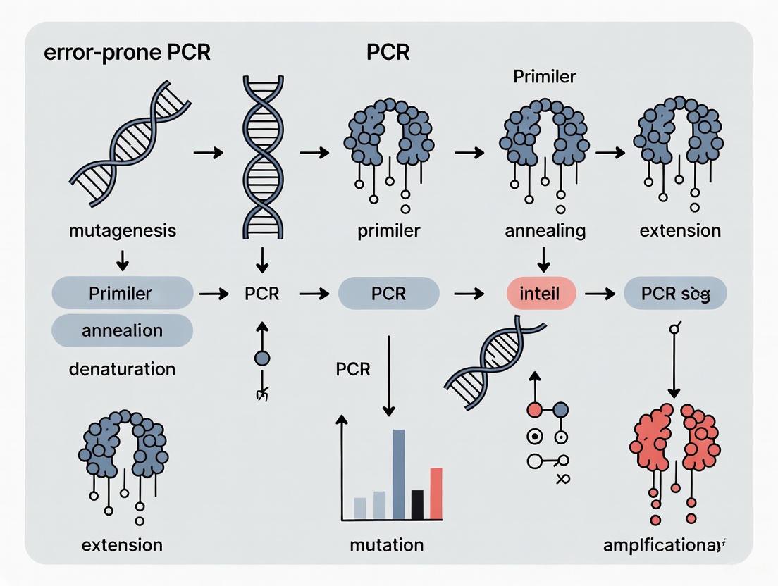
This article provides a comprehensive guide to error-prone PCR (epPCR), a cornerstone technique in directed protein evolution.
Error-Prone PCR for Random Mutagenesis: A Complete Guide from Principle to Library Construction
Abstract
This article provides a comprehensive guide to error-prone PCR (epPCR), a cornerstone technique in directed protein evolution. Tailored for researchers and drug development professionals, it covers the foundational principles of creating genetic diversity, detailed step-by-step protocols, and advanced methodologies for library construction. It also delivers systematic troubleshooting strategies to overcome common pitfalls and a critical evaluation of epPCR against other mutagenesis methods, empowering scientists to effectively engineer proteins with novel functions for therapeutic and industrial applications.
The Principles of Random Mutagenesis: Building Diversity with Error-Prone PCR
Directed evolution is a powerful protein engineering methodology that mimics the principles of natural selection in a laboratory setting to optimize proteins for human-defined applications. This forward-engineering process involves iterative cycles of genetic diversification and functional selection, compressing geological timescales of evolution into weeks or months [1]. The profound impact of directed evolution was recognized with the 2018 Nobel Prize in Chemistry awarded to Frances H. Arnold for establishing this technology as a cornerstone of modern biotechnology and industrial biocatalysis [1]. The primary strategic advantage of directed evolution lies in its capacity to deliver robust solutions—such as enhanced stability, novel catalytic activity, or altered substrate specificity—without requiring detailed a priori knowledge of a protein's three-dimensional structure or catalytic mechanism [1]. This capability allows it to bypass the inherent limitations of rational design, which relies on a predictive understanding of sequence-structure-function relationships that is often incomplete [1].
Within the directed evolution toolkit, random mutagenesis serves as a fundamental approach for generating genetic diversity. By creating large libraries of protein variants through techniques like error-prone PCR (epPCR), researchers can explore vast sequence landscapes to identify improved variants through screening or selection [2] [1]. This review provides a comprehensive examination of directed evolution methodologies with particular emphasis on random mutagenesis techniques, their applications, and experimental protocols relevant to error-prone PCR research.
The Directed Evolution Cycle
At its core, directed evolution functions as a two-part iterative engine that drives a protein population toward a desired functional goal through repeated cycles of diversity generation and selection [1]. This process consists of four key stages that form an evolutionary feedback loop, systematically accumulating beneficial mutations across successive generations.
The Directed Evolution Workflow
Figure 1: The Directed Evolution Cycle. This workflow illustrates the iterative process of diversity generation and selection that drives protein optimization.
The directed evolution workflow begins with a parent gene encoding a protein that possesses a basal level of the desired activity. This gene is subjected to mutagenesis to create a large and diverse library of variants, which are then expressed as proteins [1]. The population is challenged with a screen or selection that identifies individuals with improved performance [1]. The genes from the most improved variants are isolated and serve as templates for subsequent rounds of mutagenesis and screening at increasingly stringent conditions [1]. This iterative process continues until the desired performance target is met or no further improvements can be identified. The success of any directed evolution campaign hinges on two critical factors: the quality and diversity of the initial library, and the effectiveness of the screening method to identify rare improved variants among predominantly neutral or deleterious mutations [1].
Random Mutagenesis Techniques
Random mutagenesis aims to introduce mutations across the entire length of a gene without pre-selecting specific sites, creating diverse libraries that serve as the raw material for evolutionary optimization [1]. Several methods have been developed to introduce genetic variation, each with distinct advantages, limitations, and inherent biases that shape evolutionary trajectories.
Error-Prone PCR (epPCR)
Error-prone PCR represents the most established and widely used method for random mutagenesis [1]. This technique is a modified PCR that intentionally reduces the fidelity of DNA polymerase, thereby introducing errors during gene amplification. The methodological foundation of epPCR involves deliberate alteration of standard PCR conditions to promote misincorporation of nucleotides [3].
Table 1: Key Components and Conditions for Error-Prone PCR
| Component/Condition | Standard PCR | Error-Prone PCR | Function in Mutagenesis |
|---|---|---|---|
| DNA Polymerase | High-fidelity (e.g., Pfu) | Low-fidelity (e.g., Taq) | Reduced proofreading increases error rate |
| Mn²⁺ ions | Absent | Present (0.1-1.0 mM) | Promotes misincorporation of nucleotides |
| dNTP Concentration | Balanced | Imbalanced | Increases misincorporation probability |
| Mg²⁺ Concentration | Standard (1.5-2.0 mM) | Elevated (3.0-7.0 mM) | Further reduces polymerase fidelity |
| Mutation Rate | Minimized | 1-5 mutations/kb | Controlled introduction of point mutations |
The strategic implementation of epPCR involves carefully tuning the mutation rate, typically targeting 1-5 base mutations per kilobase, resulting in an average of one or two amino acid substitutions per protein variant [1]. This controlled mutation rate is crucial—too few mutations limit diversity, while excessive mutations generate predominantly non-functional proteins. Despite its power and straightforward implementation, epPCR is not truly random [1]. DNA polymerases exhibit intrinsic bias favoring transition mutations (purine-to-purine or pyrimidine-to-pyrimidine) over transversion mutations (purine-to-pyrimidine or vice versa) [1]. Combined with the degeneracy of the genetic code, this bias means epPCR can only access an average of 5-6 of the 19 possible alternative amino acids at any given position, constraining the accessible sequence space [1].
Advanced Random Mutagenesis Methods
Beyond standard epPCR, several advanced techniques have been developed to address specific challenges in diversity generation:
Inosine-Mediated epPCR utilizes deoxyinosine triphosphate (dITP) as a universal base during PCR amplification [4]. Inosine preferentially pairs with guanine or cytosine in subsequent amplifications, increasing GC content and introducing focused mutations that enhance thermal stability and structural rigidity in aptamer libraries [4].
Segmental Error-Prone PCR (SEP) addresses limitations in evolving large genes by dividing them into small fragments that are independently mutagenized in vitro before reassembly in Saccharomyces cerevisiae [5]. This approach ensures even distribution of beneficial mutations across large genes and minimizes negative mutations that often plague traditional epPCR of large sequences [5].
Circular Polymerase Extension Cloning (CPEC) represents a significant advancement in library construction by eliminating the need for restriction enzymes and DNA ligase [3]. CPEC uses high-fidelity DNA polymerase to extend overlapping regions between the insert and vector, forming circular molecules. This technique demonstrates superior efficiency compared to traditional Ligation-Dependent Cloning Process (LDCP), enabling acquisition of greater numbers of gene variants and accelerating cloning processes in gene library generation [3].
Table 2: Comparison of Random Mutagenesis Techniques
| Method | Mechanism | Advantages | Limitations | Best Applications |
|---|---|---|---|---|
| Error-Prone PCR | Low-fidelity PCR with Mn²⁺ and imbalanced dNTPs | Simple, widely applicable, tunable mutation rate | Transition bias, limited amino acid accessibility | General protein engineering, initial diversification |
| Inosine-Mediated epPCR | Incorporation of dITP as universal base | Increases GC content, enhances thermal stability | Specific to aptamer development | SELEX starting libraries, aptamer engineering |
| Segmental epPCR (SEP) | Fragments large genes before mutagenesis | Even mutation distribution in large genes, reduces negative mutations | Requires recombination in yeast | Large proteins, multi-domain engineering |
| DNA Shuffling | DNaseI fragmentation + reassembly | Recombines beneficial mutations, mimics natural evolution | Requires sequence homology (>70%) | Combining hits from multiple parents |
Experimental Protocols
Standard Error-Prone PCR Protocol
The following protocol for error-prone PCR mutagenesis is adapted from established methodologies with an average mutation rate of 2-4 mutations per kilobase [3] [1]:
Reagents and Materials:
- Template DNA (10-50 ng/μL in purified form)
- Taq DNA polymerase (without proofreading activity)
- 10× reaction buffer (without Mg²⁺)
- MgCl₂ stock solution (50 mM)
- MnCl₂ stock solution (10 mM)
- dNTP mix (ultrapure, 10 mM each)
- Primers specific to target gene (10 μM each)
- Sterile molecular biology grade water
- Thermocycler
- Agarose gel electrophoresis equipment
Procedure:
- Prepare the epPCR reaction mix on ice:
- 5.0 μL 10× reaction buffer
- 2.0 μL MgCl₂ (50 mM) - final concentration 2 mM
- 1.0-5.0 μL MnCl₂ (10 mM) - titrate for desired mutation rate (start with 2.0 μL for ~3 mutations/kb)
- 2.0 μL dNTP mix (10 mM each) - final concentration 0.4 mM each
- 2.0 μL forward primer (10 μM)
- 2.0 μL reverse primer (10 μM)
- 1.0 μL template DNA (10-50 ng)
- 0.5 μL Taq DNA polymerase (5 U/μL)
- Sterile water to 50 μL total volume
Mix gently by pipetting and centrifuge briefly to collect contents.
Run the PCR with the following cycling conditions:
- Initial denaturation: 94°C for 2 minutes
- 30 cycles of:
- Denaturation: 94°C for 15 seconds
- Annealing: 55-65°C (primer-specific) for 30 seconds
- Extension: 68°C for 1 minute per kb of template
- Final extension: 68°C for 5 minutes
- Hold at 4°C
Verify amplification by analyzing 5 μL of product on agarose gel electrophoresis.
Purify PCR product using standard DNA clean-up kits before downstream cloning.
Critical Considerations:
- MnCl₂ concentration is the primary factor controlling mutation rate—titrate carefully (0.1-1.0 mM final concentration)
- Higher Mg²⁺ concentrations (2-7 mM) further reduce fidelity
- Imbalanced dNTP ratios (e.g., increasing dATP/dGTP while decreasing dCTP/dTTP) can enhance mutation frequency
- Limit template amount to minimize wild-type carryover
- Number of cycles affects mutation accumulation—25-35 cycles typically optimal
Circular Polymerase Extension Cloning (CPEC) Protocol
CPEC provides superior efficiency for cloning mutant libraries compared to traditional restriction enzyme-based methods [3]:
Procedure:
- Purify both the mutant insert (from epPCR) and linearized vector.
- Design primers with 15-25 bp overlapping regions between insert and vector ends.
- Set up CPEC reaction:
- 50-100 ng vector DNA
- 3:1 molar ratio of insert:vector
- 1× high-fidelity PCR buffer
- 0.2 mM dNTPs
- High-fidelity DNA polymerase (e.g., TAKARA LA Taq)
- Sterile water to 50 μL
- Run CPEC with cycling conditions:
- 94°C for 2 minutes
- 30 cycles of: 94°C for 15 seconds, 63°C for 30 seconds, 68°C for 4 minutes
- 72°C for 5 minutes
- Transform directly into competent E. coli cells without restriction digestion.
Applications and Case Studies
Directed evolution employing random mutagenesis has demonstrated remarkable success across diverse biotechnology applications, from sustainable fuel production to therapeutic development.
Engineering Hydrocarbon-Producing Enzymes
Directed evolution approaches are being applied to engineer enzymes capable of catalyzing hydrocarbon production for sustainable fuel synthesis [6]. Native activities of these enzymes often prove insufficient for industrial bioprocesses, necessitating optimization through directed evolution [6]. The application of DE to hydrocarbon-producing enzymes presents unique challenges due to the physicochemical properties of target molecules—aliphatic hydrocarbons can be insoluble, gaseous, and chemically inert, complicating their detection in vivo and dynamic coupling to cellular fitness [6]. Despite these challenges, enzymes such as the cytochrome P450 OleTJE from Jeotgalicoccus sp., which catalyzes fatty acid decarboxylation to produce alkenes, represent promising targets for evolutionary optimization [6].
Machine Learning-Enhanced Directed Evolution
Recent advances integrate machine learning with directed evolution to navigate complex fitness landscapes more efficiently. Active Learning-assisted Directed Evolution (ALDE) represents an iterative machine learning workflow that leverages uncertainty quantification to explore protein sequence space more effectively than traditional DE methods [7]. In one application, ALDE optimized five epistatic residues in the active site of a protoglobin from Pyrobaculum arsenaticum (ParPgb) for a non-native cyclopropanation reaction [7]. Through just three rounds of wet-lab experimentation, ALDE improved the yield of the desired product from 12% to 93%, demonstrating remarkable efficiency in navigating challenging epistatic landscapes where standard DE approaches typically fail [7].
Figure 2: Comparison of Traditional DE and Machine Learning-Assisted Workflows. ALDE incorporates predictive modeling to prioritize variants more efficiently.
Enhancing Enzyme Activity and Stability
The SEP and Directed DNA Shuffling (DDS) approach has been successfully applied to simultaneously improve both the activity of β-glucosidase and its tolerance to organic acids [5]. This method minimized negative mutations and reduced revertant mutations while facilitating integration of positive mutations across the entire gene sequence [5]. Traditional directed evolution approaches for large genes often resulted in high frequencies of negative and reverse mutations, but the segmental approach guaranteed even distribution of mutation sites, generating robust variants with enhanced multiple functionalities [5].
The Scientist's Toolkit: Research Reagent Solutions
Table 3: Essential Research Reagents for Directed Evolution with Random Mutagenesis
| Reagent/Category | Specific Examples | Function/Application | Key Considerations |
|---|---|---|---|
| Low-Fidelity Polymerases | Taq polymerase, Mutazyme II | Introduces random mutations during epPCR | Lack 3'→5' proofreading; fidelity controlled by reaction conditions |
| Mutation Rate Modulators | MnCl₂, unbalanced dNTPs, elevated Mg²⁺ | Fine-tune mutation frequency in epPCR | Mn²⁺ concentration primary controller (0.1-1.0 mM typical) |
| Cloning Systems | CPEC, restriction enzyme-based cloning, yeast recombination | Vector insertion of mutant libraries | CPEC offers superior efficiency over traditional methods |
| Host Organisms | E. coli, S. cerevisiae, P. pastoris | Expression of variant libraries | E. coli: prokaryotic proteins; S. cerevisiae: eukaryotic proteins, high recombination |
| Selection/Screening Platforms | Microtiter plates, FACS, biosensors, growth coupling | Identify improved variants | Throughput must match library size; "you get what you screen for" |
Random mutagenesis remains a foundational methodology within the directed evolution paradigm, providing critical access to diverse sequence spaces without requiring extensive structural knowledge of target proteins. Error-prone PCR and its advanced derivatives offer researchers powerful tools to initiate evolutionary trajectories toward proteins with enhanced stability, novel functions, and optimized activities for industrial and therapeutic applications. Recent methodological innovations—including segmental epPCR for large proteins, circular polymerase extension cloning for improved library construction, and machine learning integration for navigating epistatic landscapes—continue to expand the capabilities and applications of random mutagenesis in protein engineering. As these technologies mature, directed evolution employing strategic random mutagenesis will undoubtedly continue to drive innovations across biotechnology, sustainable energy, and pharmaceutical development.
Error-prone polymerase chain reaction (epPCR) is a foundational technique in directed evolution that enables researchers to rapidly generate genetic diversity from a single parent sequence. Unlike conventional PCR, which aims for perfect fidelity in amplification, epPCR deliberately introduces random nucleotide mutations throughout the amplified gene, creating libraries of variants that can be screened for desired functional properties. This method has proven invaluable for protein engineering, vaccine development, and functional genomics, allowing scientists to mimic and accelerate natural evolutionary processes in laboratory settings. The core mechanism relies on compromising the inherent proofreading capabilities of DNA polymerase systems, creating a mutagenic environment that generates a broad spectrum of mutations with varying frequencies and distributions.
Core Mechanisms of Mutagenesis
The strategic introduction of random mutations in epPCR occurs through several biochemical interventions that reduce the fidelity of DNA replication:
Low-Fidelity DNA Polymerases: The use of polymerases lacking 3′→5′ proofreading exonuclease activity, such as Taq polymerase, provides a foundation for misincorporation. Engineered mutant polymerases with even lower fidelity, such as Mutazyme II, further enhance error rates while generating less biased mutational spectra [8].
Manganese Ions: The addition of Mn2+ to reaction buffers is a key strategy to reduce polymerase fidelity. Unlike Mg2+ (the natural cofactor), Mn2+ promotes misincorporation by decreasing the enzyme's ability to discriminate against incorrect nucleotides during synthesis [8] [9].
Unbalanced dNTP Concentrations: Creating non-equimolar ratios of deoxynucleotide triphosphates in the reaction mixture increases the likelihood of incorporation mismatches when the correct nucleotide is depleted or limited at the polymerase active site [8] [9].
Nucleotide Analogs: The incorporation of mutagenic base analogs like 8-oxo-dGTP and dPTP can lead to even higher error rates by forming non-standard base pairings during replication [8].
The combination of these approaches can achieve error rates ranging from approximately 1 mutation per 103 nucleotides to as high as 33 mutations per kilobase for specialized applications [8]. The mutation frequency can be controlled by adjusting the number of amplification cycles and the starting template concentration, with lower template amounts and higher cycle numbers generally producing greater mutational loads [8] [9].
Mutational Spectrum and Distribution
The mutations introduced through epPCR generate a diverse mutational landscape encompassing:
Point Mutations: Single nucleotide substitutions represent the most common type of mutation, potentially leading to amino acid changes when occurring in coding regions.
Insertions and Deletions (Indels): While less frequent than substitutions, small insertions or deletions can occur, particularly under conditions promoting high error rates.
The distribution of mutations across the target sequence generally follows a non-Poisson distribution that depends on PCR experimental parameters rather than a purely random distribution [9]. This distribution directly influences the fraction of proteins retaining function after mutation, with higher mutation rates producing more unique sequences but fewer functional clones [9]. Recent modeling approaches based on actual PCR processes provide more accurate predictions of mutational distributions and functional retention rates than previous Poisson-based models [9].
Table 1: Key Biochemical Factors in Error-Prone PCR and Their Mechanisms
| Factor | Mechanism of Action | Typical Implementation |
|---|---|---|
| Low-Fidelity Polymerase | Lacks 3′→5′ proofreading capability; reduced nucleotide discrimination | Taq polymerase; Mutazyme II; other engineered mutants |
| Manganese Ions | Promotes misincorporation by reducing polymerase discrimination | 0.5 mM MnCl₂ added to standard PCR buffer |
| Unbalanced dNTPs | Increases probability of incorporation errors when correct dNTP is limited | Non-equimolar ratios (e.g., 0.2 mM dGTP, 1.35 mM dTTP) |
| Nucleotide Analogs | Forms non-standard base pairings during replication | 8-oxo-dGTP, dPTP added to dNTP mixture |
| Increased Cycle Number | Provides more opportunities for errors to accumulate | 30-50 cycles instead of standard 25-35 |
Quantitative Analysis of Mutation Rates
Controlling and Measuring Mutational Load
The mutational load in epPCR libraries can be precisely controlled through reaction parameters and accurately measured through sequencing analysis:
Table 2: Mutation Rates and Their Effects on Protein Function
| Average Mutations per Gene | Fraction Functional (%) | Library Characteristics | Primary Applications |
|---|---|---|---|
| 1-5 | ~10-50% | High functional retention, limited diversity | Fine-tuning existing functions; stability improvement |
| 5-10 | ~1-10% | Balance of diversity and function | Broad property enhancement (e.g., thermostability) |
| 10-15 | ~0.1-1% | High diversity, reduced function | Exploring distant sequence space; major functional shifts |
| 15-30 | <0.1% | Extreme diversity, rare functional variants | Novel function discovery; antibody engineering |
The relationship between mutation rate and functional retention follows a predictable trend, with the fraction of functional proteins declining as the average number of mutations increases [9]. However, the distribution is broader than a Poisson distribution, leading to an excess of functional clones at high error rates compared to theoretical expectations [9]. This phenomenon explains why high-error-rate libraries can be enriched with improved proteins despite the overall decline in functional sequences [9].
The optimal mutation rate represents a balance between uniqueness and retention of function. While very low mutation rates produce many functional sequences, they offer limited diversity. Conversely, very high mutation rates generate mostly unique sequences but few functional clones [9]. For a standard-sized protein, the generally optimal range falls between 5-15 amino acid substitutions per gene, though this varies depending on the specific protein and selection system [9].
Research Reagent Solutions
Essential reagents and their functions for implementing error-prone PCR:
Table 3: Essential Research Reagents for Error-Prone PCR
| Reagent | Function | Examples & Notes |
|---|---|---|
| Low-Fidelity Polymerase | Catalyzes DNA amplification with reduced fidelity | Taq polymerase (no proofreading); Mutazyme II (commercial high-error variant) |
| Mutagenic Buffer | Creates chemical environment promoting misincorporation | Typically contains Mn²⁺ and unbalanced dNTP concentrations |
| Primers with Restriction Sites | Enables subsequent cloning of mutated fragments | Include artificial restriction sites (e.g., EcoRI, BamHI) compatible with plasmids |
| Cloning Vector | Host for mutated inserts for expression and screening | Gateway plasmids; standard expression vectors with appropriate resistance |
| Competent Cells | For transformation and library amplification | E. coli TOP10 (electrocompetent); other high-efficiency strains |
Experimental Protocols
Standard Error-Prone PCR Protocol
The following protocol represents a generalized approach to error-prone PCR that can be modified based on specific application requirements:
Step 1: Reaction Setup
- Prepare a 50μL reaction mixture containing:
- 1X mutagenic PCR buffer (typically including MnCl₂)
- Unbalanced dNTP mixture (concentrations vary by protocol)
- 0.1-10 ng template DNA
- 0.5 μM forward and reverse primers
- 1-2 U low-fidelity DNA polymerase
Step 2: Thermal Cycling
- Initial denaturation: 94°C for 2 minutes
- 25-35 cycles of:
- Denaturation: 94°C for 15-30 seconds
- Annealing: 50-65°C for 30 seconds (primer-specific)
- Extension: 68-72°C for 1 minute per kb of amplicon
- Final extension: 72°C for 5-10 minutes
Step 3: Product Analysis
- Verify amplification by agarose gel electrophoresis
- Purify PCR product using standard methods (e.g., column purification)
- Quantitate DNA concentration by spectrophotometry
Step 4: Library Construction
- Digest purified PCR product and vector with appropriate restriction enzymes
- Ligate insert into vector using T4 DNA ligase
- Transform competent E. coli cells
- Plate on selective media to assess library size and diversity
This protocol can yield error rates of approximately 1-10 mutations per kilobase, depending on specific conditions and cycling parameters [10] [8].
Specialized Protocol for Small Amplicons
For targeting small regions (<100 bp) such as ribosome binding sites or specific protein domains, a modified approach is necessary to achieve sufficient mutational density:
Key Modifications:
- Implement iterative dilution/reamplification cycles to increase mutation frequency
- Use touchdown PCR to prevent accumulation of incorrect products
- Employ extreme template dilution (e.g., billion-fold dilution) to minimize wild-type carryover
- Increase cycle numbers (up to 50 cycles) in each amplification round
This specialized approach can achieve high mutational loads of approximately 33 mutations/kb (1.2 mutations on average for a 36-bp amplicon), which would be impossible with standard epPCR protocols [8].
Diagram 1: Experimental workflow for error-prone PCR and library generation.
Advanced Methodological Considerations
Cloning Strategies for Mutant Libraries
The efficiency of cloning mutated PCR products significantly impacts library quality and diversity. Traditional restriction enzyme-based approaches (Ligation-Dependent Cloning Process) often lead to substantial loss of potential mutants:
Circular Polymerase Extension Cloning (CPEC): This restriction-free method uses high-fidelity DNA polymerase to extend overlapping regions between insert and vector, forming circular molecules. CPEC accelerates cloning and yields more variants than restriction-based methods [3].
Gateway Technology: This recombination-based system offers high cloning efficiency but traditionally requires multiple steps (BP and LR reactions). A streamlined one-step method eliminates the BP reaction, better preserving original library complexity [11].
Addressing Mutational Bias
Different epPCR conditions can produce distinct mutational spectra with specific nucleotide substitution biases. To create higher-quality libraries:
- Combine multiple mutagenesis conditions to achieve more balanced mutation types
- Use engineered mutator polymerases that produce less biased mutational spectra
- Consider incorporating DNA shuffling after epPCR to recombine beneficial mutations
These approaches help create more comprehensive mutant libraries that better sample sequence space [10] [8].
Diagram 2: Core mechanism of random mutation introduction in error-prone PCR.
Applications in Biotechnology and Research
Protein Engineering and Directed Evolution
epPCR serves as a cornerstone technique in directed evolution pipelines for optimizing protein properties:
Thermostability Enhancement: Multiple studies have successfully improved enzyme thermostability through epPCR-based evolution, including maltogenic amylase, phytase, and Bacillus licheniformis alpha amylase [8].
Solubility Improvement: Directed evolution using epPCR libraries has solved protein solubility challenges, as demonstrated by the evolution of a more soluble Tobacco Etch Virus protease variant [11].
Activity Optimization: The method has been applied to optimize de novo evolved proteins for improved folding stability, solubility, and ligand-binding affinity [10].
Vaccine Development
epPCR has proven valuable in vaccine seed strain development:
- Influenza Vaccine Candidates: Researchers have integrated epPCR with site-directed mutagenesis and reverse genetics to rapidly generate high-yield influenza vaccine candidates. This approach produced six high-yield candidate strains for influenza A(H1N1)pdm09 virus, with two providing complete protection in mouse challenge models [12].
Functional Characterization of Viral Proteins
Random mutagenesis helps map functional domains in viral proteins:
- Morbillivirus Research: epPCR has been used to functionally probe the receptor-binding site of peste des petits ruminants virus (PPRV) hemagglutinin protein, confirming conservation of this region across morbilliviruses [13].
Troubleshooting and Optimization
Common Challenges and Solutions
- Insufficient Mutation Rate: Increase cycle number, reduce template amount, optimize Mn2+ concentration, or incorporate nucleotide analogs
- Excessive Mutation Rate: Reduce cycle number, increase template amount, or use more balanced dNTP ratios
- Low Library Diversity: Improve cloning efficiency through CPEC or Gateway systems, increase transformation efficiency
- Biased Mutational Spectrum: Combine different mutagenesis conditions or use engineered mutator polymerases
Quality Assessment
- Sequence Verification: Randomly pick and sequence 10-20 clones to determine actual mutation rate and spectrum
- Functional Assessment: Test a subset of clones to determine the fraction retaining wild-type function
- Diversity Analysis: Ensure library contains sufficient unique variants for screening purposes
The strategic application of error-prone PCR continues to enable advances across biotechnology, from therapeutic development to fundamental biological research. By understanding and optimizing its core mechanisms, researchers can harness this powerful technique to explore sequence-function relationships and engineer biomolecules with novel properties.
In vitro selection coupled with directed evolution represents a powerful method for generating nucleic acids and proteins with desired functional properties, where creating high-quality random mutant libraries is a critical first step [10]. Error-prone PCR (epPCR) serves as a cornerstone technique for introducing random mutations into a gene of interest by exploiting reduced-fidelity DNA polymerases during amplification. The choice of DNA polymerase directly influences mutation rate, spectrum, and bias, thereby fundamentally impacting library quality and diversity. This application note provides a structured comparison of key low-fidelity DNA polymerases and detailed protocols for their effective use in random mutagenesis, framed within the context of optimizing epPCR for protein engineering and drug development research.
Enzyme Toolkit: A Comparative Analysis of Low-Fidelity DNA Polymerases
Selecting the appropriate polymerase is crucial for balancing mutational load with experimental feasibility. The table below summarizes key enzymes used in error-prone PCR.
Table 1: Characteristics of DNA Polymerases for Error-Prone PCR
| Polymerase | Proofreading Activity | Typical Error Rate (errors/bp/duplication) | Fidelity Relative to Taq | Key Features and Mutations |
|---|---|---|---|---|
| Taq Polymerase | No | 1.0 x 10⁻⁵ to 2.0 x 10⁻⁵ [14] | 1x (Baseline) | Standard enzyme for basic epPCR; fidelity can be reduced with Mn²⁺ and unbalanced dNTPs [15] [8]. |
| AccuPrime-Taq HF | No | ~1.0 x 10⁻⁵ [14] | ~9x better than Taq | A proprietary formulation designed for high-fidelity amplification, included here for contrast. |
| Mutazyme II | No | Varies with conditions | N/A | Commercial mutant polymerase known for less biased mutational spectra [8]. |
| Pfu Polymerase (exo-) | No (Disabled) | 1.0 x 10⁻⁶ to 2.0 x 10⁻⁶ [14] | 6-10x better than Taq | Engineered from wild-type Pfu; proofreading activity is abolished (e.g., D215A mutation) [15]. |
| Mutant Pfu Variants | No (Disabled) | Can be very high | Lower than wild-type Pfu | Engineered with mutations in the fingers sub-domain (e.g., T471, Q472, D473) for enhanced low-fidelity performance under standard PCR conditions [15]. |
| KOD Hot Start | Yes | ~1.0 x 10⁻⁶ [14] | ~4-50x better than Taq (varies by source) | A high-fidelity polymerase, included for comparison. |
| Phusion Hot Start | Yes | 4.0 x 10⁻⁷ to 9.5 x 10⁻⁷ [14] | >50x better than Taq | One of the highest fidelity polymerases available, included for contrast. |
The data indicates a clear fidelity hierarchy: Taq < AccuPrime-Taq < KOD ≈ Pfu (exo-) ≈ Pwo < Phusion [14]. While Taq polymerase and its variants offer a straightforward path to mutagenesis, engineered enzymes like mutant Pfu variants can provide high mutational loads with less sequence bias and operate under standard PCR conditions [15].
Experimental Protocols for Random Mutagenesis
Standard Error-Prone PCR with Modified Reaction Conditions
This protocol is optimized for use with polymerases like Taq, where reaction conditions are manipulated to reduce fidelity.
Reagents:
- Template DNA: 0.1-10 ng of plasmid DNA containing the target gene.
- Primers: Forward and reverse primers flanking the gene to be mutated.
- Low-Fidelity DNA Polymerase: e.g., Taq polymerase.
- 10X Mutagenic Buffer:
- Unbalanced dNTPs: e.g., 0.2 mM dGTP, 0.2 mM dATP, 1.0 mM dCTP, 1.0 mM dTTP [8].
Method:
- Prepare a 50 µL PCR reaction mix on ice:
- 5 µL 10X Mutagenic Buffer
- 5 µL Unbalanced dNTP Mix
- 1 µL Forward Primer (10 µM)
- 1 µL Reverse Primer (10 µM)
- 1 µL Template DNA (diluted to 0.1-10 ng)
- 0.5 µL Taq DNA Polymerase (5 U/µL)
- Nuclease-free water to 50 µL
- Run PCR with the following cycling parameters:
- Initial Denaturation: 95°C for 2 minutes
- Amplification (30-35 cycles):
- Denature: 95°C for 30 seconds
- Anneal: 55-65°C for 30 seconds
- Extend: 72°C for 1 minute per kb
- Final Extension: 72°C for 5 minutes
- Purify the PCR product using a standard PCR cleanup kit.
- The mutated gene is now ready for cloning into an expression vector.
Iterative Error-Prone PCR for Small Amplicons
Concentrating multiple mutations into very short DNA regions (<100 bp) is challenging with standard protocols. This iterative method achieves high mutational loads [8].
Reagents:
- Template DNA: Plasmid containing the target short sequence.
- Primers: Forward and reverse primers for the small amplicon.
- Low-Fidelity DNA Polymerase Mix: e.g., Mutazyme II from Agilent.
- Commercial Mutagenic Buffer: As supplied with the enzyme.
Method:
- Initial Dilution: Perform a serial dilution of the template DNA to a final concentration of 50 attograms (ag) in a 50 µL PCR reaction [8].
- Primary Amplification:
- Set up the PCR reaction with the mutagenic polymerase and primers.
- Use a Touchdown PCR protocol to prevent spurious product accumulation:
- Initial denaturation: 95°C for 2 minutes.
- 5 cycles of: 95°C for 20s, 60°C for 30s, 72°C for 20s.
- 5 cycles of: 95°C for 20s, 58°C for 30s, 72°C for 20s.
- 25 cycles of: 95°C for 20s, 55°C for 30s, 72°C for 20s.
- Final extension: 72°C for 5 minutes.
- Dilution and Re-amplification:
- Dilute the primary PCR product 1000-fold.
- Use 1 µL of this dilution as the template for a second, identical PCR amplification.
- Repeat the dilution and re-amplification step for a third cycle.
- After three total cycles, purify the final product. This iterative process can achieve mutation frequencies as high as 33 mutations/kbp (approximately 1.2 mutations in a 36-bp amplicon) [8].
One-Step Random Mutagenesis by Error-Prone Rolling Circle Amplification (epRCA)
epRCA is a ligation-independent method that simplifies library generation, using φ29 DNA polymerase under mutagenic conditions [17].
Reagents:
- Template DNA: Supercoiled plasmid containing the target gene.
- φ29 DNA Polymerase
- Exonuclease-resistant Random Hexamers
- RCA Buffer: 50 mM Tris-HCl (pH 7.5), 10 mM MgCl₂, 10 mM (NH₄)₂SO₄, 200 ng/µL BSA, 4 mM DTT.
- dNTPs: 0.2 mM each.
- MnCl₂: 1.5 mM (added to reduce fidelity).
Method:
- Mix 0.5 µL of template plasmid (or a bacterial colony resuspended in TE buffer) with 5 µL of sample buffer containing random hexamers.
- Heat the mixture at 95°C for 3 minutes, then cool to room temperature.
- Add a premix containing RCA buffer, dNTPs, φ29 DNA polymerase, and MnCl₂.
- Incubate at 30°C for 6-18 hours, then heat-inactivate at 65°C for 10 minutes.
- Purify the high-molecular-weight RCA product.
- Use 1-5 µL of the purified product directly to transform electrocompetent E. coli. The host machinery processes the tandemly repeated RCA product into circular plasmids, yielding a mutant library with 3-4 mutations per kilobase [17].
Workflow Visualization
Diagram 1: Error-Prone PCR Workflow Selection. This diagram outlines three primary methodological pathways for random mutagenesis, categorized by research goal. LDCP: Ligation-Dependent Cloning Process; CPEC: Circular Polymerase Extension Cloning.
Research Reagent Solutions
A successful error-prone PCR experiment relies on a core set of reagents, each fulfilling a specific function.
Table 2: Essential Reagents for Error-Prone PCR
| Reagent | Function | Examples & Notes |
|---|---|---|
| Low-Fidelity DNA Polymerase | Catalyzes DNA amplification while introducing misincorporated nucleotides. | Taq polymerase, mutant Pfu variants (e.g., Pfu exo- with loop mutations), Mutazyme II, φ29 (for RCA) [15] [17] [8]. |
| Mutagenic Buffer Additives | Reduces polymerase fidelity to increase error rate. | MnCl₂: A key divalent cation that promotes misincorporation [8] [16]. Elevated MgCl₂: Can also decrease fidelity. |
| Unbalanced dNTPs | Creates a pool of incorrect nucleotides, increasing misincorporation likelihood. | e.g., Increasing concentration of dCTP and dTTP relative to dATP and dGTP [8]. |
| Template DNA | The genetic template to be mutated. | Purified plasmid or a bacterial colony. For high mutational load, use minimal amounts (e.g., 0.1-10 ng for PCR, 50 ag for iterative small amplicon PCR) [8]. |
| Primers | Define the start and end points of the DNA fragment to be amplified. | Standard sequencing primers; for CPEC cloning, may require 5' extensions homologous to the vector [3]. |
| Cloning System | Inserts the mutated PCR product into a plasmid for expression and screening. | LDCP: Uses restriction enzymes and DNA ligase [3]. CPEC: A ligase-free method that can improve library coverage by circular polymerase extension [3]. |
The strategic selection of low-fidelity DNA polymerases and optimization of accompanying protocols are fundamental to generating high-quality random mutagenesis libraries. Researchers can choose from traditional options like Taq polymerase, with conditions manipulated to enhance error rates, or opt for modern engineered solutions like mutant Pfu variants that offer high mutational loads with reduced bias under standard conditions. Furthermore, advanced techniques such as iterative epPCR for small amplicons and ligation-free epRCA provide powerful alternatives to overcome specific experimental limitations. By applying the comparative data and detailed methodologies outlined in this application note, scientists can systematically approach enzyme selection and protocol design to advance their directed evolution and protein engineering projects.
In random mutagenesis, the "mutational spectrum" describes the nature and frequency of nucleotide changes introduced into a DNA sequence. A fundamental distinction within this spectrum lies between transitions and transversions. A transition is a point mutation that changes a purine to another purine (A G) or a pyrimidine to another pyrimidine (C T). In contrast, a transversion swaps a purine for a pyrimidine or vice versa (A C, A T, G C, G T). Transitions generally occur more frequently than transversions in many biological systems. However, mutational bias—the non-random preference for certain types of mutations over others—is a critical feature of all random mutagenesis techniques, including error-prone PCR (epPCR). This bias directly influences the diversity and quality of mutant libraries, shaping the available sequence space for directed evolution experiments [18] [19] [20].
Understanding and controlling this bias is essential for effective protein engineering. A biased protocol may repeatedly generate the same subset of mutations, limiting functional diversity and reducing the probability of discovering unique and beneficial enzyme variants. This application note details the sources and types of mutational bias in epPCR and provides validated protocols for analyzing mutational spectra to engineer superior biocatalysts.
Quantitative Analysis of Mutational Spectra
Different random mutagenesis methods produce distinct mutational spectra, characterized by varying frequencies of transitions vs. transversions and different nucleotide substitution preferences. The following table summarizes the performance parameters of several common methods as analyzed in a comparative study [18].
Table 1: Comparison of Random Mutagenesis Methods and Their Mutational Spectra
| Mutagenesis Method | Mutation Frequency (bp⁻¹) | Transition vs. Transversion Ratio | Key Characteristics and Biases |
|---|---|---|---|
| epPCR (Standard Taq) | High / Adjustable | Favors transitions | A/T-biased mutation rate; biased nucleotide substitutions [18] [20]. |
| epPCR (Mutazyme II) | High / Adjustable | More transversions | Designed to counterbalance Taq bias, creating a more "balanced" library [20]. |
| Hydroxylamine Treatment | Low | Narrow range | Chemical method; specific bias toward A/T to G/C transitions [18]. |
| E. coli Mutator Strain | Low | Narrow range | Biological in vivo method; exhibits a specific, narrow mutational repertoire [18]. |
The mutational bias of standard epPCR using Taq polymerase is further illustrated by its preference for specific nucleotide changes. The table below breaks down a representative mutational spectrum, highlighting the non-uniform distribution of substitutions [19].
Table 2: Detailed Mutational Spectrum and Bias in Standard Error-Prone PCR
| Mutation Type | Specific Substitution | Relative Frequency | Notes on Bias |
|---|---|---|---|
| Transition | A → G | High | A significant contributor to overall bias, leading to over-representation. |
| G → A | High | ||
| C → T | High | ||
| T → C | High | ||
| Transversion | A → T / C | Low | All transversions are typically under-represented compared to transitions. |
| G → T / C | Low | ||
| C → A / G | Low | ||
| T → A / G | Low | ||
| Other Bias | A/T Nucleotides | Higher mutation rate | Polymerase-specific bias toward mutating A and T base pairs [19]. |
Experimental Protocol: Analyzing Your Mutational Spectrum
This protocol describes how to generate a mutant library via epPCR and subsequently sequence the resulting variants to analyze the mutational spectrum.
Error-Prone PCR and Cloning
Materials:
- Template DNA: Plasmid containing the target gene.
- Primers: Specific for amplifying the target gene.
- epPCR Kit: Commercial kit (e.g., GeneMorph II Random Mutagenesis Kit) or individual components.
- epPCR Reaction Mix (50 μL):
- 10-100 ng of template DNA
- 1X Mutazyme II reaction buffer (or standard buffer with MgCl₂)
- 0.2 mM each dATP and dGTP
- 1 mM each dCTP and dTTP (for dNTP imbalance)
- 0.5 mM MnCl₂
- 5 U of Mutazyme II or Taq DNA polymerase
- Forward and reverse primers (0.2-0.5 μM each)
- Nuclease-free water to 50 μL
- Cloning Reagents: Restriction enzymes, T4 DNA ligase, and a suitable plasmid vector OR CPEC reagents (see below) [3].
Procedure:
- PCR Setup: Prepare the reaction mix on ice. Include a control PCR with high-fidelity polymerase if desired.
- Thermocycling:
- 94°C for 2 min (initial denaturation)
- 30 cycles of:
- 94°C for 15 s (denaturation)
- 55-68°C for 30 s (annealing)
- 72°C for 60 s/kb (extension)
- 72°C for 5-10 min (final extension)
- Product Purification: Verify the PCR product on an agarose gel and purify it using a commercial PCR purification kit.
- Cloning:
- Ligation-Dependent Cloning (Traditional): Digest the purified epPCR product and plasmid vector with appropriate restriction enzymes. Ligate the insert and vector using T4 DNA ligase [3].
- Circular Polymerase Extension Cloning (CPEC - Recommended): To avoid the inefficiencies of ligation, use CPEC. Mix the purified epPCR product and a linearized vector with overlapping ends. Perform a PCR-like reaction with a high-fidelity polymerase to extend the overlaps, forming circular plasmid molecules ready for transformation [3].
- Transformation: Transform the ligated or CPEC-assembled products into competent E. coli cells (e.g., TOP10) via electroporation. Plate on selective media and incubate overnight.
Sequencing and Data Analysis
Materials:
- Colony picker (optional, for HTS)
- Plasmid miniprep kit
- Sanger sequencing reagents or facilities for Next-Generation Sequencing (NGS)
Procedure:
- Library Sampling: Randomly pick a statistically significant number of colonies (e.g., 50-100 for initial analysis, or thousands for NGS) from the transformation plates.
- DNA Preparation: Grow cultures and isolate plasmid DNA from each chosen clone.
- Sequencing: Sequence the entire inserted mutant gene for each clone using Sanger or NGS methods.
- Data Analysis:
- Align the sequenced variants to the original wild-type gene sequence.
- Catalog every mutation, recording the position, original nucleotide, and new nucleotide.
- Categorize each mutation as a transition or transversion.
- Calculate the overall Transition:Transversion (Ti:Tv) ratio.
- Generate a histogram showing the frequency of each specific nucleotide substitution (A→G, A→C, etc.).
The Scientist's Toolkit: Key Research Reagents
Table 3: Essential Reagents for Error-Prone PCR and Mutational Spectrum Analysis
| Reagent / Solution | Function / Application | Key Characteristics |
|---|---|---|
| Mutazyme II / Genemorph II Kit | Low-fidelity polymerase blend for epPCR | Reduces the bias of traditional Taq by promoting a broader range of transversions and transitions [20]. |
| Manganese Chloride (MnCl₂) | Critical additive for epPCR | Increases error rate by promoting misincorporation of nucleotides by the polymerase [21] [20]. |
| Unbalanced dNTP Mixtures | Increases mutation frequency | Using skewed concentrations of dNTPs (e.g., elevated dCTP/dTTP) forces polymerase misincorporation [20]. |
| Circular Polymerase Extension Cloning (CPEC) Reagents | Ligation-free cloning of epPCR products | High-fidelity polymerase and a linearized vector; avoids the significant library bias and efficiency loss of traditional restriction-ligation cloning [3]. |
| E. coli Mutator Strain (e.g., XL1-Red) | In vivo random mutagenesis | A genetically engineered strain deficient in DNA repair pathways; generates a different mutational spectrum from epPCR, useful for combinatorial approaches [18] [21]. |
Workflow and Strategic Application
The following diagram illustrates the core decision-making workflow for managing mutational bias, from method selection to library analysis.
Diagram 1: Managing mutational bias in library generation.
A deep understanding of mutational spectra is not merely an academic exercise; it is a practical necessity for successful enzyme engineering. The inherent biases in methods like epPCR can constrain the explored evolutionary landscape. By quantitatively analyzing these spectra—comparing Transition/Transversion ratios and specific nucleotide changes—researchers can make informed decisions. Strategically combining methods with complementary biases, such as using Mutazyme-based epPCR followed by a mutator strain, provides a powerful approach to generating high-diversity, comprehensive mutant libraries. This rigorous, data-driven strategy maximizes the probability of discovering novel and enhanced biocatalysts for drug development and other industrial applications.
In vitro selection coupled with directed evolution represents a powerful method for generating nucleic acids and proteins with desired functional properties, with the creation of high-quality random mutant libraries serving as a critical step in this process [10]. Error-prone PCR (epPCR) stands as a fundamental technique for introducing random nucleotide mutations into a defined DNA sequence, enabling researchers to explore sequence-function relationships and evolve proteins with enhanced characteristics such as improved folding stability, solubility, and ligand-binding affinity [10]. This Application Note details the methodologies for implementing epPCR and advanced mutagenesis techniques, providing structured quantitative data, detailed protocols, and visualization tools to assist researchers in assessing diversity from nucleotide changes to amino acid substitutions.
Techniques for Random Mutagenesis
Random mutagenesis techniques provide diverse pathways for generating genetic diversity. Error-prone PCR utilizes the inherent low fidelity of DNA polymerases under optimized buffer conditions to introduce random base substitutions during amplification [22]. This method allows control over mutation frequency by adjusting the number of gene-doubling events and reaction components such as Mn2+ concentration, Mg2+ concentration, and unequal dNTP concentrations [10] [22].
More recently, Deaminase-Driven Random Mutation (DRM) has emerged as an alternative strategy that employs engineered cytidine deaminase (A3A-RL) and adenosine deaminase (ABE8e) to introduce a broad spectrum of mutations (C-to-T, G-to-A, A-to-G, T-to-C) across both DNA strands within a single mutagenesis round [23]. This enzyme-driven approach demonstrates a 14.6-fold higher DNA mutation frequency and produces a 27.7-fold greater diversity of mutation types compared to traditional epPCR, enabling more comprehensive exploration of sequence space [23].
Table 1: Comparison of Random Mutagenesis Techniques
| Technique | Mechanism | Key Mutations | Mutation Frequency | Key Advantages |
|---|---|---|---|---|
| Error-Prone PCR (epPCR) | Low-fidelity PCR with biased nucleotide incorporation | All possible base substitutions | Controllable via cycle number and buffer conditions | Well-established, controllable mutagenesis rate |
| Deaminase-Driven Random Mutation (DRM) | Engineered deaminases acting on DNA | C-to-T, G-to-A, A-to-G, T-to-C | 14.6× higher than epPCR | Broader mutation spectrum, higher diversity in single round |
| Combined epPCR + CPEC | epPCR with efficient Circular Polymerase Extension Cloning | All possible base substitutions | Improved library coverage | Enhanced library diversity and representation |
Quantitative Analysis of Mutagenesis Efficiency
The efficiency of random mutagenesis techniques directly impacts library quality and screening outcomes. Traditional epPCR generates mutation rates appropriate for many directed evolution experiments, typically introducing 1-10 amino acid substitutions per protein depending on the number of PCR doublings and target gene length [10] [22]. However, studies demonstrate that cloning methodology significantly affects library representation, with Circular Polymerase Extension Cloning (CPEC) outperforming traditional ligation-dependent cloning by capturing a greater diversity of variants from the same epPCR product pool [3].
Deep mutational scanning approaches enable comprehensive analysis of mutation effects, as demonstrated in studies of SARS-CoV-2 Receptor Binding Domain (RBD) where all possible amino acid mutations were experimentally measured for their effects on protein folding and ACE2-binding affinity [24]. Such datasets provide quantitative fitness landscapes, identifying constrained protein regions desirable for vaccine targeting while revealing tolerated mutations that could emerge during viral evolution.
Table 2: Quantitative Metrics for Mutagenesis Techniques
| Parameter | epPCR | DRM | epPCR + CPEC |
|---|---|---|---|
| Mutation Frequency | Baseline | 14.6× higher than epPCR [23] | Similar to epPCR, but better representation |
| Mutation Type Diversity | Limited by polymerase bias | 27.7× greater than epPCR [23] | Similar to epPCR |
| Library Coverage | Moderate | High | Enhanced vs standard epPCR |
| Transition:Transversion Bias | Varies with polymerase and conditions | Defined by deaminase specificity | Similar to epPCR |
Experimental Protocols
Standard Error-Prone PCR Protocol
Materials:
- Template DNA (10-100 ng for a 400-bp fragment)
- Taq DNA polymerase (low-fidelity)
- 10× epPCR buffer: 100 mM Tris-HCl (pH 8.3), 500 mM KCl, 0.1% gelatin
- Additional MgCl₂ (to final 5-7 mM)
- MnCl₂ (0-0.5 mM)
- Unequal dNTP mix (e.g., 0.2 mM dATP, 0.2 mM dGTP, 1 mM dCTP, 1 mM dTTP)
- Target-specific forward and reverse primers
Procedure:
- Prepare 50 μL reaction mixture containing template DNA, 1× epPCR buffer, additional MgCl₂ (final concentration 5-7 mM), MnCl₂ (0.1-0.5 mM), unequal dNTP concentrations, primers (0.1-1 μM each), and 2.5 U Taq DNA polymerase.
- Perform thermal cycling: initial denaturation at 94°C for 2 min; 25-40 cycles of denaturation at 94°C for 30 s, annealing at 50-60°C for 30 s, extension at 72°C for 1 min/kb; final extension at 72°C for 5 min.
- Control mutation rate by modulating cycle number: more cycles increase mutation frequency.
- Purify PCR product using standard methods (e.g., column purification, gel extraction).
- Clone mutated fragments into expression vector using restriction enzyme-based ligation or CPEC method [3].
Deaminase-Driven Random Mutagenesis (DRM) Protocol
Materials:
- Target DNA in appropriate vector
- Engineered cytidine deaminase A3A-RL
- Engineered adenosine deaminase ABE8e
- Reaction buffer: 20 mM HEPES (pH 7.5), 100 mM NaCl, 1 mM DTT
- STOP buffer: 500 mM NaCl, 50 mM EDTA, 0.1% Triton X-100, 2 mg/mL proteinase K
Procedure:
- Prepare 50 μL reaction mixture containing 1 μg target DNA, 1× reaction buffer, A3A-RL (0.5-2 μM), and ABE8e (0.5-2 μM).
- Incubate at 37°C for 2-4 hours with gentle mixing.
- Add STOP buffer and incubate at 50°C for 30 min to terminate reaction.
- Purify DNA using column purification or ethanol precipitation.
- Transform mutated plasmid library into appropriate expression host for screening [23].
Workflow Visualization
Random Mutagenesis Workflow
Advanced Detection and Analysis Methods
Accurate assessment of mutational diversity requires sophisticated detection and analysis methods. Digital PCR platforms enable highly multiplexed detection of variants through approaches like Universal Signal Encoding PCR (USE-PCR), which combines universal hydrolysis probes, amplitude modulation, and multispectral encoding to detect numerous targets simultaneously [25]. USE-PCR demonstrates 92.6% ± 10.7% mean target identification accuracy at high template copy and 97.6% ± 4.4% accuracy at low template copy, with a dynamic range spanning four orders of magnitude [25].
For rare allele detection in applications like circulating tumor DNA analysis, methods like SPIDER-seq enable error correction in PCR-derived libraries by reconstructing parental and daughter strand information through cluster identifier (CID)-based consensus generation [26]. This approach detects mutations at frequencies as low as 0.125% after only two consecutive general PCR cycles, facilitating high-sensitivity variant detection [26].
Color-coded detection strategies further enhance multiplexing capabilities by utilizing unique two-color combinations for target identification, dramatically expanding the number of distinguishable targets without requiring additional fluorescence channels [27]. This principle enables identification of 15 different targets using just six distinguishable fluorophores through combinatorial color coding [27].
Research Reagent Solutions
Table 3: Essential Reagents for Random Mutagenesis Studies
| Reagent/Category | Specific Examples | Function and Application Notes |
|---|---|---|
| Polymerases | Taq DNA polymerase (low-fidelity), GeneMorph II Random Mutagenesis kit | Introduces random mutations during PCR amplification; fidelity varies by enzyme |
| Deaminase Systems | Engineered cytidine deaminase A3A-RL, adenosine deaminase ABE8e | Enzyme-based mutagenesis creating C-to-T and A-to-G mutations in DRM method |
| Cloning Systems | T7 ligase, Circular Polymerase Extension Cloning (CPEC) | Vector ligation and assembly; CPEC enhances library coverage vs traditional methods |
| Vectors | pDsRed2, pCDF1b expression vector | Expression of mutated genes with selection markers |
| Host Strains | E. coli TOP10 | Electrocompetent cells for library transformation |
| Detection Probes | Molecular beacons, TaqMan probes, universal hydrolysis probes | Fluorescent detection of specific variants in multiplex assays |
| Library Prep Kits | NEBNext Ultra II DNA Library Prep Kit | Preparation of sequencing libraries from mutated DNA pools |
Application Examples
Probing Viral Protein Function
epPCR has proven valuable for functionally characterizing domains within viral proteins. In studies of peste des petits ruminants virus (PPRV) Haemagglutinin (H) protein, researchers employed epPCR to target the putative receptor binding site for SLAMF1 interaction [13]. By generating a library of increasingly mutagenized PCR products and screening for cell-cell fusion activity, they identified mutations that inhibited fusion and confirmed functional conservation of this region across morbilliviruses [13]. This unbiased mutagenic screening approach provided an alternative to classical gain-of-function experiments for studying viral host-range determinants.
Protein Engineering and Evolution
Deep mutational scanning of the SARS-CoV-2 receptor binding domain (RBD) exemplifies comprehensive sequence-function analysis, where all possible amino acid mutations were measured for effects on protein expression (folding) and ACE2-binding affinity [24]. This approach identified structurally constrained surface regions ideal for targeting by vaccines and antibody therapeutics, while revealing that mutations enhancing ACE2 affinity exist but were not selected in pandemic isolates to date [24]. Such datasets provide fundamental insights for anticipating viral evolution and designing robust countermeasures.
The continuous advancement of random mutagenesis technologies, from optimized epPCR protocols to novel deaminase-driven approaches, provides researchers with powerful tools for assessing diversity from nucleotide changes to amino acid substitutions. The integration of these mutagenesis methods with high-throughput screening platforms and sophisticated detection systems enables comprehensive exploration of sequence-function relationships across diverse applications from protein engineering to viral evolution studies. By implementing the detailed protocols, quantitative frameworks, and visualization tools presented in this Application Note, researchers can design effective mutagenesis strategies to address their specific experimental needs.
A Step-by-Step epPCR Protocol and Advanced Library Construction
Error-prone PCR (epPCR) is a foundational technique in random mutagenesis, enabling directed evolution and functional genomics by creating diverse mutant libraries from a single gene template [28] [21]. The core principle involves reducing the fidelity of DNA polymerase during amplification, thereby introducing random base substitutions [17] [21]. The success of this method critically depends on the precise optimization of reaction components and concentrations to achieve a mutational load that is both substantial and viable for protein function. This application note provides a detailed, optimized setup for epPCR, framing it within a robust random mutagenesis workflow to support researchers in drug development and protein engineering.
Critical Reaction Components and Optimization Strategies
The standard components of a PCR reaction must be carefully manipulated to promote misincorporation of nucleotides. The table below summarizes the key components and their optimized concentrations for random mutagenesis.
Table 1: Core Reaction Components for Error-Prone PCR
| Component | Standard PCR Concentration | Error-Prone PCR Optimization | Function & Optimization Rationale |
|---|---|---|---|
| DNA Polymerase | 1–2 units/50 µL reaction [29] | Use of low-fidelity polymerases (e.g., Mutazyme II, GeneMorph II) [3] [21] | Engineered or selected for low fidelity to increase misincorporation rate [21]. |
| MgCl₂ | 1.5–2.0 mM | Increased to 3–7 mM [21] | Stabilizes DNA and enzyme; higher concentrations decrease replication fidelity and promote non-specific priming [21]. |
| MnCl₂ | Not typically added | Added at 0.1–1.0 mM [17] [21] | A potent mutagen; Mn²⁺ ions can be added to drastically increase error rate, especially with Taq polymerase [17]. |
| dNTPs | 0.2 mM each [29] | Biased concentrations (e.g., unequal ratios) [21] | Imbalanced dNTP pools lead to misincorporation by unbalancing the substrate availability for the polymerase [29] [21]. |
| Primers | 0.1–1.0 µM [29] | 0.3–1.0 µM [29] | Higher concentrations may be needed for long templates; however, excess can cause mispriming [29]. |
| Template DNA | 0.1–50 ng (varies by type) [29] | 4–5 µg for high mutation rates [28] | High template amounts can be used in specific protocols to control mutation frequency [28]. |
The following workflow diagram illustrates the strategic decision-making process for setting up and optimizing an error-prone PCR experiment.
Detailed Experimental Protocols
Protocol 1: Standard Error-Prone PCR UsingTaqPolymerase
This protocol is adapted from established methodologies [17] [21] and utilizes common laboratory reagents to introduce random mutations.
Principle: The fidelity of Taq DNA polymerase is reduced by supplementing the reaction with Mn²⁺ ions and utilizing imbalanced dNTP concentrations, leading to misincorporation during amplification [17] [21].
Materials:
- Template DNA: 10–100 ng of plasmid DNA containing the gene of interest.
- Primers: Forward and reverse primers, 0.3–1.0 µM each.
- Polymerase: Standard Taq DNA polymerase (1–2 units/50 µL).
- 10X Reaction Buffer: (typically supplied with enzyme).
- MgCl₂: 50 mM stock solution.
- MnCl₂: 10 mM stock solution.
- dNTP Mix: 10 mM total dNTPs, prepared with biased ratios (e.g., 0.2 mM dATP, 0.2 mM dGTP, 1.0 mM dCTP, 1.0 mM dTTP).
Procedure:
- Prepare Master Mix: Assemble the following components on ice in a nuclease-free microcentrifuge tube for a single 50 µL reaction:
- Nuclease-free water: to 50 µL final volume
- 10X Taq Reaction Buffer: 5 µL
- MgCl₂ (50 mM): 2.5 µL (Final: 2.5 mM. Note: The final Mg²⁺ concentration must account for that present in the 10X buffer)
- MnCl₂ (10 mM): 1.0 µL (Final: 0.2 mM)
- Biased dNTP Mix (10 mM total): 1.0 µL (Final: 0.2 mM total, with biased ratios)
- Forward Primer (10 µM): 1.5 µL (Final: 0.3 µM)
- Reverse Primer (10 µM): 1.5 µL (Final: 0.3 µM)
- Taq DNA Polymerase (5 U/µL): 0.3 µL (Final: 1.5 units)
- Add Template: Add 1–5 µL of template DNA to the reaction mix.
- Amplify: Place the tube in a thermal cycler and run the following program:
- Initial Denaturation: 95°C for 2 min
- Amplification (25–35 cycles):
- Denaturation: 95°C for 30 sec
- Annealing: 55–60°C (primer-specific) for 30 sec
- Extension: 72°C for 1 min/kb
- Final Extension: 72°C for 5–10 min
- Hold: 4°C ∞
- Analyze Product: Verify amplification and size of the product by agarose gel electrophoresis.
- Purify and Clone: Purify the PCR product using a standard kit and clone into an appropriate vector for downstream screening.
Protocol 2: High-Efficiency Cloning of epPCR Products Using CPEC
A major bottleneck in library generation is the ligation efficiency. Circular Polymerase Extension Cloning (CPEC) offers a highly efficient, ligation-independent alternative [3].
Principle: CPEC uses a high-fidelity DNA polymerase to assemble and extend overlapping ends of the insert (mutated PCR product) and linearized vector, forming a circular plasmid in a single PCR-like reaction [3].
Materials:
- Insert: Purified epPCR product (gene of interest with mutations).
- Vector: Linearized plasmid backbone (50–100 ng).
- High-Fidelity DNA Polymerase: (e.g., TAKARA LA Taq).
- PCR reagents: dNTPs, buffer.
Procedure:
- Prepare Fragments: Gel-purify the epPCR insert and the linearized plasmid vector. The primers for epPCR must be designed with 15–25 bp overhangs that are homologous to the ends of the linearized vector.
- Set Up CPEC Reaction: Combine in a PCR tube:
- Linearized Vector: 50–100 ng
- epPCR Insert: 50–100 ng (Use a 1:1 to 3:1 molar ratio of insert:vector)
- 10X PCR Buffer: 5 µL
- dNTPs (2.5 mM each): 4 µL
- High-Fidelity DNA Polymerase: 1 unit
- Nuclease-free water: to 50 µL
- Run CPEC Program:
- Initial Denaturation: 94°C for 2 min
- Assembly (30 cycles):
- Denaturation: 94°C for 15 sec
- Annealing/Extension: 63°C for 4–6 min (1–2 min/kb of total plasmid size)
- Final Extension: 72°C for 5–10 min
- Hold: 4°C ∞
- Transform: Directly transform 2–5 µL of the CPEC reaction into competent E. coli cells.
Table 2: Comparison of Cloning Methods for Mutant Library Generation
| Method | Principle | Key Steps | Relative Efficiency | Advantages |
|---|---|---|---|---|
| Ligation-Dependent Cloning (LDCP) [3] | Restriction digestion and ligation of insert/vector. | 1. Digest insert and vector with restriction enzymes.2. Purify fragments.3. Ligate with T4 DNA ligase.4. Transform. | Lower | Widely known; many available vectors. |
| Circular Polymerase Extension Cloning (CPEC) [3] | Polymerase-driven overlap extension. | 1. Mix insert and vector with homologous ends.2. Single-tube polymerase extension.3. Transform. | Higher [3] | No restriction sites needed; faster; higher transformation efficiency. |
The Scientist's Toolkit: Research Reagent Solutions
Table 3: Essential Reagents for Error-Prone PCR and Mutant Library Construction
| Reagent / Kit | Supplier Examples | Function in Workflow |
|---|---|---|
| GeneMorph II Random Mutagenesis Kit | Agilent | Provides an optimized system (polymerase, buffer, dNTPs) for controlled mutation frequencies [3]. |
| XL1-Red Mutator Strain | Agilent | An E. coli strain deficient in DNA repair, used for in vivo random mutagenesis of plasmids [17] [21]. |
| Phusion High-Fidelity DNA Polymerase | Thermo Fisher Scientific | Used for high-accuracy amplification steps, such as CPEC and vector preparation, to avoid unwanted background mutations [3]. |
| T4 DNA Ligase | New England Biolabs, Thermo Fisher Scientific | Essential for traditional ligation-dependent cloning of mutant libraries [28] [3]. |
| Gibson Assembly Master Mix | New England Biolabs | An alternative ligation-independent cloning method for assembling multiple DNA fragments with homologous ends [30]. |
| DpnI Restriction Enzyme | New England Biolabs, Thermo Fisher Scientific | Digests the methylated template plasmid post-PCR, enriching for newly synthesized mutant DNA in site-directed mutagenesis [30]. |
The meticulous optimization of component concentrations—particularly Mg²⁺, Mn²⁺, dNTPs, and the choice of DNA polymerase—is paramount for generating high-quality, diverse mutant libraries via error-prone PCR. Furthermore, coupling this optimized amplification with advanced cloning techniques like CPEC significantly enhances library coverage and efficiency. The protocols and data summarized in this application note provide a reliable framework for researchers to implement and refine random mutagenesis strategies, accelerating efforts in protein engineering and therapeutic development.
Thermal Cycling Conditions for Controlled Mutagenesis Rates
Error-prone polymerase chain reaction (EP-PCR) is a foundational technique in directed evolution, enabling researchers to create diverse libraries of protein or nucleic acid variants for functional screening and selection. The core principle involves introducing random nucleotide mutations during the PCR amplification process, which are then translated into amino acid substitutions. While the biochemical conditions of the reaction—such as the use of low-fidelity DNA polymerases and biased dNTP concentrations—are well-established factors influencing mutagenesis rates, the role of thermal cycling conditions is equally critical yet often less emphasized. Proper thermal management is not merely a procedural requirement but a key parameter for controlling both the frequency and spectrum of introduced mutations. This application note details how thermal cycling parameters can be systematically manipulated to achieve precise control over mutagenesis rates, thereby optimizing the quality and diversity of EP-PCR libraries for protein engineering and drug development applications.
The Role of Thermal Cycling in Error Accumulation
The mutation frequency in an EP-PCR experiment is a composite result of errors introduced by the DNA polymerase during enzymatic copying and errors caused by thermal damage to the DNA template. Thermal cycling parameters directly influence both processes.
DNA Polymerase-Mediated Errors
The fidelity of a DNA polymerase is not a static property but is influenced by reaction kinetics, which are, in part, governed by temperature. The average nucleotide insertion time is a key kinetic parameter that affects fidelity [31]. During the extension phase of PCR, the polymerase catalyzes the addition of nucleotides to the growing DNA chain. The rate of this extension, and consequently the time the polymerase spends deliberating at each nucleotide position, can influence the probability of an incorrect nucleotide being incorporated. While high-fidelity polymerases possess proofreading (3'→5' exonuclease) activity to correct misincorporations, the error-prone polymerases typically employed in EP-PCR, such as Taq DNA polymerase, lack this function, making initial insertion fidelity and post-insertion extension critical [31] [32].
Thermally Induced DNA Damage
Prolonged exposure of DNA to elevated temperatures during thermal cycling leads to significant damage, which constitutes a major source of mutations. The primary mechanisms of thermal damage include [31]:
- Depurination (A+G): The hydrolysis of the glycosidic bond, releasing adenine or guanine from the deoxyribose sugar backbone. This creates an abasic site that can cause the polymerase to stall or incorporate an incorrect nucleotide during the subsequent amplification cycle.
- Cytosine Deamination: The hydrolytic deamination of cytosine to uracil. During PCR, this conversion leads to a G→A mutation in the complementary strand, as the polymerase reads uracil as thymine.
- Oxidative Damage: For instance, the oxidation of guanine to 8-oxoguanine (8-oxoG), which can mispair with adenine, leading to a G→T transversion.
These reactions occur at rates that are highly dependent on temperature and the duration of exposure, with single-stranded DNA being particularly vulnerable during the denaturation steps [31]. Therefore, a standard PCR protocol employing conservatively long temperature holds (e.g., 1 minute at 94°C) can result in significant levels of thermal damage—up to 0.2-0.3% of bases being damaged after one hour at 72°C [31].
Table 1: Major Sources of Errors in EP-PCR and Their Dependence on Thermal Conditions
| Error Source | Molecular Mechanism | Primary Thermal Cycling Parameter | Resulting Mutation Type |
|---|---|---|---|
| Polymerase Misincorporation | Incorrect nucleotide insertion during strand elongation | Extension temperature and time | All base substitutions |
| Depurination | Loss of adenine or guanine bases from the backbone | Denaturation temperature and time | Transversions, strand breaks |
| Cytosine Deamination | Conversion of cytosine to uracil | Denaturation temperature and time | C→T (G→A in complementary strand) |
| Oxidative Damage | Conversion of guanine to 8-oxoguanine | Cumulative time at high temperatures | G→T transversion |
The following diagram illustrates how these error pathways operate within a single PCR cycle and how they are influenced by thermal parameters.
Quantitative Model of Error Accumulation
A quantitative model of error accumulation over a PCR cycle provides a framework for understanding the interplay of these factors. The model can segment the PCR cycle into small time intervals (e.g., 10 ms) and, for each segment, calculate the number of nucleotides added by the polymerase and the degree of DNA melting at the current temperature [31].
The model predicts that the cumulative errors ((E_{total})) after (N) cycles can be conceptualized as:
(E{total} ≈ N × (E{polymerase} + E_{thermal}))
Where:
- (E_{polymerase}) is the average number of polymerase errors introduced per cycle.
- (E_{thermal}) is the average number of errors resulting from thermal damage per cycle.
The polymerase error frequency is intrinsically linked to its average nucleotide insertion time ((t{ave})), which itself depends on template composition, dNTP pool composition, and temperature [31]. The thermal error frequency is a function of the rate constants for depurination ((k{dp})), deamination ((k{dc})), and oxidative damage ((k{ox})), all of which are highly temperature-sensitive. For example, the rate of cytosine deamination increases approximately four-fold for every 10°C rise in temperature [31].
Table 2: Key Parameters in a Quantitative Model of PCR Error Accumulation
| Parameter | Description | Formula/Model Component | Influence on Mutagenesis Rate |
|---|---|---|---|
| t̅ᵢ (Insertion Time) | Average time polymerase spends per nucleotide | (t{ave} = \frac{1}{N}\sum{i=A,C,T,G} Ni \frac{[xi \tau/PS + (1-xi)\tauI/PS]}{xi + (1-xi)P_{SI}/PS}) [31] | Longer (t_{ave}) may increase fidelity |
| k_dp | Depurination rate constant | Arrhenius equation: (k = A e^{-E_a/RT}) | Increases exponentially with temperature |
| k_dc | Cytosine deamination rate constant | Arrhenius equation: (k = A e^{-E_a/RT}) | Increases exponentially with temperature |
| λ (PCR Efficiency) | Fraction of templates duplicated per cycle | Model parameter (0 < λ ≤ 1) | Affects distribution of mutations in library [9] |
| Mutation Distribution | Probability of a sequence having (m) mutations | (Pr(m) = \frac{(nλ)^{m-nλ}}{(m-nλ)!}x^{m}e^{-x}) (Non-Poisson) [9] | Governed by cycles ((n)) and efficiency ((λ)) |
This model underscores that thermal management is not solely about minimizing damage. Instead, it is about achieving a balance between polymerase-mediated mutations (the primary goal of EP-PCR) and unwanted thermal damage that can skew the mutational spectrum and reduce the yield of functional variants.
Optimized Experimental Protocols
Core Error-Prone PCR Protocol with Thermal Optimization
This protocol is adapted from established methods [10] [33] with a specific focus on thermal parameters for controlled mutagenesis.
Research Reagent Solutions
Table 3: Essential Reagents for Error-Prone PCR
| Reagent | Function | Notes for Mutagenesis Control |
|---|---|---|
| Taq DNA Polymerase | Low-fidelity polymerase for primer extension | Lacks 3'→5' proofreading activity. Source of polymerase-mediated errors. [32] [33] |
| MgCl₂ | Cofactor for polymerase activity | Elevated concentrations (e.g., 2.5-7 mM) can increase error rate by stabilizing non-complementary base pairing. [9] [12] |
| MnCl₂ | Divalent cation | Introduces base misincorporations; often used at 0.1-1.0 mM. A key driver of mutagenesis. [9] |
| Unbalanced dNTPs | Nucleotide substrates | Using unequal concentrations of dATP, dCTP, dGTP, dTTP biases the nucleotide incorporation error rate. [9] [12] |
| Mutagenic Primers | Amplification of target gene | Primers designed with homology to the ends of the gene of interest. |
Procedure:
- Reaction Setup: Assemble a 50 µL PCR mixture containing:
- 1x Standard Taq Reaction Buffer
- MgCl₂ to a final concentration of 2.5 - 7.0 mM
- MnCl₂ to a final concentration of 0.1 - 0.5 mM
- Unequal dNTP mixtures (e.g., 0.2 mM dGTP, 0.2 mM dATP, 1.0 mM dCTP, 1.0 mM dTTP)
- 10 - 100 ng of plasmid DNA template
- 10 - 50 pmol of each primer
- 1.25 - 2.5 units of Taq DNA Polymerase
Thermal Cycling: Perform amplification in a thermocycler using the following optimized protocol:
- Initial Denaturation: 95°C for 2 minutes.
- Cycling (25-35 cycles):
- Denaturation: 95°C for 10-30 seconds. Minimize this time to reduce depurination and deamination. [31]
- Annealing: 45-60°C for 20-40 seconds. (Temperature is primer-specific.)
- Extension: 72°C for 1-2 minutes per kb. While longer times may be necessary for full-length product, they also increase cumulative thermal exposure.
- Final Extension: 72°C for 5-10 minutes.
Product Analysis: Analyze the amplified DNA by agarose gel electrophoresis, purify the product, and clone into an appropriate expression vector for functional screening.
Protocol for Generating High-Yield Vaccine Candidates
This applied protocol, validated for influenza A(H1N1)pdm09 virus, integrates EP-PCR with reverse genetics to rapidly generate high-yield vaccine seed strains [12]. It demonstrates the practical application of controlled mutagenesis under a defined thermal profile.
Procedure:
- Gene Fragment Amplification: Use EP-PCR to amplify the gene segments of interest (e.g., the hemagglutinin (HA) and neuraminidase (NA) genes of influenza virus).
- Thermal Profile: The study employed a specific thermal cycling profile for EP-PCR [12]:
- 30 cycles of:
- 94°C for 40 seconds
- 55°C for 40 seconds
- 72°C for 2 minutes 30 seconds
- 30 cycles of:
- Cloning and Selection: Clone the mutated gene fragments into a reverse genetics plasmid system. Transfect cells to recover live virus and screen for high-yield candidate vaccine strains.
- Validation: Assess the efficacy and immunogenicity of the candidate strains in animal models (e.g., mouse lethal challenge model) [12].
The workflow for this integrated strategy is summarized below.
Discussion and Concluding Remarks
The strategic management of thermal cycling conditions provides a powerful and often underutilized lever for fine-tuning mutagenesis rates in EP-PCR. By moving beyond standardized "one-size-fits-all" PCR protocols, researchers can exert greater control over the mutational load and spectrum in their libraries.
The key recommendations for optimizing thermal conditions are:
- Minimize Duration of High-Temperature Holds: Shorten denaturation and extension times as much as possible to reduce thermal damage without compromising product yield or integrity [31].
- Utilize Fast-Cycling Platforms: The use of fast thermocyclers, which minimize the time DNA spends at elevated temperatures, is an optimum strategy for reducing thermal error accumulation [31].
- Balance Error Sources: Understand that the total mutation rate is a sum of polymerase and thermal errors. Adjusting thermal parameters allows for the modulation of the thermal component, potentially enabling the use of slightly more faithful polymerases while still achieving the desired overall mutagenesis rate.
- Consider the Entire Thermal History: The cumulative exposure to temperatures above 70°C across all cycles is a critical determinant of DNA damage. Protocols should be designed with this cumulative effect in mind.
In conclusion, an optimized EP-PCR protocol is a carefully balanced system where biochemical components and physical thermal parameters are co-optimized. The integration of a quantitative understanding of error accumulation with practical thermal management strategies enables the generation of high-quality, diverse mutant libraries. This approach is essential for advancing directed evolution campaigns in academic research and industrial drug development, ultimately accelerating the engineering of novel proteins and enzymes with tailored functions.
In random mutagenesis research, the construction of high-quality mutant libraries is a critical step for probing genotype-phenotype relationships and engineering proteins with improved functions. Error-prone PCR (epPCR) is a widely adopted technique for introducing random mutations across a gene of interest, generating vast populations of genetic variants [21]. However, the overall success and diversity of a mutant library depend critically on the subsequent cloning method used to ligate these mutated PCR products into plasmid vectors for expression and screening [3].
The choice of cloning strategy directly impacts key performance metrics, including the number of transformants obtained, the functional diversity of the library, and the operational efficiency of the workflow. This application note provides a detailed comparison between the traditional Ligation-Dependent Cloning Process (LDCP) and the modern Circular Polymerase Extension Cloning (CPEC) method, offering structured protocols and data to guide researchers in selecting the optimal technique for their mutagenesis projects.
Comparative Analysis: LDCP vs. CPEC
Table 1: Quantitative Comparison of LDCP and CPEC for Mutant Library Construction
| Parameter | Traditional Restriction/Ligation (LDCP) | Circular Polymerase Extension Cloning (CPEC) |
|---|---|---|
| Core Principle | Restriction enzyme digestion and T4 DNA ligase-mediated ligation [3] | Polymerase extension of overlapping homologous regions in a single PCR reaction [34] [3] |
| Key Enzymes | Two restriction enzymes, T4 DNA Ligase [3] | Single high-fidelity DNA polymerase [34] |
| Cloning Time | Multi-step process requiring several hours (digestion, inactivation, ligation) [3] | Single-step reaction; protocol can be completed in approximately 2 hours [34] |
| Cost Implications | Higher cost due to use of multiple enzymes [34] | Lower cost due to use of a single enzyme [34] |
| Mutant Library Efficiency | Lower; significant loss of potential mutants, reducing library diversity [3] | Higher; enables acquisition of a greater number of gene variants [3] |
| Experimental Evidence | In a direct comparison, yielded a lower number of fluorescent colonies from a DsRed2 mutant library [3] | In a direct comparison, yielded a higher number of fluorescent colonies from a DsRed2 mutant library [3] |
| Handling of epPCR Products | Requires incorporation of restriction sites in primers, potentially introducing unwanted sequences [3] | Truly sequence-independent; uses homologous overlaps, offering maximum flexibility [34] |
| Primary Limitation | Ligation efficiency is a bottleneck, limiting library size and diversity [3] | Potential for polymerase-derived mutations if low-fidelity polymerases are used [34] |
Workflow and Mechanism
The following diagram illustrates the fundamental procedural and mechanistic differences between the two cloning methods.
Detailed Experimental Protocols
Protocol 1: Traditional Restriction/Ligation Cloning (LDCP)
This protocol is adapted from the methodology used to clone a DsRed2 mutant library, as described in Scientific Reports [3].
Step 1: Vector Preparation
- Digest 1-2 µg of the plasmid vector (e.g., pDsRed2) with the appropriate restriction enzymes (e.g., BamHI-HF and EcoRI-HF).
- Reaction Setup: Combine plasmid DNA, 1x restriction enzyme buffer, 10 U of each enzyme, and nuclease-free water to a final volume of 50 µL.
- Incubation: 2 hours at 37°C.
- Enzyme Inactivation: 20 minutes at 65°C. Purify the linearized vector using a commercial DNA clean-up kit.
Step 2: Insert Preparation
- Digest the epPCR product (the "mutant insert") with the same restriction enzymes.
- Use the same reaction conditions and purification steps as for the vector.
Step 3: Ligation
- Reaction Setup: Combine the purified, linearized vector and digested insert in a 1:1 molar ratio. Add 1x T4 DNA Ligase Buffer and 400 U of T4 DNA Ligase (e.g., from New England Biolabs, Cat. No M0318). Adjust the volume to 20 µL with nuclease-free water.
- Incubation: 30 minutes at room temperature or 16°C for 2-16 hours.
Step 4: Transformation
- Transform 1 µL of the ligation product into 40-50 µL of electrocompetent E. coli TOP 10 cells via electroporation (0.2 cm cuvette, 2.5 kV/cm, 25 µF, 200 Ω, 1 pulse).
- Recover cells in 480 µL of SOC medium for 1.5 hours at 37°C with shaking.
- Plate the entire volume onto LB agar plates containing the appropriate antibiotic (e.g., spectinomycin at 100 µg/mL). Incubate overnight at 37°C [3].
Protocol 2: Circular Polymerase Extension Cloning (CPEC)
This protocol synthesizes the core CPEC method with specific application notes for mutant library construction [34] [3].
Step 1: Vector and Insert Preparation
- Vector: Linearize the plasmid vector (e.g., pCDF1b) by restriction digestion or PCR amplification.
- Insert: Amplify the epPCR product using primers that add 25-base pair homologous overlaps to the vector ends. The melting temperature (Tm) of these overlapping regions should be similar and fall within the range of 55°C to 70°C for specific annealing [34].
Step 2: CPEC Reaction Assembly
- Reaction Setup: In a standard PCR tube, combine:
- 50-100 ng of linearized vector.
- A molar equivalent of the purified insert(s).
- 1x PCR buffer (supplied with the polymerase).
- 0.25 mM dNTPs.
- 1 U of a high-fidelity DNA polymerase without strand displacement activity (e.g., TAKARA LA Taq).
- Nuclease-free water to a final volume of 50 µL.
- Critical Note: Do not add external primers to the reaction [34].
- Reaction Setup: In a standard PCR tube, combine:
Step 3: Thermocycling
- Run the following program in a thermal cycler:
- Initial Denaturation: 94°C for 2 minutes.
- Cycling (25-30 cycles):
- Denaturation: 94°C for 15 seconds.
- Annealing: 63-66°C for 30 seconds.
- Extension: 68°C for 1-4 minutes (allow 1-2 minutes per kb of total plasmid size).
- Final Extension: 72°C for 5-10 minutes [3].
- Run the following program in a thermal cycler:
Step 4: Transformation
- Test 5 µL of the CPEC product on an agarose gel to confirm assembly.
- Transform 5-10 µL of the CPEC reaction directly into competent E. coli cells without purification. The nicks remaining in the extended product are repaired in vivo by cellular machinery [34].
The Scientist's Toolkit: Research Reagent Solutions
Table 2: Essential Reagents for Mutant Library Construction
| Reagent / Kit | Function / Application | Example Product / Note |
|---|---|---|
| Error-Prone PCR Kit | Introduces random mutations during gene amplification. | GeneMorph II Random Mutagenesis Kit (Agilent) [3]. |
| High-Fidelity DNA Polymerase | Essential for CPEC; extends homologous overlaps with high accuracy. | TAKARA LA Taq [3]; KAPA HiFi HotStart [35]. |
| Restriction Enzymes | Linearizes vector and digest inserts for traditional LDCP. | EcoRI-HF, BamHI-HF (New England Biolabs) [3]. |
| DNA Ligase | Joins digested vector and insert fragments in LDCP. | T7 DNA Ligase (New England Biolabs, Cat. No M0318) [3]. |
| Cloning Vector | Plasmid for harboring and expressing mutant gene inserts. | pCDF1b expression vector (Novagen) [3]. |
| Electrocompetent Cells | High-efficiency transformation of large plasmid libraries. | E. coli TOP 10 strain [3]. |
For constructing mutant libraries via error-prone PCR, CPEC offers a compelling advantage over traditional restriction/ligation cloning. Its simplicity, speed, cost-effectiveness, and superior efficiency in preserving library diversity make it the recommended method for most high-throughput mutagenesis applications. By adopting the CPEC protocol outlined in this document, researchers can minimize the loss of valuable mutants and accelerate the process of protein engineering and functional screening.
Within the broader scope of a thesis on random mutagenesis, this case study exemplifies the practical application of error-prone PCR (EP-PCR) to simultaneously enhance two critical protein properties: solubility and ligand-binding affinity. Directed evolution, mimicking natural selection in a laboratory setting, allows researchers to improve biomolecules without requiring prior structural knowledge [36]. As a cornerstone technique of directed evolution, error-prone PCR introduces random mutations across a gene sequence, creating diverse libraries from which superior variants can be selected [10] [37]. This document provides a detailed protocol and application notes for using EP-PCR to address a common challenge in protein engineering: achieving a balanced improvement in both expression (via solubility) and function (via binding affinity).
Experimental Design and Workflow
The following workflow outlines the complete experimental process, from library generation to the identification of improved variants.
Diagram 1: A high-level overview of the key stages in a directed evolution campaign for improving protein solubility and ligand-binding affinity.
Key Considerations for a Successful EP-PCR Campaign
- Defining Selection Pressure: A crucial first step is designing a screening assay that can effectively distinguish improved variants. For solubility, this may involve measuring total versus soluble protein yield. For binding affinity, techniques like ELISA, surface plasmon resonance (SPR), or native mass spectrometry can be employed [38] [36].
- Mutation Rate Optimization: The mutation rate is a critical parameter. A very low rate produces many functional but similar sequences, while a very high rate produces mostly non-functional proteins. An optimal rate balances the creation of unique, functional variants [9]. Modern EP-PCR protocols can achieve mutagenicity in the range of 0.6-2.0% in a single reaction [37].
- Library Quality Over Size: A common misconception is that larger libraries are always better. A high-quality library with a well-controlled mutation rate and good diversity is more valuable than an excessively large one with a high proportion of non-functional clones.
Detailed Error-Prone PCR Protocol
This protocol is adapted from established methodologies for random mutagenesis using EP-PCR [39] [10] [37].
Reagents and Equipment
Table 1: Research Reagent Solutions and Essential Materials
| Item | Function/Description | Example/Note |
|---|---|---|
| Template DNA | The gene of interest to be mutated. | Use a high-quality plasmid prep. |
| Taq DNA Polymerase | Thermostable polymerase with no proofreading activity, essential for introducing errors. | Standard for EP-PCR. |
| Mutagenic dNTP Mix | Imbalanced dNTP concentrations to promote misincorporation. | e.g., 0.2 mM dGTP, 1.35 mM dTTP [9]. |
| MgCl₂ & MnCl₂ | Divalent cations that increase polymerase error rate. | MgCl₂ (2.5-7 mM), MnCl₂ (0-0.5 mM) [39] [9]. |
| Gene-Specific Primers | Forward and reverse primers flanking the cloning site. | Ensure they are high-performance liquid chromatography (HPLC) purified. |
| Thermal Cycler | Instrument for performing PCR. | Standard equipment. |
Step-by-Step Procedure
Reaction Setup: Prepare a 50 µL EP-PCR reaction mixture on ice.
Table 2: A standard Error-Prone PCR reaction setup
Component Final Concentration/Amount 10X PCR Buffer (with Mg²⁺) 1X Additional MgCl₂ (25 mM) 2.5 mM (final) MnCl₂ (10 mM) 0.15 mM (final) dATP (10 mM) 0.35 mM dCTP (10 mM) 0.40 mM dGTP (10 mM) 0.20 mM dTTP (10 mM) 1.35 mM Forward Primer (10 µM) 0.5 µM Reverse Primer (10 µM) 0.5 µM Template DNA (10-50 ng/µL) 10-100 ng Taq DNA Polymerase 1.25 U Nuclease-Free Water To 50 µL Thermal Cycling: Run the following PCR program in a thermal cycler.
Table 3: Standard thermal cycling conditions for error-prone PCR
Cycle Step Temperature Time Cycles Initial Denaturation 95 °C 2 min 1 Denaturation 95 °C 30 sec Annealing 55-65 °C* 30 sec 25-30 Extension 72 °C 1 min/kb Final Extension 72 °C 5 min 1 Hold 4 °C ∞ 1 *Note: The annealing temperature should be optimized for your specific primer-template system.
Post-PCR Processing: Analyze 5 µL of the PCR product by standard agarose gel electrophoresis to confirm successful amplification. Purify the remaining product using a PCR purification kit. The purified product can then be cloned into an expression vector using standard molecular biology techniques.
Critical Protocol Parameters and Troubleshooting
The distribution of mutations in an EP-PCR library is not always Poisson; it is influenced by PCR efficiency and the number of doublings [9]. Controlling these factors is key to generating a high-quality library.
Table 4: Key parameters for controlling mutagenesis rates in error-prone PCR
| Parameter | Effect on Mutation Rate | Recommendation |
|---|---|---|
| MgCl₂ Concentration | Increasing concentration can raise error rate. | Titrate between 2.5 - 7.0 mM. |
| MnCl₂ Concentration | Significantly increases misincorporation. | Use 0.15 - 0.5 mM; higher concentrations can be inhibitory. |
| dNTP Imbalance | Depleting dATP and dGTP increases misincorporation. | Follow Table 2 or use a commercial kit. |
| Number of Thermal Cycles | More cycles lead to more cumulative errors. | 25-30 cycles is typical. |
| Amount of Template DNA | Less template forces more doublings, increasing mutations. | Use 10-100 ng of plasmid DNA. |
| Polymerase Choice | Taq has inherent error rate; some kits use specialized mutator polymerases. | Taq is standard; kits can offer higher and more biased rates. |
Downstream Screening and Analysis
Following transformation, the mutant library must be screened for the desired traits. A tiered screening approach is often most efficient.
Diagram 2: A tiered screening strategy for efficiently identifying improved protein variants from a large library.
Quantitative Analysis of Improved Variants
For hits identified through screening, precise quantitative measurements are essential for validation.
Table 5: Key metrics for validating improved protein variants
| Protein Variant | Soluble Yield (mg/L) | Binding Affinity (Kd, nM) | Key Mutations Identified |
|---|---|---|---|
| Wild-Type | 5.0 | 100.0 | N/A |
| Mutant A1 | 45.5 | 12.5 | V12A, F88S |
| Mutant B4 | 32.0 | 5.5 | L34P, H102R, K155E |
| Mutant D7 | 60.2 | 45.0 | A45T, D99G |
This application note demonstrates that error-prone PCR is a powerful and accessible method for improving protein solubility and ligand-binding affinity. The success of a directed evolution campaign hinges on a well-optimized mutagenesis protocol to generate a high-quality library and robust screening assays to identify improved variants. By following the detailed protocols and considerations outlined herein, researchers can effectively employ this technique to overcome challenges in protein engineering as part of a comprehensive thesis on random mutagenesis. The iterative nature of this process—using a selected improved variant as a template for subsequent rounds of EP-PCR—can further refine and enhance protein properties to meet specific application needs [36].
The directed evolution of proteins through random mutagenesis represents a powerful strategy in modern biotherapeutics development. Error-prone PCR (epPCR) serves as a cornerstone technique in this process, enabling researchers to create diverse mutant libraries from parent sequences for screening improved variants [10] [40]. This application note details integrated experimental protocols for implementing epPCR in engineering therapeutic enzymes and antibodies, framed within a broader thesis context on random mutagenesis methodologies. We present optimized procedures that have demonstrated success in enhancing critical therapeutic properties, including catalytic efficiency, binding affinity, and thermal stability.
The biotechnology and pharmaceutical industries increasingly rely on engineered biological macromolecules to address challenging therapeutic targets. Therapeutic enzymes such as IdeZ (Immunoglobulin G-degrading enzyme from Streptococcus zooepidemicus) require optimization for clinical applications including gene therapy and autoimmune disease treatment [41]. Similarly, engineered antibodies including bispecific formats and antibody-drug conjugates (ADCs) demand sophisticated protein engineering approaches to achieve desired specificity, stability, and effector functions [42] [43]. The protocols described herein provide a systematic framework for advancing such therapeutic proteins through iterative cycles of mutagenesis and screening.
Error-Prone PCR Mutagenesis: Core Principles and Reagents
Theoretical Basis
Error-prone PCR utilizes modified reaction conditions to reduce the fidelity of DNA polymerase, thereby introducing random point mutations throughout the amplified gene sequence. Unlike standard PCR protocols optimized for accuracy, epPCR deliberately enhances error rates through several biochemical approaches: increased magnesium concentrations (up to 7 mM), partial substitution of Mg²⁺ with Mn²⁺, and use of unbalanced dNTP ratios [40]. These conditions exploit the natural error rate of non-proofreading enzymes like Taq polymerase (typically 10⁻⁴ to 10⁻⁵ errors per base), elevating it to a practically useful range of 0.6–2.0% [40]. This controlled randomization enables the creation of comprehensive mutant libraries from which improved protein variants can be isolated.
Essential Research Reagents
Table 1: Key reagents for error-prone PCR and their functions
| Reagent | Function | Example/Note |
|---|---|---|
| DNA Polymerase | Catalyzes DNA synthesis with reduced fidelity | Non-proofreading enzyme (e.g., Taq Polymerase) [40] |
| Error-Prone Buffer | Creates mutagenic conditions | Contains elevated Mg²⁺ and Mn²⁺ ions [40] |
| Unbalanced dNTPs | Promotes misincorporation | Unequal concentrations of dATP, dCTP, dGTP, dTTP [40] |
| Template DNA | Gene to be mutated | 2-50 ng per 50 μL reaction [40] |
| Primers | Target-specific amplification | 20-100 pmol per reaction; flank gene of interest [40] |
Experimental Protocols
Standard Error-Prone PCR Protocol
The following optimized protocol for random mutagenesis is adapted from the JBS Error-Prone Kit methodology and established literature procedures [10] [40]:
Reaction Setup: In a sterile 0.2 mL PCR tube, assemble the following components in order:
- 5 μL 10× Reaction Buffer (blue cap)
- 2 μL dNTP Error-prone Mix (unbalanced ratio)
- 20-100 pmol forward and reverse primers
- 2-50 ng template DNA (approximately 3-100 fmol)
- 0.4-1 μL Taq Polymerase (2-5 units)
- PCR-grade water to 45 μL total volume
Critical Step: Add 5 μL of 10× Error-prone Solution (yellow cap) last to prevent precipitation. Protect from oxidation as Mn²⁺ conversion to Mn³⁺ can inactivate the polymerase.
Thermal Cycling:
- Initial denaturation: 94°C for 2 minutes
- 30 cycles of:
- Denaturation: 94°C for 30 seconds
- Annealing: 45-68°C (primer-specific) for 30 seconds
- Extension: 72°C for 1 minute per kbp of amplified product
- Final extension: 72°C for 5 minutes
Post-Amplification Processing: Purify PCR products using standard methods (e.g., column-based purification) before cloning into appropriate expression vectors.
Diagram: Error-prone PCR experimental workflow
Mutant Library Processing and Screening
Following epPCR amplification, the mutagenized DNA fragments must be cloned into expression vectors and transformed into appropriate host cells (e.g., E. coli) to generate a mutant library. Subsequent screening approaches vary based on the target protein and desired properties:
- Therapeutic Enzymes: Screen for improved catalytic efficiency using chromogenic/fluorogenic substrates, enhanced thermal stability via temperature challenge assays, or altered substrate specificity [41] [44].
- Engineered Antibodies: Employ phage display, yeast display, or FACS-based methods to identify variants with increased affinity, altered specificity, or improved biophysical properties [42].
Positive clones identified through primary screening should be sequenced to characterize mutation profiles and subjected to secondary validation including functional assays and biophysical characterization.
Application Notes
Engineering Therapeutic Enzymes: IdeZ Case Study
IdeZ, an IgG-degrading enzyme from Streptococcus zooepidemicus, has been engineered for enhanced properties relevant to gene therapy and autoimmune disease treatment. Implementation of the epPCR protocol described above enabled isolation of IdeZ variants with improved functional characteristics:
Table 2: IdeZ enzyme properties and engineering targets
| Property | Wild-Type Value | Engineering Target | Therapeutic Application |
|---|---|---|---|
| Catalytic Efficiency (kcat/Km) | 1.5×10⁷ M⁻¹s⁻¹ | Increase >2-fold | Enhanced IgG clearance [41] |
| pH Stability | pH 4.0–9.0 | Broaden range | GI tract applications [41] |
| Thermal Stability | 37°C, ≥48 hours | Increase >10°C | Improved shelf life [41] |
| Substrate Range | IgG1/IgG2/IgG4 | Include IgG3/IgE | Expanded indications [41] |
Key applications of engineered IdeZ variants include:
- AAV Gene Therapy: Pretreatment with IdeZ (0.2 mg/kg) clears neutralizing antibodies, creating a 72-hour therapeutic window for AAV vector administration [41].
- Autoimmune Disease: Monthly IdeZ administration in rheumatoid arthritis trials significantly reduced disease activity scores (DAS28 Δ=1.8 vs. placebo Δ=0.4) [41].
- Antibody Manufacturing: IdeZ-generated F(ab')₂ fragments enable efficient bispecific antibody production with yields up to 85% compared to 30% with traditional methods [41].
Engineering Therapeutic Antibodies
Antibody engineering employs epPCR primarily for affinity maturation and stability enhancement. Critical parameters for successful antibody engineering include:
Table 3: Antibody engineering applications and methodologies
| Engineering Approach | Key Methodology | Target Outcome | Therapeutic Example |
|---|---|---|---|
| Affinity Maturation | epPCR, DNA shuffling, phage display | Enhanced target binding | Improved oncology therapeutics [42] |
| Humanization | CDR grafting, surface reshaping | Reduced immunogenicity | Reduced HAMA response [42] |
| Fc Engineering | Site-directed mutagenesis | Modulated effector function | Enhanced ADCC, extended half-life [42] |
| Bispecific Formats | Dual vector systems, knob-into-hole | Multiple target engagement | T-cell engaging therapies [43] |
Advanced antibody engineering workflows increasingly combine epPCR with computational design and AI-driven optimization to efficiently navigate the vast sequence space. For example, Fc engineering through specific mutations (M252Y/S254T/T256E) enhances FcRn binding, significantly extending antibody half-life [42]. Bispecific antibody production benefits from optimized expression systems such as single plasmid vectors containing two enhanced CMV promoters, which improve correct heavy-light chain pairing and increase protein yields [43].
Diagram: Integrated antibody engineering workflow
Troubleshooting and Technical Considerations
Optimizing Mutational Spectrum
The mutational rate and spectrum in epPCR can be fine-tuned depending on experimental goals:
- Low Mutation Frequency (0.6–1.0%): Ideal for optimizing proteins that already have substantial function, as it minimizes disruptive mutations.
- Medium Mutation Frequency (1.0–1.5%): Appropriate for general affinity maturation and stability engineering.
- High Mutation Frequency (1.5–2.0%): Best for exploring radically new functions or engineering proteins with poorly characterized regions.
If mutational bias is observed (e.g., overrepresentation of specific transitions/transversions), consider supplementing with mutagenic dNTP analogs (8-oxo-dGTP, dPTP) or employing DNA shuffling approaches to increase diversity [40].
Integration with Advanced Technologies
Contemporary protein engineering increasingly combines epPCR with complementary technologies:
- AI-Driven Optimization: Machine learning models predict stability-enhancing mutations, accelerating the Design-Make-Test-Analyze (DMTA) cycle [44].
- CRISPR Integration: CRISPR-mediated mutagenesis enables targeted diversification of specific gene regions [45].
- High-Throughput Screening: Microfluidics and automation allow screening of >10⁷ variants, dramatically improving selection efficiency [42].
These integrated approaches significantly reduce development timelines for therapeutic enzymes and antibodies, enabling rapid optimization of critical pharmaceutical properties.
Error-prone PCR remains a fundamental methodology in the therapeutic protein engineering toolkit, providing a straightforward yet powerful approach for generating molecular diversity. When implemented using the optimized protocols described herein, researchers can effectively create and screen mutant libraries to isolate improved variants of therapeutic enzymes like IdeZ and various antibody formats. The continuing integration of epPCR with computational design, AI optimization, and high-throughput screening technologies promises to further accelerate the development of novel biotherapeutics for challenging medical applications.
Solving Common epPCR Problems and Optimizing for High-Yield Diversity
Troubleshooting No Amplification or Low Yield
Within the broader scope of a thesis on developing robust error-prone PCR (epPCR) protocols for random mutagenesis, the challenge of no amplification or low yield is a critical bottleneck. The success of directed evolution campaigns in drug development and enzyme engineering hinges on the ability to generate high-quality, diverse mutant libraries. Failed or inefficient amplification reactions directly compromise library diversity and size, limiting the potential for discovering variants with improved functions. This application note provides a structured troubleshooting guide, combining foundational principles of standard PCR with specific considerations for the modified reaction conditions inherent to epPCR, to assist researchers in systematically diagnosing and resolving amplification failure.
Problem Analysis: Root Causes of Amplification Failure
Amplification failure in epPCR can stem from the same factors that affect standard PCR, compounded by the specific reagent adjustments used to force polymerase errors. The common root causes can be categorized as follows:
- Suboptimal Template Quality or Quantity: The DNA template must be of sufficient purity, integrity, and concentration to serve as a viable starting point for amplification. Impurities or degradation will prevent polymerization, while too much or too little template can lead to no yield or smeared results [46] [47].
- Incorrect Reaction Composition and Cycling Conditions: The precise concentrations of reagents—especially
Mg²⁺,Mn²⁺, dNTPs, and primers—are critical. Deviations from optimal ranges, particularly the stringent conditions required for epPCR, are a primary cause of failure [46] [48]. - Inhibition of DNA Polymerase Activity: The presence of inhibitors in the template or reaction mix can directly impede the polymerase enzyme [46] [47].
- Issues with Primer Design and Annealing: Primers with secondary structures, self-complementarity, or incorrect melting temperatures (
Tm) will not bind efficiently to the template [46] [47].
Systematic Troubleshooting Guide
The following section provides a step-by-step methodology for diagnosing and correcting amplification failure. The logical flow of this investigative process is summarized in Figure 1 below.
Figure 1. Logical troubleshooting workflow for diagnosing PCR amplification failure.
Verify Template DNA Integrity and Purity
The first step is to confirm the quality and quantity of the DNA template. Impurities such as salts, proteins, phenol, or ethanol can co-purify with DNA and inhibit polymerase activity [46] [47]. Degraded template will also result in poor or no amplification.
- Protocol: Assessing Template DNA
- Quantification: Measure the concentration of the DNA template using a spectrophotometer (NanoDrop) or, preferably, a fluorometer (Qubit) for higher accuracy. Visually check the A260/A280 ratio (ideal range: ~1.8) and A260/A230 ratio (ideal range: >2.0) to assess protein or chemical contamination [46].
- Quality Check: Run 100-200 ng of the template on an agarose gel. A clean, high-molecular-weight band should be visible without smearing, which indicates degradation.
- Troubleshooting Actions:
- If impurities are suspected: Re-purify the template using a silica-column-based cleanup kit or by ethanol precipitation [46] [47].
- If amplification fails with pure template: Perform a serial dilution of the template (e.g., 1:10, 1:100, 1:1000) and use 1 µL of each in a new PCR. This can help overcome inhibitors and determine the optimal template amount [47].
Optimize Reaction Composition for epPCR
The reagent concentrations used in error-prone PCR deliberately lower replication fidelity. However, these very modifications can also be the source of amplification failure if not properly balanced. Table 1 provides a quantitative overview of key parameters to optimize.
Table 1: Optimization of Critical epPCR Reaction Components
| Component | Standard PCR Concentration | epPCR Concentration (Range) | Function & Optimization Consideration |
|---|---|---|---|
| MgCl₂ | ~1.5 mM [48] | ~7 mM [48] | Cofactor for polymerase activity. Higher concentrations stabilize non-complementary base pairs, increasing error rate but can also promote non-specific binding. |
| MnCl₂ | Not typically added | ~0.5 mM [48] | Greatly increases error rate by promoting misincorporation of nucleotides. Can be inhibitory if concentration is too high. |
| dNTPs | Balanced (e.g., 200 µM each) | Unbalanced (e.g., 0.35 mM dATP, 0.40 mM dCTP, 0.20 mM dGTP, 1.35 mM dTTP) [9] [48] | Unbalanced dNTP pools force the polymerase to incorporate incorrect nucleotides. Ensure final concentration is not limiting for polymerization. |
| Polymerase | As per manufacturer | 1.25-2.5 U/50 µL reaction | The enzyme drives the reaction. Hot-start polymerases are recommended to prevent primer-dimer formation and non-specific amplification at room temperature [46]. |
| Primers | 0.1-1 µM | 0.1-1 µM | High primer concentrations can promote mispriming and primer-dimer formation, consuming reaction resources [46]. |
- Protocol: Titrating
Mg²⁺andMn²⁺for epPCR- Prepare a master mix containing all standard PCR components except
MgCl₂,MnCl₂, and the template. - Aliquot the master mix into several tubes.
- Add
MgCl₂to final concentrations of 5, 7, and 9 mM. - To each
MgCl₂condition, addMnCl₂to final concentrations of 0.1, 0.3, and 0.5 mM. - Add template and run the PCR with optimized cycling conditions.
- Analyze results on an agarose gel to identify the
Mg²⁺/Mn²⁺combination that provides the strongest specific yield.
- Prepare a master mix containing all standard PCR components except
Refine Thermal Cycling Conditions
The PCR cycling program must be tailored to the specific template and primer set.
- Protocol: Optimizing Annealing Temperature and Cycle Number
- Annealing Temperature Gradient: Use a thermal cycler with a gradient function. Set a temperature range that spans 5-10°C below and above the calculated
Tmof the primers. A typical range might be 55°C to 70°C. The correct temperature will produce a single, strong band of the expected size [47]. - Cycle Number: If the yield is low but a specific product is visible, increase the number of cycles by 3-5, up to a maximum of 40 cycles [47]. For epPCR, note that more cycles also increase the total mutational load [10].
- Extension Time: Ensure the extension time is sufficient for the polymerase to fully copy the template. A general guideline is 1 minute per kilobase, but this should be verified with the polymerase's manufacturer [47].
- Annealing Temperature Gradient: Use a thermal cycler with a gradient function. Set a temperature range that spans 5-10°C below and above the calculated
Assess and Mitigate PCR Inhibition
Inhibition is a common, often overlooked, cause of failure.
- Protocol: Testing for and Overcoming Inhibition
- Positive Control: Always include a positive control reaction using a known, high-quality template and primer set that is known to work. Failure of the positive control indicates a problem with the core PCR reagents themselves.
- Additive Supplementation: Add potential enhancing agents to the reaction. Bovine Serum Albumin (BSA) at a final concentration of 0.1-0.4 µg/µL can bind to and neutralize common inhibitors [46]. Betaine (0.5-1.5 M) can help destabilize secondary structures in GC-rich templates [46].
- Template Dilution: As mentioned in Section 3.1, diluting the template can reduce the concentration of inhibitors to a level that no longer affects the polymerase.
Evaluate and Redesign Primers
Faulty primers are a primary cause of failed PCR.
- Protocol: Primer Quality Control and Redesign
- In silico Analysis: Use software to check for self-complementarity (which can lead to primer-dimer formation) and secondary structures [46]. Verify specificity by performing a BLAST analysis against the template sequence.
- Empirical Testing: If primers are suspected, test them with a positive control template. If they fail, redesign and synthesize new primers.
- Hot-Start Polymerase: To prevent mispriming at low temperatures during reaction setup, switch to a hot-start polymerase. These enzymes remain inactive until a high-temperature activation step, dramatically improving specificity and yield [46].
The Scientist's Toolkit: Essential Research Reagents
Table 2: Key Reagent Solutions for epPCR and Troubleshooting
| Item | Function in epPCR | Example & Notes |
|---|---|---|
| Low-Fidelity Polymerase | Introduces random mutations during amplification. | Taq DNA Polymerase is commonly used due to lack of proofreading activity [8]. Commercial kits like GeneMorph II (Agilent) use engineered enzymes for less biased mutational spectra [3] [8]. |
| MgCl₂ & MnCl₂ | Key divalent cations for modulating error rate. | MgCl₂ is a standard PCR cofactor used at higher concentrations in epPCR. MnCl₂ is a critical additive that significantly increases misincorporation [48]. |
| Unbalanced dNTPs | Creates nucleotide pool imbalances to force incorporation errors. | Prepared by mixing individual dNTPs in non-equimolar ratios [9] [48]. |
| Hot-Start Polymerase | Suppresses non-specific amplification and primer-dimer formation prior to thermal cycling. | Available as antibody-inactivated or chemically modified versions. Essential for improving yield in difficult amplifications [46]. |
| PCR Additives | Mitigate specific reaction challenges. | BSA: Neutralizes inhibitors [46]. Betaine: Destabilizes secondary structure in GC-rich templates [46]. DMSO: Can improve amplification of complex templates. |
| High-Fidelity Cloning Kit | For efficient downstream cloning of mutant libraries. | Circular Polymerase Extension Cloning (CPEC) is a ligation-independent method shown to produce libraries with greater diversity than traditional methods [3]. |
Advanced epPCR Protocol for Small Amplicons
Achieving a high mutational load in small amplicons (<100 bp), such as those encoding ribosome binding sites, is particularly challenging. Standard epPCR protocols often result in mostly wild-type sequences. The following iterative protocol is designed to concentrate mutations into small regions.
- Protocol: Iterative epPCR for High Mutational Load [8]
- Initial Dilution: Perform a serial dilution of the template DNA to an extremely low concentration (e.g., a billion-fold dilution, resulting in ~50 attograms).
- Primary epPCR:
- Use a commercial epPCR kit (e.g., GeneMorph II) or a sloppy PCR mixture with added
MnCl₂. - Use a touchdown PCR program: initial denaturation at 94°C for 2 min; followed by 10 cycles of 94°C for 15 s, 65°C for 30 s (decreasing by 1°C per cycle), and 72°C for 30 s; then 20 cycles of 94°C for 15 s, 55°C for 30 s, and 72°C for 30 s; final extension at 72°C for 5 min.
- Use a commercial epPCR kit (e.g., GeneMorph II) or a sloppy PCR mixture with added
- Iterative Re-amplification: Dilute the product from the primary epPCR 1000-fold and use it as the template for a new round of epPCR under the same conditions. Repeat this dilution/reamplification cycle 2-3 times.
- Cloning: Clone the final mutant library using an efficient method like CPEC to maximize the recovery of variants [3]. This method can achieve mutation rates as high as 33 mutations/kbp for a 36-bp amplicon [8].
Resolving the issue of no amplification or low yield in error-prone PCR requires a systematic approach that begins with verifying fundamental reaction components like template and primers before moving to the specific optimization of mutagenic conditions. The protocols and data tables provided here offer a comprehensive roadmap for researchers to diagnose failures and implement effective solutions. Success in this foundational step is paramount, as it directly dictates the quality and diversity of the mutant library, thereby underpinning the entire directed evolution workflow for drug development and protein engineering.
Eliminating Non-Specific Products and Primer-Dimers
In error-prone PCR (epPCR) for random mutagenesis, the success of creating a high-quality mutant library is critically dependent on the specificity of the amplification reaction. The formation of non-specific products and primer-dimers presents a major technical obstacle, consuming reaction reagents, reducing the yield of the desired mutant gene, and complicating downstream cloning and screening processes [49]. This application note details validated protocols and novel technologies designed to suppress these artifacts, thereby enhancing the efficiency and fidelity of library generation for drug development and protein engineering research.
Understanding Primer-Dimers and Non-Specific Amplification
Primer-dimers are short, artifactual double-stranded DNA fragments formed when PCR primers anneal to each other via complementary regions, rather than to the intended target DNA sequence [49]. Their formation is facilitated by:
- High primer concentrations
- Low annealing temperatures
- Primers with self-complementary or cross-complementary sequences [49]
Once formed, primer-dimers are efficiently amplified in subsequent PCR cycles, competing with the target amplicon for enzymes, nucleotides, and primers. This can lead to false-negatives due to signal dampening or false-positives in downstream detection assays [50]. In the context of epPCR, where mutant fragments must be cloned into plasmid vectors, these artifacts significantly reduce the functional diversity of the resulting library [3].
Optimized Experimental Protocols
Protocol 1: Standardized Error-Prone PCR with Hot-Start
This protocol is designed to introduce random mutations while minimizing off-target amplification.
Materials:
- Template DNA: Purified plasmid or genomic DNA containing the target gene.
- Primers: Designed for high specificity (see Table 2).
- Error-Prone PCR Kit: Commercial kit (e.g., GeneMorph II Random Mutagenesis Kit) or a custom mix [3] [21].
- Hot-Start DNA Polymerase: Reduces non-specific activity at room temperature.
- MgCl₂ and MnCl₂: MgCl₂ is often elevated, and MnCl₂ is added to reduce polymerase fidelity [9] [21].
- Unbalanced dNTPs: Utilizing unequal concentrations of nucleotides to promote misincorporation [21].
Method:
- Reaction Setup:
- Assemble the following reaction on ice:
- Thermal Cycling:
- Initial Denaturation: 94°C for 2 minutes (activates hot-start polymerase).
- Amplification (30 cycles):
- Denature: 94°C for 15–30 seconds
- Anneal: Optimized temperature (typically 55–65°C) for 30 seconds. A temperature gradient is recommended to determine the optimum for each primer pair.
- Extend: 72°C for 1–2 minutes per kb of amplicon.
- Final Extension: 72°C for 5–10 minutes.
- Hold: 4°C.
- Post-Amplification Analysis:
- Analyze 5 µL of the PCR product by agarose gel electrophoresis.
- A single, sharp band of the expected size should be visible. A smear or lower molecular weight bands indicate non-specific amplification or primer-dimer formation.
Protocol 2: Advanced Cloning of Mutant Libraries Using CPEC
Traditional Ligation-Dependent Cloning Process (LDCP) using restriction enzymes is inefficient and leads to significant loss of mutant diversity [3]. Circular Polymerase Extension Cloning (CPEC) offers a highly efficient, ligation-independent alternative for library construction.
Materials:
- epPCR Product: Purified using a kit (e.g., Illustra GFX PCR DNA and Gel Band Purification Kit).
- Expression Vector: Linearized, compatible with CPEC.
- High-Fidelity DNA Polymerase: (e.g., TAKARA LA Taq).
- Primers for CPEC: Designed with overlapping regions homologous to the linearized vector ends [3].
Method:
- Purify the epPCR product (mutant insert) from Protocol 1 to remove enzymes, salts, and primers.
- Prepare the Vector by linearizing the plasmid, if not already available.
- CPEC Reaction:
- Mix the following:
- Purified mutant insert (50–100 ng)
- Linearized vector (molar ratio of insert:vector ~ 3:1)
- 1X PCR buffer
- 0.25 mM dNTPs
- 1.25 U high-fidelity DNA polymerase
- Water to 25 µL
- Run the following program:
- 94°C for 2 minutes
- 30 cycles of:
- 94°C for 15 seconds
- 63°C for 30 seconds
- 68°C for 4 minutes (or 2–3 min per kb of total fragment + vector size)
- Final extension at 72°C for 5–10 minutes [3]
- Mix the following:
- Transform 1–5 µL of the CPEC reaction directly into competent E. coli cells via electroporation for highest efficiency [3].
The following workflow diagram illustrates the key steps in this optimized process for generating a mutagenesis library, from PCR to cloning.
Quantitative Data and Performance Comparison
The effectiveness of optimization strategies is quantified in the table below, comparing traditional methods with advanced techniques.
Table 1: Comparative Performance of Strategies to Reduce Non-Specific Amplification
| Method / Technology | Key Principle | Reported Efficacy / Improvement | Key Advantages |
|---|---|---|---|
| Standard Hot-Start PCR [49] | Polymerase is inactive until high temperature is reached, preventing primer-dimer formation during setup. | Common best practice; reduces but does not prevent propagation of existing dimers. | Easy to implement; available in many commercial kits. |
| Optimized Primer Design [49] | Designing primers without self-complementarity or 3'-end complementarity. | Foundational step; drastically reduces the potential for dimer initiation. | Low-cost, in-silico method that prevents the problem at its source. |
| Cooperative Primers [50] | A novel primer technology that chemically prevents the propagation of primer-dimers after they form. | 2.5 million–fold improvement: Amplified 60 template copies amidst 150 million primer-dimers without signal dampening. | Unprecedented specificity; essential for highly multiplexed or sensitive applications. |
| Circular Polymerase Extension Cloning (CPEC) [3] | Ligation-independent cloning using polymerase to fuse insert and vector. | Yields a "greater number of gene variants" compared to restriction-enzyme based methods. | Streamlines workflow; avoids loss of diversity during ligation; increases library coverage. |
The Scientist's Toolkit: Essential Reagents and Materials
Table 2: Key Research Reagent Solutions for epPCR Optimization
| Item | Function / Application | Example Products / Notes |
|---|---|---|
| Hot-Start DNA Polymerase | Reduces non-specific amplification and primer-dimer formation by remaining inactive until the initial denaturation step. | Various commercial kits (e.g., from Stratagene, Clontech, Takara). |
| Error-Prone PCR Kits | Provide optimized buffer conditions and low-fidelity polymerases to introduce random mutations at a controlled rate. | GeneMorph II Random Mutagenesis Kit (Agilent). |
| Cooperative Primers [50] | Specialized primers that dramatically reduce the propagation of primer-dimers, enabling highly specific amplification even in complex backgrounds. | Technology described by DNA Logix Inc. |
| High-Fidelity DNA Polymerase | Essential for the CPEC cloning step to ensure accurate fusion of the mutant insert and vector without introducing additional errors. | TAKARA LA Taq DNA Polymerase. |
| Electrocompetent E. coli | High-efficiency bacterial cells for transforming CPEC reaction products or plasmid libraries to ensure maximum library size. | e.g., TOP 10 strain. |
Advanced Techniques and Future Directions
For particularly challenging applications, consider these advanced methods:
- High-Resolution Melting Analysis (HRM): This technique can differentiate specific target amplification from primer-dimer products based on their distinct melting curves, providing a powerful post-PCR validation tool [49].
- Modified Bases: Incorporating bases like Locked Nucleic Acids (LNAs) or Peptide Nucleic Acids (PNAs) into primers can enhance their specificity and reduce self-complementarity, thereby minimizing dimer formation [49].
- Deep Learning for Primer Design: Emerging deep learning models (e.g., 1D-CNNs) are being developed to predict sequence-specific amplification efficiencies, which could revolutionize the design of homogeneous amplicon libraries by identifying primers prone to artifacts before synthesis [51].
The rigorous elimination of non-specific products and primer-dimers is not merely a technical refinement but a critical determinant for the success of random mutagenesis campaigns. By integrating meticulous primer design, the use of hot-start enzymes, and adopting advanced cloning technologies like CPEC, researchers can dramatically improve the quality and diversity of their mutant libraries. For the most demanding applications, novel technologies such as cooperative primers offer a transformative leap in specificity. Adopting these optimized protocols and reagents empowers scientists in drug development and protein engineering to construct superior libraries, thereby maximizing the probability of isolating enzymes with novel, desired functions.
Optimizing Mg2+ and dNTP Concentrations to Control Mutation Frequency
Error-prone PCR (epPCR) is a cornerstone technique in directed evolution, enabling researchers to mimic natural evolution in a laboratory setting by creating diverse libraries of protein variants. Unlike conventional PCR, which aims to replicate DNA with high fidelity, epPCR deliberately introduces random mutations during amplification by exploiting and manipulating the error-prone nature of DNA polymerases. The core objective in optimizing any epPCR protocol is to exert control over the mutation frequency—the average number of mutations incorporated per kilobase of amplified DNA. An optimal mutation frequency is critical; too low a frequency yields insufficient diversity for screening, while too high a frequency generates an abundance of non-functional variants, overwhelming the screening process with deleterious mutations.
The manipulation of Mg2+ and dNTP concentrations represents one of the most fundamental and effective strategies for controlling the error rate of the polymerase. These key reaction components directly influence enzyme fidelity and the accuracy of nucleotide incorporation. This application note provides a structured comparison of established epPCR protocols, detailing specific experimental methods for modulating Mg2+ and dNTPs to achieve desired mutagenesis outcomes for random mutagenesis research.
Foundational Principles and Comparative Protocol Analysis
The fidelity of DNA polymerases is not absolute, and this inherent imperfection is the engine of epPCR. Taq DNA polymerase, commonly used in epPCR, possesses a natural error rate on the order of 10−4 to 10−5 errors per base pair [52]. This error rate can be significantly enhanced by creating non-physiological reaction conditions that further compromise the polymerase's accuracy. The two primary chemical strategies involve:
- Altering Divalent Cation Balance: Mg2+ is an essential cofactor for DNA polymerase activity. The addition of Mn2+, particularly MnCl2, is a classic method to reduce fidelity. Mn2+ can substitute for Mg2+ in the polymerase active site but promotes misincorporation by increasing the error rate, even without dNTP imbalance [53] [54].
- Unbalancing dNTP Concentrations: Providing unequal concentrations of the four dNTPs (dATP, dTTP, dGTP, dCTP) creates a biased nucleotide pool. When one or more dNTPs are depleted, the polymerase is more likely to misincorporate an incorrect but more abundant nucleotide during synthesis [53] [55].
These strategies are often used in concert in well-established protocols, primarily the pioneering Leung method and the refined Cadwell method, which differ in their specific conditions and resulting mutation profiles.
Table 1: Comparative Analysis of Key epPCR Protocols
| Feature | Leung et al. (1989) Protocol | Cadwell & Joyce (1992) Protocol | dATP Reduction Method (Gao et al., 2014) |
|---|---|---|---|
| Core Mutagenic Strategy | Mn2+ addition + unbalanced dNTPs + elevated Mg2+ | Optimized Mg2+ + lower Mn2+ + balanced dNTPs | Severe imbalance of a single dNTP (dATP) |
| MgCl2 Concentration | Elevated (e.g., 7 mM) [53] | Increased (e.g., 5 mM) [53] | Standard concentration (not a key variable) [55] |
| MnCl2 Concentration | ~0.5 mM [53] | ~0.2 - 0.5 mM [53] | Not used [55] |
| dNTP Concentrations | Unbalanced (e.g., dATP/dGTP: 1 mM; dCTP/dTTP: 0.2 mM) [53] | Balanced (e.g., 0.2 mM each) [53] | Highly unbalanced dTTP/dCTP/dGTP : dATP (20:1 to 40:1) [55] |
| Typical Mutation Rate | High (~2-4 mutations/kb) [53] | Moderate (~0.5-2 mutations/kb) [53] | ~14-18 mutations/kb (1.4%-1.8%) [55] |
| Mutation Spectrum | Biased towards A•T → G•C transitions [53] | More balanced spectrum of transitions and transversions [53] | Highly biased towards A•T → G•C transitions [55] |
| Primary Application | Generating high diversity for initial exploration [53] | Producing functional variants for screening [53] | Targeted increase of GC content; simple setup [55] |
Workflow for Protocol Selection and Optimization
The following diagram outlines a logical decision pathway for selecting and optimizing an epPCR protocol based on project goals.
Detailed Experimental Protocols
Protocol 1: Leung et al. Method for High Mutation Frequency
This protocol is designed to introduce a high rate of random mutations, making it suitable for the initial diversification of a gene when broad exploration of sequence space is desired [53].
Materials:
- Template DNA: 10-100 ng of purified plasmid or genomic DNA.
- Primers: Forward and reverse primers flanking the target gene.
- 10X Standard Taq Reaction Buffer
- MgCl2: 50 mM stock solution.
- MnCl2: 5 mM stock solution.
- dNTPs: 100 mM stock solutions of dATP, dGTP, dCTP, and dTTP.
- Taq DNA Polymerase (e.g., 5 U/μL).
- Nuclease-free water.
Step-by-Step Methodology:
- Prepare Reaction Mixture: Assemble the following components on ice in a sterile PCR tube:
- Nuclease-free water: to 50 μL final volume.
- 10X Taq Reaction Buffer: 5 μL.
- dATP (100 mM): 0.5 μL (final 1 mM).
- dGTP (100 mM): 0.5 μL (final 1 mM).
- dCTP (100 mM): 0.1 μL (final 0.2 mM).
- dTTP (100 mM): 0.1 μL (final 0.2 mM).
- MgCl2 (50 mM): 1.4 μL (final 7 mM – note: additional to buffer Mg2+).
- MnCl2 (5 mM): 5 μL (final 0.5 mM).
- Forward Primer (10 μM): 2.5 μL (final 0.5 μM).
- Reverse Primer (10 μM): 2.5 μL (final 0.5 μM).
- Template DNA: X μL (10-100 ng).
- Taq DNA Polymerase: 0.5 μL (2.5 U).
- Thermal Cycling: Run the following PCR program:
- Initial Denaturation: 94°C for 2–5 minutes.
- Cycling (25–30 cycles):
- Denaturation: 94°C for 30 seconds.
- Annealing: 45–60°C for 30 seconds (optimize based on primer Tm).
- Extension: 72°C for 1 minute per kilobase of target.
- Final Extension: 72°C for 5–10 minutes.
- Hold: 4°C.
- Post-Amplification Analysis:
- Verify successful amplification by analyzing 5 μL of the product via agarose gel electrophoresis.
- Purify the PCR product using a standard PCR purification kit.
- Quantify Mutation Frequency: This is a critical quality control step. Clone the purified PCR product into a suitable vector, sequence 10-20 individual clones, and align the sequences with the wild-type gene to calculate the average number of mutations per kilobase.
Protocol 2: Cadwell & Joyce Method for Controlled Mutagenesis
This protocol offers a more balanced mutation spectrum and a moderate mutation rate, increasing the likelihood of generating functional, improved variants for downstream screening [53].
Materials:
- As in Protocol 1, with modifications to dNTP and cation concentrations.
Step-by-Step Methodology:
- Prepare Reaction Mixture: Assemble the following components on ice:
- Nuclease-free water: to 50 μL final volume.
- 10X Taq Reaction Buffer: 5 μL.
- dATP (100 mM): 0.1 μL (final 0.2 mM).
- dGTP (100 mM): 0.1 μL (final 0.2 mM).
- dCTP (100 mM): 0.1 μL (final 0.2 mM).
- dTTP (100 mM): 0.1 μL (final 0.2 mM).
- MgCl2 (50 mM): 1.0 μL (final 5 mM – note: additional to buffer Mg2+).
- MnCl2 (5 mM): 2 μL (final 0.2 mM).
- Primers and Template: As in Protocol 1.
- Taq DNA Polymerase: 0.5 μL (2.5 U).
- Thermal Cycling: Use the same cycling conditions as described in Protocol 1.
- Post-Amplification Analysis: Proceed with gel analysis, purification, and mutation frequency quantification as in Protocol 1.
The Scientist's Toolkit: Essential Research Reagents
Table 2: Key Reagents for epPCR Library Construction
| Reagent / Material | Function in epPCR | Considerations for Use |
|---|---|---|
| Taq DNA Polymerase | The workhorse enzyme; has a naturally lower fidelity compared to high-fidelity polymerases, making it ideal for epPCR [52]. | Lacks proofreading (3'→5' exonuclease) activity. Consider "hot-start" versions to reduce non-specific amplification during reaction setup [52]. |
| MnCl2 (Manganese Chloride) | The primary mutagenic agent. Substitutes for Mg2+ in the active site, dramatically increasing error rate across all sequence contexts [53] [54]. | Concentration is critical; too much can inhibit PCR amplification entirely [54]. Titrate between 0.1-0.5 mM. |
| MgCl2 (Magnesium Chloride) | Essential cofactor for polymerase activity. Elevated concentrations can stabilize non-complementary base pairing, contributing to increased error rates [53] [56]. | Total Mg2+ concentration (from buffer + addition) must be optimized. Acts synergistically with Mn2+. |
| Unbalanced dNTPs | Creates a biased nucleotide pool, forcing the polymerase to misincorporate nucleotides when the correct one is limiting [53] [55]. | The type of imbalance (e.g., low dATP) dictates a biased mutation spectrum (e.g., A•T→G•C) [55]. |
| High-Fidelity Polymerase (e.g., Q5, Pfu) | Used for downstream cloning steps, such as amplifying the vector backbone or in CPEC, to avoid introducing unwanted mutations outside the target gene [3]. | Possesses proofreading activity, resulting in significantly higher replication fidelity than Taq [52]. |
Advanced Optimization and Practical Considerations
Quantitative Effects of Reaction Components
Understanding the individual and synergistic effects of each component is key to fine-tuning mutation frequency.
Table 3: Titration Guide for Key epPCR Parameters
| Component | Effect on Mutation Frequency | Effect on PCR Yield | Recommended Titration Range |
|---|---|---|---|
| [Mn2+] | Strong positive correlation; primary driver of mutagenesis [53] [54]. | High concentrations (>0.8 mM) can be inhibitory [54]. | 0.05 - 0.5 mM |
| [Mg2+] (Total) | Positive correlation; stabilizes DNA duplexes and non-standard base pairs [53] [56]. | Bell-shaped curve; too low or too high can reduce yield [56]. | 3 - 8 mM |
| dNTP Ratio (Imbalance) | Positive correlation; specific to the type of imbalance [53] [55]. | Severe imbalance can lead to polymerase stalling and reduced yield. | Ratio of 1:5 to 1:20 for the limiting dNTP [55] |
| Polymerase Type | Lower-fidelity polymerases (Taq) yield higher rates than high-fidelity counterparts (Pfu, Q5) [52]. | Varies by enzyme; follow manufacturer's recommendations. | N/A |
| Cycle Number | Positive correlation; more cycles allow for accumulation of mutations [57]. | Plateaus after a certain number of cycles; excessive cycles can increase spurious products. | 25 - 35 cycles |
Calculation of Mutation Frequency
After sequencing a representative number of clones (e.g., 10-20), the mutation frequency is calculated as follows:
Mutation Frequency (mutations/kb) = (Total number of mutations observed / Total number of base pairs sequenced) x 1000
For example, if you sequenced 15 clones of a 1-kb gene (total of 15,000 bp) and observed 22 mutations, your mutation frequency would be (22 / 15,000) * 1000 = 1.47 mutations/kb.
Critical Considerations for Library Construction
- Cloning Efficiency: The traditional method of cloning epPCR products using restriction enzymes and ligation (Ligation-Dependent Cloning Process, LDCP) is inefficient and can lead to significant loss of library diversity. Circular Polymerase Extension Cloning (CPEC) is a highly efficient, ligation-independent alternative that can produce a greater number of transformants and better preserve library diversity [3].
- Avoiding Bottlenecks: Every step in the workflow, from PCR amplification to bacterial transformation, represents a potential bottleneck where diversity can be lost. Using adequate amounts of starting template and ensuring highly efficient transformation are crucial for maintaining a representative library.
- Mutation Spectrum vs. Goal: The choice of protocol inherently biases the types of mutations you will obtain. Consider if a bias (like the A•T→G•C bias of the Leung protocol) is beneficial or detrimental to your specific protein engineering goal [53] [55].
Addressing Template GC-Richness and Secondary Structures
In random mutagenesis research, error-prone PCR (epPCR) serves as a fundamental technique for generating genetic diversity, enabling protein evolution and functional genomics studies. However, the presence of GC-rich sequences and stable secondary structures in DNA templates presents a significant technical challenge. These elements can impede polymerase progression, reduce amplification efficiency, and drastically lower mutation rates, compromising library quality and diversity. This Application Note provides detailed, experimentally validated methodologies to overcome these obstacles, ensuring successful epPCR outcomes even with challenging templates, framed within the broader context of optimizing random mutagenesis protocols for drug development and basic research.
Key Challenges and Optimization Strategies
GC-rich regions and secondary structures hinder epPCR primarily by causing polymerase stalling, premature dissociation, and non-uniform mutation incorporation. The table below summarizes the core challenges and corresponding strategic solutions.
Table 1: Summary of Challenges and Strategic Mitigations for GC-rich Templates in epPCR
| Challenge | Impact on epPCR | Primary Mitigation Strategy |
|---|---|---|
| High Thermostability of GC-rich Duplexes | Reduced polymerase efficiency and low yield; increased false-priming [58]. | Use of specialized PCR additives and co-solvents. |
| Formation of Stable Secondary Structures | Polymerase pausing, truncated products, and mutation bias [58]. | Incorporation of denaturing agents and optimized thermal cycling. |
| Stringency of Primer Annealing | Low efficiency and specificity with conventional methods [58]. | Adoption of advanced primer design with 3'-overhangs. |
Reagent and Solution Formulations
This section details the specific chemical compositions and working concentrations for the optimized reagents mentioned in the strategic table.
Table 2: Optimized Reagent Formulations for GC-Rich epPCR
| Reagent / Solution | Final Concentration | Function & Mechanism | Considerations |
|---|---|---|---|
| Dimethyl Sulfoxide (DMSO) | 5-10% (v/v) | Disrupts hydrogen bonding in secondary structures, lowering DNA melting temperature. | Higher concentrations may inhibit polymerase activity. |
| Betaine (Trimethylglycine) | 0.5 - 1.5 M | Equalizes the thermodynamic stability of GC- and AT-rich regions, promoting uniform amplification. | Compatible with most commercial polymerases. |
| 7-Deaza-dGTP | Substitute for 50-100% of dGTP | Analog incorporated into DNA, reducing Hoogsteen base pairing and secondary structure stability. | Requires adjustment of nucleotide mix; may affect downstream applications. |
| MnCl₂ | 0.1 - 0.5 mM | Introduces point mutations by reducing polymerase fidelity; essential for mutagenesis in epPCR [54] [21]. | Titration is critical as excess Mn²⁺ strongly inhibits PCR [54]. |
| High-Fidelity Polymerase Blends | As per manufacturer | Engineered enzymes with enhanced processivity to traverse through challenging DNA structures. | Often proprietary blends; consult supplier for GC-rich protocol adjustments. |
Detailed Experimental Protocols
Protocol 1: Standardized epPCR with GC-Rich Additives
This protocol is designed to effectively amplify GC-rich templates (≥70% GC content) for random mutagenesis applications.
Materials:
- Template DNA: 1-10 ng of plasmid DNA or 10-100 ng of genomic DNA.
- Primers: Standard or specialized primers targeting the region of interest.
- Nucleotides: 1mM dNTP solution (or a mix with 7-Deaza-dGTP).
- 10X epPCR Buffer: 500 mM KCl, 100 mM Tris-HCl (pH 8.3), 25 mM MgCl₂, 1% Triton X-100.
- Additives: 100% DMSO, 5M Betaine solution, 50 mM MnCl₂.
- Polymerase: Blend of Taq and a high-fidelity polymerase (e.g., Q5).
Procedure:
- Prepare a 50 µL reaction mix on ice:
- 5 µL 10X epPCR Buffer
- 2.5 µL DMSO (5% final)
- 7.5 µL Betaine (1.5 M final)
- 1 µL dNTP mix (0.2 mM final each)
- 0.5 µL MnCl₂ (0.5 mM final)
- 1 µL Forward Primer (10 µM)
- 1 µL Reverse Primer (10 µM)
- 1 µL Template DNA
- 0.5 µL Polymerase Blend (e.g., 0.25 µL Taq + 0.25 µL Q5)
- Nuclease-free water to 50 µL
- Run the following thermal cycling program:
- Initial Denaturation: 98°C for 2 min (to fully melt secondary structures).
- Amplification (30-35 cycles):
- Denature: 98°C for 20 sec
- Anneal: 65-72°C for 30 sec (temperature must be optimized for primers).
- Extend: 72°C for 1 min/kb
- Final Extension: 72°C for 7 min.
- Hold: 4°C.
- Post-PCR Analysis: Purify the PCR product using a standard kit. Analyze 5 µL by agarose gel electrophoresis to confirm amplification success and product size. Clone the mutagenized library for screening.
Protocol 2: P3 Site-Directed Mutagenesis for Problematic Templates
For targeted mutagenesis on difficult plasmids, the P3 method, which uses primers with 3'-overhangs, has demonstrated high efficiency where traditional methods like QuickChange fail, including on large (7.0-13.4 kb) mammalian expression vectors [58].
Materials:
- Template: Supercoiled plasmid DNA (50-100 ng).
- P3 Primers: Phosphorylated, complementary primers containing the desired mutation, designed with 12-16 bp overlapping 3'-ends.
- Enzyme: High-fidelity Pfu DNA polymerase.
- Buffer: Appropriate 10X polymerase buffer.
Procedure:
- Primer Design: Design a pair of complementary primers that are 25-45 bases long, with the mutation in the middle. The key is to ensure the 3'-ends have 12-16 complementary bases.
- PCR Setup: In a 50 µL reaction:
- 5 µL 10X Pfu Buffer
- 1 µL dNTP mix (0.2 mM final)
- 2.5 µL DMSO (5% final)
- 1 µL of each P3 primer (10 µM)
- 50-100 ng plasmid template
- 1 µL Pfu polymerase
- Nuclease-free water to 50 µL
- Thermal Cycling:
- 95°C for 2 min.
- 25 cycles of: 95°C for 20 sec, 55-60°C for 30 sec, 68°C for 2 min/kb.
- 68°C for 7 min.
- DpnI Digestion: Post-PCR, add 1 µL of DpnI restriction enzyme (cuts methylated parental DNA) directly to the PCR tube and incubate at 37°C for 1-2 hours.
- Transformation: Transform 2-5 µL of the DpnI-treated reaction into competent E. coli cells. The reported efficiency for this method is approximately 50%, with some cases approaching 100% [58].
The Scientist's Toolkit: Essential Research Reagents
Table 3: Key Research Reagent Solutions for epPCR
| Reagent / Kit | Supplier Examples | Primary Function |
|---|---|---|
| Commercial epPCR Kits | Stratagene, Clontech (Takara Bio) | Provide pre-optimized buffers with Mn²⁺ and biased nucleotide ratios for controlled random mutagenesis [21]. |
| XL1-Red Mutator Strain | Agilent Technologies | An E. coli strain deficient in DNA repair pathways (mutS, mutD, mutT) to propagate random mutations in plasmids over multiple generations [21]. |
| 7-Deaza-2'-deoxyguanosine | Merck (Sigma-Aldrich) | Nucleotide analog used to replace dGTP in PCR, effectively suppressing secondary structure formation in GC-rich regions. |
| Pfu DNA Polymerase | New England Biolabs (NEB), Stratagene | High-fidelity polymerase used in the P3 mutagenesis method for its efficiency in amplifying from primers with 3'-overhangs [58]. |
Workflow and Pathway Visualizations
Balancing Mutation Rate with Library Quality and Protein Function
Error-prone PCR (epPCR) serves as a fundamental technique in directed evolution for generating protein diversity. Achieving a balance between mutation rate, library quality, and functional protein output is a central challenge. This application note provides a consolidated framework for designing epPCR experiments that optimize this balance, detailing theoretical principles, practical protocols, and advanced library construction methods to maximize the recovery of unique, functional variants for drug discovery and protein engineering.
In vitro selection coupled with directed evolution represents a powerful method for generating nucleic acids and proteins with desired functional properties, functioning as a cornerstone for modern drug development and enzyme engineering [10]. The creation of high-quality libraries of random sequences is a critical step in this pipeline, enabling the generation of numerous variants from a single parent sequence for subsequent screening of novel or improved phenotypes [10] [48].
Error-prone PCR (epPCR) is a widely adopted method for introducing random nucleotide mutations into a parent sequence. Its utility hinges on the ability to control the mutational load, thereby influencing both the diversity of the library and the probability of retaining protein function. A key insight from recent research is that libraries created with high error rates often show a surprising enrichment in functional and even improved proteins, contrary to the expectation that function declines exponentially with increasing mutations [9]. This occurs because epPCR produces a broader, non-Poisson distribution of mutations, leading to a greater number of unique, functional clones at optimal error rates, thus enhancing the probability of discovering variants with enhanced properties [9].
Theoretical Foundation: Mutation Rate Optimization
The relationship between mutation rate and protein function is not linear. While very low mutation rates produce many functional sequences, they offer limited diversity. Conversely, very high mutation rates generate mostly unique sequences, but few retain function [9]. An optimal mutation rate therefore exists that maximizes the number of unique, functional clones.
The Paradox of High-Error-Rate Libraries
The fraction of proteins retaining wild-type function after mutation was historically thought to decline exponentially as the average number of mutations per gene increases. However, libraries with 15 to 30 mutations per gene, on average, have demonstrated orders of magnitude more functional proteins than this trend would predict [9]. This apparent paradox is explained by the specific mutational distribution generated by epPCR. The distribution is not Poisson; instead, it is better modeled by accounting for the actual PCR process, including variables like the number of thermal cycles and PCR efficiency [9]. This non-Poisson distribution directly leads to an excess of functional clones at higher error rates.
Calculating the Optimal Mutation Rate
The optimal mutation rate balances the retention of protein function with the exploration of novel sequence space. A simple measure of optimality can be used to evaluate this, demonstrating that the most improved proteins are often isolated from libraries with mutation rates near this calculated optimum [9]. The model shows that while low mutation rates yield many functional sequences, they are often redundant. High mutation rates produce unique sequences but with low functionality. The optimum balances these factors.
Table 1: Key Parameters Influencing Mutation Rate and Library Outcomes in epPCR
| Parameter | Impact on Mutation Rate | Effect on Library | Considerations |
|---|---|---|---|
| MgCl₂ Concentration | Increases error rate by stabilizing non-complementary base pairs [48]. | Higher diversity but potential for increased non-functional clones. | Typical concentration is ~7 mM [48]. |
| MnCl₂ Addition | Significantly increases error-rate [48]. | Can lead to a broader distribution of mutations [9]. | Often used in conjunction with MgCl₂. |
| dNTP Ratios | Imbalanced dNTP pools enhance misincorporation by polymerase [48]. | Allows fine-tuning of the mutation frequency. | Varying ratios can achieve 0.11 to 2% mutation rates [48]. |
| Template Amount | Lower initial template increases the number of effective doublings, raising mutations [10] [48]. | Increases the likelihood of multiple mutations per gene. | ~2 fmol (~10 ng of an 8-kb plasmid) is a typical starting point [48]. |
| Number of Cycles | More cycles increase the total number of doublings and accumulated errors [10]. | Directly correlates with higher mutational load. | Often 35-50 cycles [48]. |
Experimental Protocols for Error-Prone PCR
Standard Error-Prone PCR Protocol
This protocol is designed to reduce mutational bias and allows control over the degree of mutagenesis by managing the number of gene-doubling events [10] [48].
Research Reagent Solutions:
- Polymerase: Standard Taq polymerase is commonly used for its lower fidelity compared to high-fidelity polymerases [48].
- Buffer System: A modified 10X PCR buffer, often with supplemental MgCl₂ and sometimes MnCl₂ [48].
- dNTP Mix: A 50X dNTP mix can be used, with imbalanced ratios to further promote errors [48].
- Primers: Standard primers targeting the gene of interest, typically 30 pmol per 100 µL reaction [48].
- Template DNA: ~2 fmol (approximately 10 ng of an 8-kb plasmid) of the target gene [48].
Procedure:
- Reaction Setup: In a PCR tube, combine the following components to a final volume of 100 µL:
- 10 µL of 10X normal error-prone PCR buffer
- 10 µL of 55 mM MgCl₂ (optional, for increased rate)
- 10 µL of 55 mM MnCl₂ (optional, for increased rate)
- 2 µL of 50X dNTP mix (additional dNTPs can be added to alter ratios)
- 30 pmol of each primer
- ~2 fmol template DNA
- 1 µL Taq polymerase (5 U)
- Nuclease-free H₂O to 100 µL [48]
- Thermocycling: Run the following PCR program:
- Initial Denaturation: 94°C for 30 seconds
- Cycling (35-50 cycles):
- Denaturation: 94°C for 30 seconds
- Annealing: 30 seconds at the primer-specific temperature
- Extension: 72°C for 1 minute (for a ~1 kb gene)
- Final Extension: 72°C for 5 minutes
- Hold: 4°C [48]
- Product Analysis: Verify the amplicon size and yield using agarose gel electrophoresis and purify the product using a commercial PCR purification kit.
Application in Drug Target Identification: A Case Study
A novel approach for drug target identification in Streptococcus pneumoniae utilized an ordered genomic library of PCR amplicons generated under error-prone conditions.
Methodology:
- Library Design: An ordered library of overlapping ~4 kb amplicons, spanning the entire S. pneumoniae R6 chromosome, was generated.
- Mutagenesis: Error-prone PCR was performed using a commercial random mutagenesis kit.
- Transformation & Selection: The mutagenized amplicon pools were transformed directly into the highly competent S. pneumoniae. Transformation with an amplicon containing a mutated drug target gene resulted in a significant increase in drug-resistant transformants over the background spontaneous resistance rate.
- Target Identification: The genetic content of amplicons conferring resistance was analyzed to identify candidate drug target genes. This method successfully identified known targets like fusA (elongation factor G) for fusidic acid resistance [59].
Advanced Library Construction Techniques
A major bottleneck in epPCR is the efficient cloning of mutated PCR products into plasmid vectors for library generation. Traditional Ligation-Dependent Cloning Process (LDCP) has limited efficiency, leading to inevitable loss of potential mutants [3].
Circular Polymerase Extension Cloning (CPEC)
CPEC is a ligase- and restriction enzyme-free method that can significantly improve the coverage of random mutagenesis libraries [3].
Principle: CPEC uses a high-fidelity DNA polymerase to extend the overlapping regions between the insert (the mutated PCR product) and the linearized vector, forming a circular recombinant molecule [3].
Procedure:
- Prepare Insert and Vector: Generate the mutated gene insert via epPCR and a linearized vector plasmid via PCR with primers containing 5'-overhangs complementary to the insert ends.
- CPEC Reaction: Mix the insert and vector in a single tube with a high-fidelity DNA polymerase (e.g., TAKARA LA Taq). The PCR conditions are:
- 94°C for 2 minutes (initial denaturation)
- 30 cycles of:
- 94°C for 15 seconds
- 63°C for 30 seconds (annealing/extension)
- 68°C for 4 minutes (extension)
- Final extension at 72°C for 5 minutes [3]
- Transformation: The CPEC product is directly transformed into competent E. coli.
Advantage: Studies comparing CPEC to LDCP for cloning a mutated DsRed2 gene found that CPEC accelerates the cloning process and yields a greater number of gene variants, thereby capturing more diversity from the epPCR [3].
Table 2: Comparison of Cloning Methods for epPCR Libraries
| Feature | Ligation-Dependent Cloning (LDCP) | Circular Polymerase Extension Cloning (CPEC) |
|---|---|---|
| Principle | Restriction enzyme digestion and ligation [3]. | Polymerase-mediated overlap extension [3]. |
| Efficiency | Lower; loss of potential mutants is unavoidable [3]. | Higher; enables acquisition of more gene variants [3]. |
| Steps | Multiple, involving digestion, purification, and ligation. | Single-tube reaction. |
| Cost & Time | Higher cost and longer time due to multiple enzymes and steps. | More economical and faster. |
| Flexibility | Requires incorporation of restriction sites in primers. | No restriction sites needed; requires overlapping primers. |
Workflow and Strategic Balance
The following diagram illustrates the core experimental workflow for generating an epPCR library and the critical strategic balance between mutation rate and functional output.
Successful directed evolution campaigns rely on the careful balancing of mutation rate with library quality and function retention. By leveraging optimized epPCR conditions, such as controlled divalent cation concentrations and dNTP ratios, and pairing them with high-efficiency cloning methods like CPEC, researchers can construct high-quality libraries that are maximally enriched for diversity. Understanding the non-Poisson distribution of mutations in epPCR allows for the strategic design of experiments that probe distant regions of sequence space, increasing the likelihood of isolating dramatically improved proteins for therapeutic and industrial applications.
Validating Your Library and Comparing Mutagenesis Methods
Sequencing Strategies to Determine Mutation Frequency and Spectrum
In random mutagenesis research, techniques like error-prone PCR (epPCR) are powerful for generating genetic diversity by creating libraries of gene variants. However, the full potential of this approach is only realized with robust strategies to sequence these libraries and accurately determine the mutation frequency (the average number of mutations per gene) and mutation spectrum (the types and locations of these mutations). These parameters are critical for assessing library quality, diversity, and its suitability for downstream functional screens. This Application Note details integrated methodologies for generating mutant libraries via epPCR and employing next-generation sequencing (NGS) to characterize them, providing a comprehensive protocol for researchers in protein engineering and drug development.
Table 1: Key Sequencing Methods for Mutation Characterization
| Method Category | Key Technique(s) | Best Detection Limit (VAF) | Primary Application in Mutagenesis |
|---|---|---|---|
| Standard NGS | Illumina Sequencing | ~0.5% (5x10⁻³) [60] [61] | Initial library spectrum analysis for higher-frequency mutations. |
| Ultrasensitive NGS | Duplex Sequencing, Safe-SeqS, SiMSen-Seq [60] [61] | ~10-5 [60] [61] | Detecting very rare mutations; accurate baseline mutation frequency. |
| Digital PCR | Droplet Digital PCR (ddPCR) | Absolute quantification, not VAF-based [62] [63] | Validating specific low-frequency mutations found by NGS. |
| Allele-Specific PCR | qPCR with blocking oligos [64] [65] | ~0.001% (10-5) [65] | Targeted quantification of a specific known mutation. |
Mutagenesis and Library Generation Protocol
Error-Prone PCR (epPCR)
The goal of this initial step is to introduce random mutations into the target gene.
- Principle: epPCR reduces the fidelity of DNA polymerase by manipulating reaction conditions, such as using Mn2+ ions, unequal dNTP concentrations, or low-fidelity polymerases, to introduce random errors during amplification [10] [3].
- Detailed Workflow:
- Reaction Setup: Prepare a PCR mixture containing:
- Template DNA (e.g., plasmid containing the target gene).
- Gene-specific primers with appropriate overhangs for subsequent cloning.
- Mutagenic buffer: Often includes MnCl2.
- Unbalanced dNTPs: e.g., elevated concentrations of dATP and dTTP.
- Low-fidelity DNA polymerase (e.g., from the GeneMorph II Random Mutagenesis kit) [3].
- Thermocycling: Perform PCR with standard cycling conditions (e.g., 30 cycles of 94°C for 15s, 60-68°C for 30s, 72°C for 1-2 min/kb) [3].
- Product Purification: Verify the amplified product on an agarose gel and purify it using a commercial PCR cleanup kit.
- Reaction Setup: Prepare a PCR mixture containing:
Library Construction via Circular Polymerase Extension Cloning (CPEC)
Traditional, restriction-enzyme-based cloning can lead to significant loss of mutant diversity. CPEC offers a highly efficient, ligation-independent alternative.
- Principle: CPEC uses a high-fidelity DNA polymerase to extend overlapping regions between the insert (the mutated PCR product) and the linearized vector, forming a circular recombinant plasmid [3].
- Detailed Workflow:
- Prepare Vector: Amplify the plasmid vector using primers that have 5' overhangs complementary to the ends of your epPCR product.
- CPEC Reaction: Mix the purified epPCR product (insert) and the linearized vector. Use a high-fidelity DNA polymerase (e.g., TAKARA LA Taq) under the following conditions:
- 94°C for 2 min (initial denaturation)
- 30 cycles of:
- 94°C for 15s
- 63-66°C for 30s (annealing/extension)
- 68°C for 4 min (extension per kb of combined vector + insert size)
- Final extension at 72°C for 5-10 min [3].
- Transformation: Directly transform the CPEC reaction product into competent E. coli cells via electroporation or heat shock. Plate on selective media and incubate overnight.
- Library Harvesting: Pool a representative number of colonies (aim for >10x library diversity) and isolate the plasmid library using a midi- or maxi-prep kit for sequencing.
Sequencing and Analytical Protocols
Next-Generation Sequencing (NGS) Strategies
Standard NGS is sufficient for general characterization, but for a precise measurement of very low-frequency mutations, ultrasensitive methods are required.
Standard NGS Workflow:
- Library Prep & Sequencing: Prepare an NGS library from the plasmid library DNA and sequence on a platform like Illumina, aiming for high coverage (>1000x per base) to reliably detect low-frequency variants.
- Bioinformatic Analysis:
- Alignment: Map sequencing reads to the reference (wild-type) gene sequence.
- Variant Calling: Use variant callers (e.g., GATK) to identify positions that differ from the reference.
- Calculate Mutation Frequency:
Mutation Frequency (MF) = (Total number of mutations called) / (Total number of bases sequenced). - Determine Mutation Spectrum: Categorize mutations by type (A→C, A→G, C→T, etc.) and analyze sequence context (e.g., 3-mer subtypes like AAA→ATA) [66].
Ultrasensitive NGS Workflow (e.g., Duplex Sequencing):
- Tagging: Label each individual DNA molecule with a unique barcode before amplification.
- Sequencing: Sequence to high depth.
- Consensus Building: Group reads originating from the same original molecule. A true mutation is only reported if it is found in both strands of the original DNA molecule, effectively eliminating errors introduced by PCR or sequencing [60] [61]. This allows detection of mutations with a Variant Allele Frequency (VAF) as low as 10-5 to 10-7 per nucleotide [60].
Validation Using Digital PCR (ddPCR)
For absolute quantification of specific low-frequency mutations identified by NGS, use ddPCR.
- Principle: The PCR reaction is partitioned into thousands of nanoliter-sized droplets. The fraction of negative droplets is used to absolutely quantify the target DNA without a standard curve, providing high sensitivity and precision [62] [63].
- Workflow:
- Assay Design: Design a fluorescent probe assay (e.g., TaqMan) specific for the mutant allele.
- Partitioning and Amplification: Generate droplets from the sample and PCR mix, then run the PCR to endpoint.
- Analysis: Read the droplets on a droplet reader. The concentration of the mutant target is calculated using Poisson statistics based on the ratio of positive to negative droplets [62].
The Scientist's Toolkit: Research Reagent Solutions
Table 2: Essential Reagents and Their Functions
| Reagent / Kit | Function in Protocol |
|---|---|
| GeneMorph II Random Mutagenesis Kit | Provides optimized buffers and enzymes for performing controlled error-prone PCR [3]. |
| High-Fidelity DNA Polymerase | Used in CPEC for efficient, seamless assembly of inserts and vectors without restriction enzymes [3]. |
| Electrocompetent E. coli Cells | For high-efficiency transformation of the assembled plasmid library to ensure maximum diversity capture. |
| Ultrasensitive NGS Kit (e.g., Duplex Seq) | Library preparation kits that incorporate unique molecular identifiers (UMIs) for error-suppressed sequencing [60]. |
| ddPCR Supermix & Assays | Reagents for partitioning and amplifying target DNA for absolute quantification of specific mutations [63]. |
Workflow and Data Analysis Diagrams
Diagram 2: Ultrasensitive vs Standard NGS Principle
Statistical Tools for Analyzing Library Diversity (e.g., MAP Program)
In the field of protein and promoter engineering, the creation and analysis of diverse mutant libraries is a fundamental process for attaining new functions in microbial and protein engineering efforts [67]. Random mutagenesis serves as a powerful tool for generating thousands to millions of genetic variants, enabling researchers to explore vast sequence spaces for optimized or novel functionalities [21]. The MAP program—an acronym for Mutagenesis Analysis Protocol—provides a standardized framework for statistically robust characterization of these libraries, ensuring that researchers can accurately quantify diversity and identify functional variants.
The quality of a mutant library directly influences the success of downstream screening and selection processes. A well-characterized library exhibits high diversity with minimal bias, increasing the probability of discovering rare variants with desired phenotypes, such as altered enzyme activity, substrate specificity, or ligand binding affinity [10]. Within the broader context of error-prone PCR research, statistical tools for library analysis are indispensable for validating library quality before committing resources to high-throughput screening, thereby optimizing research efficiency and experimental outcomes for drug development professionals [67].
Essential Statistical Concepts and Data Presentation
Quantitative Metrics for Library Diversity
Analyzing library diversity requires tracking specific quantitative metrics that collectively describe the composition and quality of a mutant library. The table below summarizes the key parameters, their descriptions, and calculation methods that form the core of the MAP program analytical suite.
Table 1: Key Statistical Metrics for Mutagenesis Library Analysis
| Metric | Description | Calculation Method | Optimal Range |
|---|---|---|---|
| Mutation Frequency | Average number of mutations per gene | Total mutations / Total sequences analyzed | 1-5 mutations/kb [67] |
| Mutation Spectrum | Distribution of transition vs. transversion mutations | (AG, CT) / (AC, AT, GC, GT) | Varies by method |
| Diversity Coverage | Percentage of possible amino acid changes achieved | (Observed changes / Possible changes) × 100 | >70% for robust libraries |
| Functional Retention | Percentage of clones maintaining base function | (Functional clones / Total clones) × 100 | Dependent on selection pressure |
| Library Size | Total number of independent transformants | Count of colony-forming units | 10⁴-10⁷ variants [67] |
These metrics enable researchers to make data-driven decisions about library quality. For instance, mutation frequency must be carefully balanced—too low reduces diversity, while too high may eliminate functional variants through disruptive changes [21]. The mutation spectrum indicates mutational bias, which varies between different mutagenesis methods such as error-prone PCR, mutator strains, or chemical mutagenesis [10].
Data Visualization for Library Characterization
Effective visualization transforms raw data into actionable insights. For categorical data like amino acid substitutions, bar charts and pie charts best display the distribution of changes across different residue types [68]. For continuous data like expression levels or activity measurements, box plots effectively show the central tendency, spread, and outliers of library populations compared to wild-type controls [69].
Table 2: Data Visualization Selection Guide for Library Analysis
| Data Type | Visualization Format | Application Example | Interpretation Guidance |
|---|---|---|---|
| Categorical | Bar Chart | Distribution of mutation types | Taller bars indicate more frequent mutation types |
| Categorical | Pie Chart | Proportion of functional vs. non-functional clones | Larger sectors represent greater proportions |
| Continuous | Box Plot | Enzyme activity distribution across library | Whiskers show range, box shows IQR, line shows median |
| Continuous | Histogram | Mutation frequency distribution | Peaks indicate most common mutation counts |
| Relationship | Scatter Plot | Correlation between mutation count and activity | Correlation coefficient indicates strength of relationship |
When presenting categorical data, such as the distribution of mutation types, researchers should include both absolute frequencies (counts) and relative frequencies (percentages) to provide comprehensive information [68]. For continuous data like fitness measurements, displaying the distribution through histograms or box plots is crucial, as summary statistics alone can obscure important patterns such as bimodal distributions or outliers [69].
Experimental Protocol: MAP Program Workflow
Library Construction and Quality Control
The initial phase of the MAP program focuses on generating a high-quality mutant library through error-prone PCR with rigorous quality control measures. The following protocol outlines the key steps for library construction and initial characterization:
Step 1: Error-Prone PCR Setup
- Prepare a 50μL reaction containing: 10-100 ng DNA template, 5μL 10× error-prone buffer (70 mM MgCl₂, 5 mM MnCl₂, 1 mM dCTP, 1 mM dTTP, 0.2 mM dATP, 0.2 mM dGTP), 2.5 U Taq polymerase, and 10 pmol of each primer [21] [10]
- Perform thermal cycling: 94°C for 3 min; 25-30 cycles of 94°C for 30s, 50-60°C for 30s, 72°C for 1 min/kb; 72°C for 5 min
- Note: Adjust cycle number to control mutation frequency—more cycles increase diversity [10]
Step 2: Purification and Cloning
- Purify PCR products using standard gel extraction or PCR cleanup kits
- Clone purified fragments into appropriate expression vector using restriction enzyme digestion and ligation or recombination-based cloning
- Transform into high-efficiency competent cells (≥10⁸ CFU/μg) to ensure adequate library size [67]
Step 3: Initial Quality Assessment
- Pick 10-20 random clones for sequence analysis to determine baseline mutation frequency and spectrum
- Verify insert size through colony PCR or restriction digest of plasmid minipreps
- Calculate library size by serial dilution and plating of transformation mixture [67]
This library construction and quality control phase typically requires 6-9 days to complete and requires basic molecular biology lab experience [67]. The critical success factors include achieving sufficient library diversity (10⁴-10⁷ variants) while maintaining a mutation frequency that preserves protein function (typically 1-5 mutations per gene) [67] [21].
High-Throughput Screening and Data Collection
Once a qualified library is established, the MAP program implements fluorescence-activated cell sorting (FACS) as a high-throughput screening method to identify variants with desired phenotypes:
Step 1: Reporter System Implementation
- Engineer an appropriate fluorescent reporter system responsive to the desired phenotype (e.g., enzyme activity, binding affinity, expression level)
- Validate reporter response using known positive and negative controls [67]
Step 2: FACS Screening
- Grow library under inducing conditions in liquid culture
- Harvest cells during mid-log phase (OD₆₀₀ ≈ 0.6-0.8)
- Resuspend in appropriate buffer for FACS analysis
- Perform initial sort using gates based on positive and negative controls
- Collect subpopulations with desired fluorescence characteristics [67]
Step 3: Iterative Enrichment
- Regrow collected fractions and repeat FACS screening for 3-5 rounds with increasingly stringent gates
- Include negative selection steps to remove false positives when applicable
- After 3-5 rounds, plate cells and pick individual clones for characterization [67]
The entire screening process typically requires 3-5 days, with the timeframe dependent on the growth characteristics of the host organism and the number of iterative rounds required for sufficient enrichment [67]. This protocol requires specific training for the FACS equipment being used.
Data Analysis and Variant Validation
The final phase of the MAP program focuses on comprehensive data analysis and validation of selected variants:
Step 1: Sequence Analysis of Enriched Variants
- Sequence 20-50 clones from the final enriched pool
- Align sequences to identify mutation patterns and potential hotspots
- Categorize mutations as silent, missense, or nonsense
Step 2: Statistical Correlation Analysis
- Correlate specific mutations with phenotypic improvements
- Identify synergistic mutation pairs or clusters through combinatorial analysis
- Calculate enrichment factors for specific mutations across sorting rounds
Step 3: Functional Validation
- Reclone selected variants as clean isolates (without background mutations)
- Measure key performance indicators (e.g., specific activity, expression level, stability)
- Compare to wild-type and intermediate variants to establish structure-function relationships
This comprehensive validation process ensures that identified improvements are reproducible and attributable to specific genetic changes rather than experimental artifacts or epigenetic effects.
Workflow Visualization
Figure 1: MAP Program Experimental Workflow
Research Reagent Solutions
Successful implementation of the MAP program requires specific reagents and tools optimized for random mutagenesis and library analysis. The following table details essential research reagents and their functions in the experimental workflow.
Table 3: Essential Research Reagents for Error-Prone PCR and Library Analysis
| Reagent/Tool | Function | Application Notes |
|---|---|---|
| Error-Prone PCR Kit (e.g., Stratagene, Clontech) | Introduces random mutations during amplification | Provides optimized buffer conditions with Mn²⁺ and unbalanced dNTPs [21] |
| Mutator Strains (e.g., XL1-Red) | Generates random mutations in vivo through defective DNA repair | Useful for secondary diversification; limited by progressive sickness [21] |
| FACS Instrument | High-throughput screening based on fluorescence | Enables sorting of 10,000+ cells/second; requires fluorescent reporter [67] |
| Fluorescent Reporter | Links desired phenotype to detectable signal | Can be transcriptional, FRET-based, or direct fusion depending on application [67] |
| High-Efficiency Competent Cells | Maximizes library size during transformation | ≥10⁸ CFU/μg essential for large libraries (>10⁶ variants) [67] |
| Next-Generation Sequencing Platform | Comprehensive diversity assessment | Provides deep sampling of library composition pre- and post-selection |
Advanced Applications and Protocol Adaptation
The MAP program framework can be adapted for various specialized applications in protein engineering and synthetic biology. For promoter engineering, targeted regions might include the -35/-10 boxes, ribosomal binding sites, or transcription factor binding sites to modulate expression levels [67]. For directed evolution of enzymes, the focus shifts to regions affecting substrate specificity, catalytic efficiency, or thermal stability.
When adapting the protocol for specific applications, consider these modifications:
- For fine-tuning gene expression: Limit mutagenesis to ribosomal binding sites and spacer regions, leaving -35/-10 regions intact [67]
- For creating sensory modules: Randomize operator regions where transcription factors bind to develop novel biosensors [67]
- For pathway optimization: Use DNA shuffling to recombine beneficial mutations from different library selections [21]
Troubleshooting common issues:
- Low mutation frequency: Increase MnCl₂ concentration (up to 0.5 mM) or number of PCR cycles
- Mutation bias: Supplement with nucleotide analogs or use commercial kits with engineered polymerases
- Limited library size: Switch to higher efficiency competent cells or use electroporation
- High proportion of non-functional clones: Reduce mutation frequency or target mutagenesis to specific domains
The adaptability of the MAP program to these diverse applications underscores its utility as a standardized yet flexible framework for analyzing library diversity in random mutagenesis research.
epPCR vs. Mutator Strains and Chemical Mutagenesis
Error-prone PCR (epPCR) serves as a fundamental technique in directed evolution, enabling researchers to engineer proteins with enhanced or novel properties without requiring prior structural knowledge. This method intentionally introduces random mutations into a gene sequence by reducing the fidelity of the PCR process. Alternative methods, such as mutator strains and chemical mutagenesis, provide different pathways for creating genetic diversity. The choice of mutagenesis strategy significantly impacts the quality and diversity of the mutant library, which is crucial for successful downstream screening and selection campaigns. This application note provides a comparative analysis of these techniques, supported by quantitative data and detailed protocols, to guide researchers in selecting the optimal approach for their protein evolution goals.
Comparative Analysis of Mutagenesis Methods
A critical evaluation of common random mutagenesis methods reveals significant differences in their operational parameters and resulting mutant libraries [70]. Error-prone PCR methods generally achieve the highest mutation frequencies and offer the widest operational range, allowing researchers precise control over the mutational load. In contrast, biological and chemical methods, such as the E. coli mutator strain and hydroxylamine treatment, typically generate a lower level of mutations and exhibit a narrower range of operation [70]. Furthermore, the repertoire of transitions versus transversions varies considerably among the methods, suggesting that a combination of techniques may be necessary for achieving full-scale, high-diversity mutagenesis [70].
Table 1: Quantitative Comparison of Random Mutagenesis Methods
| Method | Typical Mutation Frequency | Key Mutagenic Agent | Operational Range | Bias Notes |
|---|---|---|---|---|
| Error-Prone PCR | Up to ~33 mutations/kbp [8] | Mn2+, unbalanced dNTPs, nucleotide analogs [48] [71] | Wide, easily controlled [70] | AT → GC transitions and AT → TA transversions are common with Taq polymerase [71] |
| Mutator Strain (e.g., XL1-Red) | ~0.5 mutations/kbp under standard conditions [17] | Deficient DNA repair pathways (MutS, MutD, MutT) [17] | Narrow [70] | Low mutation frequency requires prolonged cultivation for multiple mutations [17] |
| Chemical Mutagenesis (e.g., Hydroxylamine) | Low level of mutations [70] | Hydroxylamine | Narrow [70] | Not specified in search results |
| Error-Prone RCA | 3–4 mutations/kbp [17] | Mn2+ in rolling circle amplification [17] | Not specified in search results | Method is simpler and more convenient than epPCR [17] |
| Heavy Water (D₂O) epPCR | Up to 1.8 × 10⁻³ errors/bp (~1.8/kbp) [71] | D₂O as solvent, often with Mn2+ [71] | Not specified in search results | Prefers AT → GC transitions; 99% D₂O with 0.6 mM Mn2+ introduced all mutation types [71] |
A novel method termed Deaminase-driven Random Mutation (DRM) has recently been developed, demonstrating a significant advancement in mutagenesis capability. This in vitro strategy uses engineered cytidine (A3A-RL) and adenosine (ABE8e) deaminases to introduce C-to-T, G-to-A, A-to-G, and T-to-C mutations across both DNA strands. When compared to a standard epPCR, the DRM strategy exhibited a 14.6-fold higher mutation frequency and produced a 27.7-fold greater diversity of mutation types, enabling a more comprehensive exploration of sequence space [23].
Detailed Experimental Protocols
Standard Error-Prone PCR Protocol
This protocol outlines a common method for epPCR using Taq polymerase and mutagenic buffers [48].
Research Reagent Solutions:
- 10X Normal Error-Prone PCR Buffer: Typically contains Tris-HCl, KCl, and higher-than-standard concentrations of MgCl₂.
- 50X dNTP Mix: A solution containing dATP, dTTP, dGTP, and dCTP at a balanced concentration.
- MgCl₂ Solution (55 mM): Used to further increase Mg2+ concentration, stabilizing non-complementary base pairs.
- MnCl₂ Solution (55 mM): A key mutagenic agent that significantly increases the error rate of the polymerase.
- Forward and Reverse Primers: Specifically designed to amplify the target gene.
- Template DNA: A small amount (e.g., 2 fmol) of the plasmid or gene to be mutated.
- Taq DNA Polymerase (5 U/μL): A polymerase lacking proofreading activity.
Procedure:
- Reaction Setup: For a 100 μL reaction, combine the following components in a PCR tube:
- 10 μL of 10X normal error-prone PCR buffer
- 2 μL of 50X dNTP mix
- 10 μL of 55 mM MgCl₂ (optional, for increased mutation rate)
- 10 μL of 55 mM MnCl₂ (optional, for increased mutation rate)
- 30 pmol of each primer
- ~10 ng (2 fmol) of template DNA
- 1 μL of Taq polymerase (5 U)
- Nuclease-free H₂O to a final volume of 100 μL [48]
- PCR Amplification: Run the following program on a thermal cycler:
- Initial Denaturation: 94°C for 30 seconds
- Cycling (35-50 cycles):
- Denature: 94°C for 30 seconds
- Anneal: 30 seconds at the primer-specific temperature
- Extend: 72°C for 1 minute (for a ~1 kb gene)
- Final Extension: 72°C for 5 minutes
- Hold: 4°C [48]
- Library Construction: The purified epPCR product must be cloned into an expression vector using techniques such as Gibson Assembly or Golden Gate Assembly, followed by transformation into a suitable host strain for screening [48].
Error-Prone PCR for Small Amplicons
For mutagenizing very short DNA regions (<100 bp), standard epPCR protocols often yield an insufficient mutational load. The following iterative method can achieve high mutation frequencies, such as ~33 mutations/kbp for a 36-bp amplicon [8].
Procedure:
- Template Dilution: Perform a serial dilution of the template DNA to a final dilution factor of 1 in a billion (e.g., three sequential 1:1000 dilutions) [8].
- Primary Amplification:
- Use 1 μL of the highly diluted template in a Touchdown PCR reaction.
- The reaction should include a mutagenic buffer (e.g., with Mn2+) and a low-fidelity polymerase like Mutazyme II [8].
- The touchdown program starts with an annealing temperature several degrees above the primer's calculated Tm and decreases the temperature incrementally each cycle to a "touchdown" temperature, then continues with several cycles at this final temperature. This prevents the accumulation of incorrect products [8].
- Secondary Amplification:
- Dilute the primary PCR product 1000-fold.
- Use 1 μL of this dilution as a template for a second round of touchdown PCR under the same conditions [8].
- Purification and Cloning: Purify the final PCR product and clone it into your desired vector for library creation.
Error-Prone Rolling Circle Amplification (RCA)
This one-step method is highly efficient for mutating plasmid DNA without the need for restriction enzymes or ligases [17].
Procedure:
- Amplification Reaction:
- Use 0.5 μL of a bacterial colony harboring the target plasmid or purified plasmid DNA as template.
- Mix with a commercial RCA sample buffer and heat at 95°C for 3 minutes to denature the DNA and lyse cells.
- Cool to room temperature.
- Add a premix containing RCA reaction buffer, φ29 DNA polymerase, and MnCl₂ (typically 1.5-2.5 mM final concentration for mutagenesis).
- Incubate at 30°C for several hours for the amplification reaction [17].
- Transformation: Inactivate the enzyme by heating at 65°C for 10 minutes. Use a small aliquot of the RCA product directly to transform electrocompetent E. coli. The RCA product re-circularizes in vivo, producing a mutant plasmid library [17].
The Scientist's Toolkit
Table 2: Essential Research Reagent Solutions for Random Mutagenesis
| Reagent / Kit | Function / Application | Example Use |
|---|---|---|
| MgCl₂ and MnCl₂ Solutions | Increase error rate of DNA polymerase by stabilizing mispaired bases and reducing fidelity. | Added to standard PCR buffer in epPCR to create mutagenic conditions [48] [71]. |
| Unbalanced dNTPs | Creating biased dNTP pools to promote misincorporation by the polymerase. | Used in various epPCR protocols to enhance mutation frequency [48]. |
| Nucleotide Analogs (8-oxo-dGTP, dPTP) | Incorporated by polymerase but cause mispairing in subsequent replication cycles. | Used in specialized, high-mutation-rate epPCR protocols [8]. |
| Low-Fidelity Polymerases (e.g., Taq, Mutazyme II) | Polymerases with inherent or engineered low fidelity for foundational epPCR. | Mutazyme II is noted for generating less biased mutational spectra [8]. |
| φ29 DNA Polymerase | High-fidelity polymerase used for isothermal Rolling Circle Amplification. | Used in error-prone RCA when combined with Mn2+ [17]. |
| Heavy Water (D₂O) | Solvent that alters enzyme kinetics and specificity when used in place of H₂O. | Used as a solvent for epPCR to increase error rate and alter mutational spectrum [71]. |
| Commercial Kits (e.g., GeneMorph II) | Provide optimized, standardized reagents for controlled random mutagenesis. | Simplifies the process of epPCR with controlled mutation frequency [48]. |
Workflow and Strategic Application
The following diagram illustrates the core decision-making workflow for selecting and applying random mutagenesis methods in a directed evolution project.
The selection of a random mutagenesis method is a critical determinant of success in directed evolution experiments. Error-prone PCR remains the most versatile and widely used technique, offering high mutation frequencies and excellent control for gene-sized targets. For specific applications, error-prone RCA provides a streamlined, cloning-free alternative for plasmid-wide mutagenesis, while iterative protocols solve the unique challenge of mutagenizing small amplicons. Although mutator strains are simple to use, their low mutation rate can be a limitation. The emergence of novel strategies, such as deaminase-driven mutagenesis, promises even greater diversity and efficiency for future protein engineering efforts. Researchers are advised to align their choice of method with the specific requirements of their project, considering the desired mutation rate, template size, and operational throughput to effectively navigate the genetic landscape and discover novel protein variants.
Error-prone PCR (epPCR) is a foundational technique in directed evolution for generating random mutant libraries. By reducing the fidelity of DNA polymerase during amplification, researchers can create diverse genetic variants from a single parent gene, enabling the selection of proteins with improved properties [3] [10]. However, the practical application of epPCR is constrained by significant technical limitations, including pronounced mutational bias and the unwanted introduction of stop codons. These factors can drastically reduce the quality and functional diversity of the mutant library, limiting the success of downstream screening efforts [35] [72]. This application note details these limitations within a standard epPCR protocol and presents quantitative analyses and alternative strategies to mitigate these challenges for researchers in enzyme engineering and drug development.
Core Limitations of epPCR
The utility of an epPCR-generated library is primarily determined by its diversity and the functional integrity of its variants. Two major limitations compromise these qualities.
Mutational Bias and Restricted Diversity
Contrary to the ideal of truly random mutagenesis, epPCR produces a highly biased and restricted spectrum of mutations. Statistical analyses reveal that instead of the 19 possible amino acid substitutions at each residue, traditional epPCR methods achieve an average of only 3.15 to 7.4 substitutions [72]. This bias stems from two main sources:
- Sequence Context Bias: The inherent error-rate of the DNA polymerase is influenced by the local sequence context.
- Codon Bias: Due to the degeneracy of the genetic code, mutations at the third nucleotide position of a codon often do not change the encoded amino acid. This results in a high fraction of silent mutations that do not contribute to protein diversity [35] [72].
The following table summarizes the restricted and biased amino acid substitution profile of a typical epPCR method.
Table 1: Characteristic Amino Acid Substitution Profile of an epPCR Library
| Metric | Value | Implication for Library Quality |
|---|---|---|
| Average Amino Acid Substitutions per Residue | 3.15 - 7.4 (out of 19 possible) | Severely restricted sequence space exploration [72]. |
| Fraction of Silent/Preserved Amino Acids | 16.2% - 44.2% | Large proportion of mutants are identical to the parent, reducing functional diversity [72]. |
| Fraction Introducing Stop Codons | 0.5% - 7% | Significant portion of variants are non-functional, truncating the protein [72]. |
| Fraction Resulting in Glycine or Proline | 4.5% - 23.9% | High risk of introducing structurally destabilizing residues [72]. |
The Stop Codon Problem
A particularly detrimental consequence of epPCR's random nucleotide substitutions is the generation of stop codons. The three stop codons—UAA (ochre), UAG (amber), and UGA (opal or umber)—signal the termination of translation [73]. When a sense codon is mutated into any of these three, it leads to the premature termination of the protein chain during synthesis.
- Impact on Library Functionality: As shown in Table 1, up to 7% of amino acid substitutions introduced by epPCR can result in a stop codon [72]. This creates a substantial fraction of truncated, non-functional proteins within the library. These variants consume screening resources without providing useful phenotypic information and can complicate assays if the truncated proteins exert dominant-negative effects [74].
- Context-Dependent Effects: The efficiency of translation termination is influenced by the stop codon's identity and its immediate nucleotide context. For example, UAA is generally the most efficient terminator, and the nucleotide immediately downstream (+1 position) influences efficiency (UAAA > UAGC) [75] [74]. This context dependence means that some stop codons generated by epPCR may lead to near-complete translational shutdown, while others might permit low levels of readthrough, adding noise to phenotypic screens.
Quantitative Analysis of Mutational Spectra
Understanding the specific nucleotide-level biases is crucial for evaluating epPCR methods. The transition/transversion (Ts/Tv) ratio is a key metric for assessing this bias. A non-biased mutational spectrum would have a Ts/Tv ratio of 0.5; however, epPCR methods consistently deviate from this ideal.
Table 2: Transition/Transversion Bias in epPCR Mutagenesis Methods
| Mutagenesis Method | Typical Ts/Tv Ratio | Key Characteristics and Biases |
|---|---|---|
| Standard epPCR (e.g., using Mn²⁺) | Often > 1.5 | Favors transitions (AG, CT), leading to a higher proportion of conservative amino acid changes and a more restricted chemical diversity [72]. |
| Ideal, Non-Biased Method | 0.5 | Equal probability of all 12 possible nucleotide substitutions, providing the most uniform coverage of sequence space [72]. |
The consequence of a high Ts/Tv bias is a library enriched for certain types of amino acid changes while lacking others. For instance, transversions are often required to mutate between certain amino acid families (e.g., from hydrophobic to charged residues), and their underrepresentation limits the chemical diversity of the library [72].
Mitigation Strategies and Advanced Methodologies
To overcome the limitations of conventional epPCR, several advanced strategies have been developed.
Alternative Cloning: CPEC
The traditional "cut-and-paste" cloning of epPCR products using restriction enzymes (Ligation-Dependent Cloning Process, LDCP) is inefficient and can lead to significant loss of library members [3]. Circular Polymerase Extension Cloning (CPEC) offers a highly efficient, ligation-independent alternative.
Protocol: Cloning epPCR Products via CPEC
- Generate Insert and Vector: Perform epPCR to generate the mutated insert. Amplify the linearized plasmid vector using primers that create ends homologous to the insert.
- Mix and Extend: Mix the insert and vector in a 1:1 to 3:1 molar ratio in a PCR tube with a high-fidelity DNA polymerase (e.g., TAKARA LA Taq) and dNTPs.
- PCR Extension Program:
98°C for 30 s(initial denaturation)30 cycles of:[3]98°C for 10-15 s(denaturation)63-66°C for 30 s(annealing of overlapping regions)68-72°C for 1-2 min/kb(polymerase extension to form a circular hybrid)
72°C for 5-10 min(final extension)
- Transform and Screen: Directly transform the CPEC reaction product into competent E. coli cells. This method has been shown to yield a greater number of functional gene variants compared to LDCP, thereby better preserving library diversity [3].
Alternative Mutagenesis: Oligo-Based Synthesis
For applications requiring precise and comprehensive coverage, chip-based oligonucleotide synthesis represents a powerful alternative to epPCR.
Principle: Instead of relying on polymerase errors, defined oligonucleotides containing the desired mutations are synthesized in parallel on a high-throughput microarray chip [35]. These oligos are then assembled into full-length genes via PCR-based methods like Gibson assembly.
Advantages:
- Precision and Control: Enables the construction of specific, pre-defined mutant libraries, such as an amber codon scanning library, achieving mutation coverages as high as 93.75% [35].
- Avoids Stop Codons: Allows for the design of libraries that systematically exclude unwanted stop codons.
- Uniform Distribution: Provides more even sampling of mutational space compared to the biased distribution of epPCR.
The Scientist's Toolkit: Essential Research Reagents
Table 3: Key Reagents for epPCR and Advanced Mutagenesis
| Reagent | Function & Rationale |
|---|---|
| Low-Fidelity DNA Polymerase (e.g., from GeneMorph II Kit) | Engineered or used under conditions (e.g., Mn²⁺, unbalanced dNTPs) to introduce errors during PCR amplification [3] [10]. |
| High-Fidelity DNA Polymerase (e.g., KAPA HiFi HotStart, Platinum SuperFi II) | Critical for downstream steps like CPEC and gene assembly from oligos to minimize the introduction of additional, unintended mutations [35] [3]. |
| Chip-Synthesized Oligo Pool | A pool of thousands of predefined, mutated oligonucleotides synthesized in parallel for the construction of high-quality, designed mutant libraries [35]. |
| Homologous Recombination System (e.g., B. subtilis SCK6 strain) | Enables efficient library construction via direct chromosomal integration of mutagenic PCR products, avoiding plasmid instability issues [76]. |
While error-prone PCR remains a accessible entry point for random mutagenesis, its inherent mutational bias and tendency to generate stop codons pose significant barriers to constructing high-quality, diverse libraries. Researchers must be aware of these limitations when interpreting screening results. For critical applications requiring broad and deep exploration of sequence space, modern alternatives like CPEC for improved cloning efficiency and chip-based oligonucleotide synthesis for precise, comprehensive mutagenesis offer superior paths to success in directed evolution campaigns.
Integrating epPCR with Other Methods for Comprehensive Protein Engineering
Error-prone PCR (epPCR) serves as a fundamental technique in protein engineering for generating diverse mutant libraries. However, its standalone application often yields biased mutational spectra and limited sequence space exploration. This application note details robust strategies for integrating epPCR with advanced methodologies—including chip-based oligonucleotide synthesis, saturation mutagenesis, and deep learning-guided prediction—to create high-quality, comprehensive protein variant libraries. These integrated approaches mitigate the inherent limitations of conventional epPCR, such as mutational bias and restricted coverage, thereby accelerating the directed evolution pipeline for researchers and drug development professionals.
Integrated Methodologies: Complementing epPCR
epPCR with Chip-Based Oligonucleotide Synthesis
The integration of epPCR with high-throughput, chip-based oligonucleotide synthesis enables the construction of precisely controlled, high-coverage mutagenesis libraries. While epPCR efficiently generates random point mutations, chip-based synthesis allows for the precise incorporation of defined mutations, such as amber stop codons at every amino acid position in a target gene like PSMD10. This hybrid strategy achieves high mutation coverage (e.g., 93.75%) and minimizes variant dropouts. The key to this integration lies in using high-fidelity DNA polymerases, such as KAPA HiFi HotStart, Platinum SuperFi II, and Hot-Start Pfu DNA Polymerase, which demonstrate higher amplification efficiency and lower chimera formation rates during the assembly of synthesized oligonucleotides into full-length genes [35].
epPCR with Saturation Mutagenesis
Saturation mutagenesis is a targeted approach for systematically replacing amino acids at specific positions. An improved two-stage PCR method, which uses a mutagenic primer and a non-mutagenic "antiprimer," is particularly effective for difficult-to-amplify templates. In the first stage, a megaprimer is generated; in the second stage, the annealing temperature is increased to favor megaprimer binding and plasmid amplification. This method overcomes challenges associated with traditional whole-plasmid amplification protocols (e.g., QuikChange) and allows for the randomization of single or multiple residues in a single reaction, irrespective of their location in the gene sequence. Combining this with epPCR-generated libraries enables broader exploration of sequence space [77].
Inosine-Mediated epPCR for Aptamer Development
Revisiting inosine-mediated epPCR provides a cost-effective strategy for generating functional starting libraries for aptamer development. Inosine acts as a universal base during PCR, preferentially converting to guanine or cytosine in subsequent amplifications. This increases the GC content of the resulting sequences, which enhances thermal stability and structural rigidity—properties correlated with successful aptamer binding. This method simplifies the creation of diverse libraries from a single template, lowering the barrier for initiating successful SELEX (Systematic Evolution of Ligands by Exponential Enrichment) campaigns and serves as a practical alternative to commercial oligo pools [4].
Deep Learning-Guided Exploration
Deep learning algorithms can dramatically enhance the efficiency of directed evolution guided by epPCR. The DeepDE algorithm, for instance, uses iterative supervised learning on a compact library of approximately 1,000 triple mutants to explore a vast sequence space efficiently. When applied to GFP evolution, this approach achieved a 74.3-fold increase in activity over just four rounds. This method demonstrates that limited, focused screening can overcome data sparsity problems in protein engineering. The algorithm's predictions help prioritize epPCR-generated variants for further characterization, optimizing resource allocation [78].
Addressing Amplification Bias with Deep Learning
A significant challenge in multi-template PCR, including epPCR library construction, is non-homogeneous amplification efficiency, which skews variant abundance. Deep learning models, specifically one-dimensional convolutional neural networks (1D-CNNs), can predict sequence-specific amplification efficiencies based on sequence data alone. Models trained on synthetic DNA pools achieve high predictive performance (AUROC: 0.88). The interpretation framework CluMo identifies motifs near adapter priming sites that cause poor amplification, such as those leading to adapter-mediated self-priming. This insight allows for the design of more homogeneous amplicon libraries, reducing the required sequencing depth to recover 99% of amplicon sequences by fourfold and minimizing coverage bias in epPCR libraries [51].
Key Experimental Protocols
High-Throughput Mutagenesis Library Construction
This protocol describes the construction of a full-length amber codon scanning mutagenesis library for the PSMD10 gene (226 amino acids) using chip-synthesized oligonucleotides and Gibson assembly [35].
- Library Design: Divide the target gene coding sequence into sub-libraries (e.g., ten segments of ~24 amino acids each). Design oligonucleotides for each segment with 16-19 bp homologous overlapping arms for recombination. Each oligonucleotide in a sub-library introduces a single TAG mutation at a specific amino acid position.
- Oligonucleotide Synthesis: Synthesize the variant oligonucleotide pool using high-throughput, chip-based oligonucleotide synthesis technology (e.g., GenTitan Oligo Pool).
- PCR Amplification: Amplify the synthesized oligonucleotide pool. A recommended 50 μL reaction includes:
- 25 µL of KAPA HiFi HotStart ReadyMix
- 1.5 µL of each 10 µM primer
- 10 ng of template oligonucleotide pool
- Nuclease-free water to 50 µL
- Cycling conditions: 1 cycle of 98°C for 30 s; 30 cycles of 98°C for 20 s, 65°C for 10 s, and 72°C for 40 s; final extension at 72°C for 1 min.
- Product Analysis and Purification: Separate PCR products by electrophoresis on a 1% agarose gel (120 V, 35 min). Purify using solid-phase reversible immobilization (SPRI) beads (e.g., VAHTS DNA Clean Beads) and elute in 15 µL of nuclease-free water.
- Gibson Assembly: Assemble the purified PCR products into the plasmid vector using Gibson assembly to create the full-length mutant library.
Two-Stage Saturation Mutagenesis for Difficult Templates
This protocol is optimized for templates that are recalcitrant to amplification by standard methods [77].
- Primer Design: Design a mutagenic primer containing the desired degenerate codon (e.g., NNK) and an antiprimer, a non-mutagenic primer that binds elsewhere on the plasmid to facilitate megaprimer formation.
- First-Stage PCR (Megaprimer Generation): Perform a limited number of cycles to generate the megaprimer.
- Reaction Setup: Use a high-fidelity polymerase like KOD Hot Start.
- Cycling Conditions: Typically 5-10 cycles with an annealing temperature suitable for both the mutagenic primer and the antiprimer.
- Second-Stage PCR (Plasmid Amplification): Amplify the plasmid using the megaprimer.
- Cycling Conditions: Increase the annealing temperature to eliminate binding of the short oligonucleotide primers. Perform ~20 cycles to amplify the mutated plasmid.
- Post-Amplification Processing: Digest the PCR product with DpnI to eliminate the methylated parental template. Transform the digested product directly into competent E. coli cells.
Error-Prone PCR for Small Amplicons
Standard epPCR protocols often fail to achieve high mutational loads in small amplicons (<100 bp). This iterative protocol solves this problem [8].
- Template Dilution: Prepare a serial dilution of the template DNA to a final concentration of 50 attograms (ag) in a 50 µL reaction. This requires a billion-fold dilution.
- Touchdown Error-Prone PCR: Set up the reaction and cycling to maximize mutations while preventing spurious amplification.
- Reaction Components:
- 1x Mutazyme II reaction buffer (Agilent)
- 0.5 µM each primer
- 50 ag template DNA
- 1 µL Mutazyme II DNA polymerase
- Cycling Conditions:
- Initial Denaturation: 95°C for 2 min.
- 5 cycles of Touchdown PCR: Denature at 95°C for 20 s, anneal starting at 50°C for 20 s (decreasing by 1°C per cycle to 46°C), extend at 72°C for 15 s.
- 25 cycles with constant annealing: Denature at 95°C for 20 s, anneal at 45°C for 20 s, extend at 72°C for 15 s.
- Final Extension: 72°C for 3 min.
- Reaction Components:
- Iterative Re-amplification: Dilute the first-round PCR product 1000-fold and use it as the template for a second round of epPCR using the same touchdown protocol. This step multiplicatively increases the mutation frequency.
Data Presentation and Analysis
Quantitative Comparison of DNA Polymerases in Library Construction
Systematic evaluation of DNA polymerases is crucial for optimizing library quality. The following table summarizes the performance of five high-fidelity polymerases in a chip-based oligonucleotide library construction project [35].
Table 1: Performance Evaluation of DNA Polymerases for High-Throughput Library Construction
| DNA Polymerase | Amplification Efficiency | Chimera Formation Rate | Relative Fidelity | Recommended Use Case |
|---|---|---|---|---|
| KAPA HiFi HotStart | High | Low | High | High-efficiency, low-bias assembly |
| Platinum SuperFi II | High | Low | High | Complex or GC-rich templates |
| Hot-Start Pfu | High | Low | High | Maximum sequence accuracy |
| Polymerase A | Medium | Medium | Medium | General purpose |
| Polymerase B | Lower | Higher | Medium | Non-critical applications |
Mutagenesis Methods and Their Characteristics
Different mutagenesis methods offer distinct advantages and limitations. The table below provides a comparative overview of several key techniques [35] [4] [21].
Table 2: Comparison of Protein Engineering Mutagenesis Methods
| Method | Key Principle | Mutational Spectrum | Control & Precision | Typical Throughput |
|---|---|---|---|---|
| Error-Prone PCR (epPCR) | Low-fidelity PCR amplification | Point mutations (substitutions predominant) | Low (random) | High |
| Chip-Based Synthesis | Array-synthesized diversified oligos | Defined substitutions (e.g., TAG), insertions | High (programmable) | Very High |
| Saturation Mutagenesis | Degenerate primers at target sites | All amino acids at chosen positions | Medium (targeted) | Medium to High |
| Inosine-epPCR | dITP incorporation as universal base | GC-biased point mutations | Low (random, biased) | High |
| DNA Shuffling | Recombination of homologous genes | Recombination of existing mutations | Low (random recombination) | Medium |
Workflow Visualization
Integrated epPCR Protein Engineering Workflow
The following diagram illustrates the synergistic integration of various methods with epPCR within a modern protein engineering pipeline.
Two-Stage Saturation Mutagenesis Workflow
This diagram details the two-stage PCR protocol for saturation mutagenesis, which is particularly useful for difficult-to-amplify templates [77].
The Scientist's Toolkit: Research Reagent Solutions
Table 3: Essential Reagents for Integrated Mutagenesis Workflows
| Reagent / Tool | Function / Principle | Key Considerations |
|---|---|---|
| KAPA HiFi HotStart Polymerase | High-fidelity PCR for assembly of oligo pools. | Low chimera formation, high efficiency for library construction [35]. |
| Mutazyme II (Agilent) | Error-prone PCR with less biased mutational spectra. | Preferred over traditional Taq for more uniform mutation distribution [8]. |
| Chip-Synthesized Oligo Pools | High-throughput synthesis of diversified oligonucleotides. | Enables precise, parallel mutation design (e.g., amber scanning) [35]. |
| Deoxyinosine Triphosphate (dITP) | Universal base for inosine-epPCR. | Increases GC content and thermal stability of aptamer libraries [4]. |
| KOD Hot Start DNA Polymerase | High-fidelity amplification for saturation mutagenesis. | Robust performance on difficult templates in two-stage PCR [77]. |
| Deep Learning Models (1D-CNN) | Predicts sequence-specific PCR efficiency. | Identifies motifs causing poor amplification; designs better libraries [51]. |
| DpnI Restriction Enzyme | Digests methylated parental plasmid template. | Critical for reducing background in site-directed mutagenesis protocols [77]. |
Conclusion
Error-prone PCR remains a powerful and accessible method for generating genetic diversity, fundamental to advancing directed protein evolution. By understanding its principles, meticulously optimizing protocols, and critically evaluating the resulting libraries, researchers can effectively navigate its inherent biases and limitations. The integration of epPCR with modern cloning techniques like CPEC and a thorough analytical approach paves the way for creating high-quality mutant libraries. Future directions will focus on combining epPCR with rational design and machine learning to predict functional variants, accelerating the development of novel enzymes, biologics, and therapeutics for biomedical and clinical research.
References
- 1. A Technical Guide to Directed Evolution for Enhancing ... [https://www.ranomics.com/a-technical-guide-to-directed-evolution-for-enhancing-protein-stability-and-function]
- 2. Directed Evolution for the Discovery of Engineered ... [https://pubmed.ncbi.nlm.nih.gov/40660468/]
- 3. Combination of error-prone PCR (epPCR) and Circular ... [https://www.nature.com/articles/s41598-024-66584-y]
- 4. Rediscovering Inosine-EpPCR to Create Starting Libraries for ... [https://pmc.ncbi.nlm.nih.gov/articles/PMC12293787/]
- 5. A novel directed evolution approach for co-evolution of β- ... [https://www.sciencedirect.com/science/article/abs/pii/S0168165625000422]
- 6. Directed evolution of hydrocarbon-producing enzymes [https://biotechnologyforbiofuels.biomedcentral.com/articles/10.1186/s13068-025-02689-4]
- 7. Active learning-assisted directed evolution [https://www.nature.com/articles/s41467-025-55987-8]
- 8. An error prone PCR method for small amplicons [https://www.sciencedirect.com/science/article/abs/pii/S0003269721001676]
- 9. Why High-error-rate Random Mutagenesis Libraries are ... [https://www.sciencedirect.com/science/article/abs/pii/S0022283605005541]
- 10. Random mutagenesis by error-prone PCR [https://pubmed.ncbi.nlm.nih.gov/20676978/]
- 11. One-step generation of error-prone PCR libraries using ... [https://microbialcellfactories.biomedcentral.com/articles/10.1186/1475-2859-11-14]
- 12. Error-prone pcr-based mutagenesis strategy for rapidly ... [https://www.sciencedirect.com/science/article/pii/S004268221500197X]
- 13. Application of error-prone PCR to functionally probe the ... [https://pmc.ncbi.nlm.nih.gov/articles/PMC8290269/]
- 14. Error Rate Comparison during Polymerase Chain Reaction ... [https://pmc.ncbi.nlm.nih.gov/articles/PMC4150459/]
- 15. Low-fidelity Pyrococcus furiosus DNA polymerase mutants ... [https://pmc.ncbi.nlm.nih.gov/articles/PMC545472/]
- 16. How Metal Ions Impact Error-prone DNA Replication [https://azurebiosystems.com/blog/how-metal-ions-impact-error-prone-dna-replication/]
- 17. One-step random mutagenesis by error-prone rolling circle ... [https://pmc.ncbi.nlm.nih.gov/articles/PMC528823/]
- 18. Critical evaluation of random mutagenesis by error-prone ... [https://www.sciencedirect.com/science/article/abs/pii/S0003269709000955]
- 19. Leveraging AI and Deep Mutational Scanning to Engineer ... [https://www.ranomics.com/leveraging-ai-and-deep-mutational-scanning-to-engineer-novel-enzymes]
- 20. Accelerating enzyme discovery and engineering with high ... [https://pubs.rsc.org/en/content/articlehtml/2025/np/d4np00031e]
- 21. Random Mutagenesis: 8 Approaches for Success [https://bitesizebio.com/252/8-approaches-to-random-mutagenesis/]
- 22. Random mutagenesis by PCR - PubMed [https://pubmed.ncbi.nlm.nih.gov/18265275/]
- 23. Deaminase-driven random mutation enables efficient DNA ... [https://pubs.rsc.org/en/content/articlelanding/2025/sc/d5sc00861a]
- 24. Deep Mutational Scanning of SARS-CoV-2 Receptor Binding ... [https://pmc.ncbi.nlm.nih.gov/articles/PMC7418704/]
- 25. Multiplexed, universal probe-based rare variant detection ... [https://www.nature.com/articles/s41598-025-08814-5]
- 26. Correcting errors in PCR-derived libraries for rare allele ... [https://www.nature.com/articles/s42003-025-08537-3]
- 27. Color-coded molecular beacons for multiplex PCR screening ... [https://journals.plos.org/plosone/article?id=10.1371/journal.pone.0213906]
- 28. Gene-targeted Random Mutagenesis to Select ... [https://pmc.ncbi.nlm.nih.gov/articles/PMC6101219/]
- 29. PCR Setup—Six Critical Components to Consider [https://www.thermofisher.com/us/en/home/life-science/cloning/cloning-learning-center/invitrogen-school-of-molecular-biology/pcr-education/pcr-reagents-enzymes/pcr-component-considerations.html]
- 30. High-throughput mutagenesis using a two-fragment PCR ... [https://www.nature.com/articles/s41598-017-07010-4]
- 31. A QUANTITATIVE MODEL OF ERROR ACCUMULATION ... [https://pmc.ncbi.nlm.nih.gov/articles/PMC1544370/]
- 32. Targeted mutagenesis: A sniper-like diversity generator in ... [https://pmc.ncbi.nlm.nih.gov/articles/PMC5636951/]
- 33. Random mutagenesis by error-prone PCR [https://asu.elsevierpure.com/en/publications/random-mutagenesis-by-error-prone-pcr/]
- 34. CPEC– a Quick and Inexpensive Cloning Strategy [https://bitesizebio.com/44113/cpec-a-quick-and-inexpensive-cloning-strategy/]
- 35. A Rapid, High-Throughput Method for the Construction of ... [https://pmc.ncbi.nlm.nih.gov/articles/PMC12649946/]
- 36. A primer to directed evolution: current methodologies and ... [https://pmc.ncbi.nlm.nih.gov/articles/PMC10074555/]
- 37. Molecular Evolution: Random Mutagenesis [https://www.jenabioscience.com/molecular-biology/cloning-and-mutagenesis/random-mutagenesis-kits]
- 38. A straightforward method for measuring binding affinities of ... [https://pubs.rsc.org/en/content/articlehtml/2025/sc/d5sc02460a]
- 39. Mutagenesis (error-prone PCR and site-directed) [https://bio-protocol.org/exchange/minidetail?id=1783793&type=30]
- 40. JBS Error-Prone Kit - Random Mutagenesis [https://www.jenabioscience.com/molecular-biology/cloning-and-mutagenesis/random-mutagenesis-kits/pp-102-jbs-error-prone-kit]
- 41. IdeZ酶:抗体工程与基因治疗中的革命性工具 [https://www.ua-bio.com/articledetail.html?nid=327]
- 42. 抗体工程: 原理, 技术, 应用及常见问题解答 [https://cn.sinobiological.com/resource/antibody-technical/antibody-engineering]
- 43. 复杂抗体表达平台化策略探析:从质粒设计到系统优化 [https://www.bioworkshops.com/cn/html/news/3732.html]
- 44. 以人工智能技术优化工程酶性质,助力加速生物药研发进程 [https://www.xtalpi.com/news-29/]
- 45. Pre-PCR Mutation-Enrichment Methods for Liquid Biopsy ... [https://pmc.ncbi.nlm.nih.gov/articles/PMC9264780/]
- 46. PCR Troubleshooting: Common Problems and Solutions [https://www.mybiosource.com/learn/pcr-troubleshooting-common-problems-and-solutions/]
- 47. Troubleshooting your PCR [https://www.takarabio.com/learning-centers/pcr/faq/troubleshooting?srsltid=AfmBOorPdNIYOWRvmp4YIrYH6Qq5Tp-w_wtlHrBWGdhUjmRB8dVlvDGD]
- 48. Error-prone PCR - iDEC Resources Wiki [https://wiki.idec.io/resources/epPCR/]
- 49. PCR Optimization: Strategies To Minimize Primer Dimer [https://genemod.net/blog/pcr-optimization]
- 50. Cooperative Primers: 2.5 Million–Fold Improvement in the ... [https://www.sciencedirect.com/science/article/pii/S1525157813002493]
- 51. Predicting sequence-specific amplification efficiency in ... [https://www.nature.com/articles/s41467-025-64221-4]
- 52. Polymerase Chain Reaction [https://www.snapgene.com/guides/polymerase-chain-reaction]
- 53. epPCR Protocols: Leung and... - The Scholar Post [https://thescholarpost.com/genetics-gene-technology/protein-engineering/eppcr-protocols-leung-and-cadwells-protocols-comparative-analysis/]
- 54. Deaminase-driven random mutation enables efficient DNA ... [https://pubs.rsc.org/en/content/articlehtml/2025/sc/d5sc00861a]
- 55. [A simple error - prone method through dATP reduction] PCR [https://pubmed.ncbi.nlm.nih.gov/24783859/]
- 56. PCR Optimization: Factors Affecting Reaction Specificity ... [https://www.mybiosource.com/learn/pcr-optimization-factors-affecting-reaction-specificity-and-efficien/]
- 57. Distinguishing low frequency mutations from RT-PCR and ... [https://bmcgenomics.biomedcentral.com/articles/10.1186/s12864-015-1456-x]
- 58. P3 site-directed mutagenesis: An efficient method based ... [https://www.sciencedirect.com/science/article/pii/S0021925825000663]
- 59. PCR-Based Ordered Genomic Libraries: a New Approach to ... [https://pmc.ncbi.nlm.nih.gov/articles/PMC127335/]
- 60. Next-Generation Sequencing Methodologies To Detect ... [https://pmc.ncbi.nlm.nih.gov/articles/PMC10843083/]
- 61. Next-generation sequencing methodologies to detect low ... [https://www.sciencedirect.com/science/article/abs/pii/S1383574223000194]
- 62. Digital PCR: from early developments to its future application ... [https://pubs.rsc.org/en/content/articlehtml/2025/lc/d5lc00055f]
- 63. Digital droplet PCR is an accurate and precise method to ... [https://www.nature.com/articles/s41598-025-20944-4]
- 64. A Quantitative Allele-Specific PCR Test for the BRAF ... [https://pmc.ncbi.nlm.nih.gov/articles/PMC5808148/]
- 65. An allele-specific quantitative PCR strategy for detection of ... [https://www.sciencedirect.com/science/article/pii/S0141813025049992]
- 66. The effect of mutation subtypes on the allele frequency ... [https://pmc.ncbi.nlm.nih.gov/articles/PMC10085755/]
- 67. Targeted mutagenesis and high-throughput screening of ... [https://pmc.ncbi.nlm.nih.gov/articles/PMC10400447/]
- 68. Presenting data in tables and charts - PMC [https://pmc.ncbi.nlm.nih.gov/articles/PMC4008059/]
- 69. Scientific Data Presentation: a Picture Is Worth a Thousand ... [https://www.aje.com/arc/scientific-data-presentation-picture-worth-thousand-words]
- 70. Critical evaluation of random mutagenesis by error-prone ... [https://pubmed.ncbi.nlm.nih.gov/19454214/]
- 71. A new method for random mutagenesis by error-prone polymerase chain reaction using heavy water - ScienceDirect [https://www.sciencedirect.com/science/article/abs/pii/S0168165611005499]
- 72. A Statistical Analysis of Random Mutagenesis Methods ... [https://www.sciencedirect.com/science/article/abs/pii/S0022283605013677]
- 73. START and STOP | News-Medical Codons [https://www.news-medical.net/life-sciences/START-and-STOP-Codons.aspx]
- 74. context influences genome-wide stimulation of termination... Stop codon [https://elifesciences.org/articles/52611]
- 75. Selective forces and mutational drive biases usage in the... stop codon [https://pmc.ncbi.nlm.nih.gov/articles/PMC4869280/]
- 76. Random Mutagenesis by Insertion of Error-Prone PCR ... [https://www.frontiersin.org/journals/microbiology/articles/10.3389/fmicb.2020.570280/full]
- 77. Improved PCR method for the creation of saturation ... [https://pmc.ncbi.nlm.nih.gov/articles/PMC7419347/]
- 78. An iterative deep learning-guided algorithm for directed ... [https://www.sciencedirect.com/science/article/pii/S2589004225015858]