Phage Display for Antibody Affinity Maturation: Strategies, Challenges, and Future Directions
This article provides a comprehensive overview of phage display technology for antibody affinity maturation, a critical process in therapeutic antibody development.
Phage Display for Antibody Affinity Maturation: Strategies, Challenges, and Future Directions
Abstract
This article provides a comprehensive overview of phage display technology for antibody affinity maturation, a critical process in therapeutic antibody development. It covers foundational principles, including library design and the role of synthetic libraries like Pioneer. Methodological sections detail practical protocols, diversification strategies, and successful applications against challenging targets such as GPCRs. The content addresses common troubleshooting issues, including expression biases and library limitations, and explores optimization techniques. Finally, it examines validation methods and comparative analyses with other display platforms, offering researchers and drug development professionals a robust guide to leveraging phage display for generating high-affinity therapeutic antibodies.
Understanding Phage Display and Affinity Maturation Fundamentals
Core Principles of Antibody Phage Display Technology
Antibody phage display is a powerful in vitro selection technique that enables the discovery and engineering of monoclonal antibodies with high specificity and affinity. First described by George P. Smith in 1985 and later developed for antibody display by McCafferty et al., this technology has revolutionized monoclonal antibody development, culminating in the 2018 Nobel Prize in Chemistry and the creation of therapeutic antibodies like adalimumab [1] [2]. The core principle establishes a physical connection between the antibody phenotype (displayed on the phage surface) and its genotype (encapsulated within the phage particle), allowing for repeated rounds of antigen-guided selection and amplification to isolate rare, antigen-specific binders from highly diverse libraries [3] [2]. This method is particularly valuable in toxinology and antivenom research, where it facilitates the development of recombinant antivenoms with enhanced efficacy and safety profiles [2].
Unlike traditional hybridoma technology, phage display allows for the in vitro selection of antibodies without animal immunization, significantly shortening development timelines and providing access to antibody repertoires that may be difficult to obtain through immune responses [1]. The technology's robustness, ease of performance, and cost-effectiveness have made it a cornerstone of modern therapeutic antibody discovery pipelines.
Fundamental Technological Principles
The M13 Bacteriophage System
The M13 filamentous bacteriophage is the most widely used vector in antibody phage display systems [2] [1]. This bacteriophage infects Escherichia coli strains expressing the F pilus and establishes a chronic, non-lytic infection, allowing continuous release of new phage particles without host cell lysis [2]. The M13 phage possesses a single-stranded DNA genome approximately 6407 base pairs in length, encoding 11 proteins—five coat proteins and six proteins involved in replication and assembly [2].
The phage structure is characterized by several coat proteins, with the capsid protein G8P being the most abundant, forming an envelope of approximately 2700 copies around the chromosomal DNA [2]. For display purposes, the minor coat proteins, particularly G3P (approximately 5 copies), are most relevant as they allow for the fusion and surface presentation of antibody fragments without significantly compromising phage infectivity [2] [1]. The infection process begins with the adsorption of the G3P coat protein to the tip of the F pilus on E. coli, followed by genome injection and subsequent replication via a rolling circle mechanism [2].
Genotype-Phenotype Linkage
The fundamental innovation of phage display technology is the physical linkage between the displayed antibody fragment (phenotype) and the genetic information encoding it (genotype). This is achieved by genetically fusing the gene encoding an antibody fragment (such as scFv or Fab) to a gene encoding a phage coat protein (typically pIII) [3] [4]. When the recombinant phage infects its bacterial host and undergoes assembly, the antibody fragment is expressed as a fusion protein on the phage surface, while the phage particle encapsulates the DNA encoding that same antibody [3]. This critical linkage enables the selection of antibodies based on their binding properties, followed by immediate access to their genetic sequence for subsequent cloning and expression.
Library Diversity and Construction
The success of phage display depends on the quality and diversity of the antibody library. Library construction begins with the isolation of B lymphocytes from sources such as peripheral blood, spleen, or lymph nodes [1]. mRNA is extracted from these cells and reverse-transcribed into cDNA, which serves as a template for PCR amplification of antibody variable heavy (VH) and variable light (VL) chain genes using defined primer sets specific for different VH and VL chain region gene families [3] [1].
These PCR products are then ligated into a phage display vector (e.g., phagemid pComb3X) and used to transform E. coli, creating a library of phage particles, each displaying a unique antibody fragment [3]. Library diversity can be enhanced through various strategies, including the use of synthetic libraries with randomized oligonucleotides or semi-synthetic approaches that combine natural framework regions with synthetic complementarity-determining regions (CDRs) [5].
Table 1: Types of Phage Display Antibody Libraries
| Library Type | Source of Diversity | Advantages | Limitations |
|---|---|---|---|
| Naïve | Natural antibody repertoires from non-immunized donors | No immunization required; broad epitope coverage | Potential for lower affinity binders |
| Immunized | Antibody repertoires from immunized animals or humans | Enriched for antigen-specific binders; higher initial affinity | Limited diversity; immune bias |
| Synthetic | Designed oligonucleotides with randomized sequences | Extremely high diversity; controlled design | May contain non-functional sequences |
| Semi-Synthetic | Combination of natural frameworks and synthetic CDRs | Balances natural stability with designed diversity | Complex construction process |
Key Methodologies and Experimental Protocols
Phage Display Vector Systems
Most modern phage display systems utilize phagemid vectors rather than full phage genomes. Phagemids, such as pComb3X, contain the antibody gene fused to a phage coat protein gene but lack other genes necessary for phage replication [3]. When E. coli harboring the phagemid is infected with a helper phage, it provides the missing proteins in trans, resulting in the production of phage particles displaying the antibody fragment [3]. This system allows for efficient library construction and easy switching between phage display and soluble antibody production.
Biopanning: Selection of Antigen-Bind Clones
The process of selecting antigen-specific antibodies from a phage library is known as biopanning. This cyclic process consists of multiple rounds of selection that enrich for phages displaying antibodies with desired binding specificities [3] [1]. The standard biopanning protocol involves these critical steps:
- Adsorption: The phage library is incubated with the immobilized target antigen, which can be coated on ELISA plates, conjugated to beads, or presented on cell surfaces [3] [1]. During this incubation, phages displaying antibodies that bind to the antigen are captured.
- Washing: Non-specifically bound or weakly associated phages are removed through repeated washing steps with appropriate buffers, often containing low concentrations of detergents like Tween-20 to reduce background binding [1].
- Elution: Specifically bound phages are recovered using elution conditions such as low pH buffers (e.g., glycine or citric acid), proteolytic cleavage (e.g., trypsin), or competitive displacement with soluble antigen [1].
- Amplification: Eluted phages are used to infect E. coli cells, which are then rescued with helper phage to produce an enriched phage population for the next round of selection [3] [1].
Typically, 3-5 rounds of panning are performed with increasing stringency (e.g., longer washing times, higher detergent concentrations) to enrich for high-affinity binders [1].
Affinity Maturation Strategies
Affinity maturation is a critical process for enhancing antibody binding properties for therapeutic applications. Phage display enables in vitro affinity maturation through various mutagenesis strategies [6] [7]:
- Error-prone PCR: Random mutations are introduced throughout the antibody variable genes during PCR amplification under conditions that promote nucleotide misincorporation [7].
- Site-directed mutagenesis: Mutations are targeted to specific regions, particularly the complementarity-determining regions (CDRs) that form the antigen-binding paratope. Techniques include small perturbation mutagenesis (SPM) using degenerate codons (NWG, NWC, NSG) to saturate candidate positions without introducing cysteine or stop codons [6].
- Chain shuffling: The heavy or light chain of a beneficial antibody is paired with a library of complementary chains to identify novel pairings with improved properties [6].
- In vivo mutagenesis: Mutator bacterial strains such as E. coli JS200, which harbor low-fidelity DNA polymerases, are used to introduce random mutations during plasmid propagation [8].
Table 2: Quantitative Analysis of Affinity Maturation Outcomes
| Mutagenesis Method | Theoretical Library Size | Mutation Rate | Affinity Improvement | Key Applications |
|---|---|---|---|---|
| Error-Prone PCR | 10^7 - 10^9 | 1-10 amino acid substitutions per gene | 10-100 fold | Broad optimization across entire antibody sequence |
| Site-Saturation Mutagenesis | 10^8 - 10^10 | Targeted to specific residues | Up to 158-fold [6] | Fine-tuning of specific CDR residues |
| In Vivo Mutagenesis (JS200 strain) | 2.19 × 10^8 [8] | Preferential mutation near ColE1 origin | ~50% reduction in sequence diversity after selection [8] | Whole-gene optimization with low technical burden |
Affinity Maturation by Phage Display
Advanced Applications in Research and Therapeutics
Antibody phage display has enabled numerous advances across biomedical research and therapeutic development:
Therapeutic Antibody Discovery
Phage display has revolutionized therapeutic antibody development, yielding multiple FDA-approved drugs. The technology enables the discovery of fully human antibodies without the need for humanization, significantly streamlining the development pipeline [3] [1]. This approach has been successfully applied to target various disease mechanisms, including oncology, autoimmune disorders, and infectious diseases [4].
Antibody Affinity Maturation
As detailed in the methodologies section, phage display provides a powerful platform for in vitro affinity maturation to enhance antibody binding properties. By creating diverse mutant libraries and applying stringent selection pressures, researchers can isolate antibody variants with dramatically improved affinities, sometimes achieving picomolar to femtomolar binding constants essential for therapeutic efficacy [6] [8].
Toxinology and Antivenom Development
Phage display is gaining increasing importance in toxinology, particularly for developing recombinant antivenoms targeting animal toxins [2]. This approach allows for the selection of human or camelid antibody fragments against snake venom components, potentially leading to safer and more effective treatments for snakebite envenoming, which the WHO has classified as a neglected tropical disease [2].
Diagnostic and Research Reagents
Beyond therapeutics, phage-derived antibodies serve as valuable research reagents and diagnostic tools [4]. Their high specificity and the availability of their genetic sequences enable consistent production and engineering for various applications, including immunohistochemistry, flow cytometry, and biosensor development.
Essential Research Reagents and Materials
Successful implementation of antibody phage display requires specific biological reagents and materials. The following table outlines key components and their functions:
Table 3: Essential Research Reagents for Antibody Phage Display
| Reagent/Material | Function | Examples/Specifications |
|---|---|---|
| Phage Display Vector | Genetic backbone for antibody fragment expression | pComb3X, pCANTAB-5E with Sfi I and Not I cloning sites [9] [3] |
| E. coli Host Strains | Library propagation and phage production | TG1 (for library amplification), ER2738 (for phage production) [9] [8] |
| Helper Phage | Provides phage proteins for virion assembly | M13K07 or VCSM13 for rescue of phagemid libraries [3] |
| Selection Antigen | Target for biopanning | Purified protein, peptide, or whole cells presenting native antigen [8] |
| Mutagenesis Reagents | Introduction of diversity for affinity maturation | Error-prone PCR kits, degenerate oligonucleotides, mutator strains (XL1-Red, JS200) [6] [8] [7] |
| Detection Reagents | Identification and characterization of binders | Anti-phage antibodies (conjugated to HRP or AP), anti-tag antibodies [9] [3] |
Phage Display Library Construction
Antibody phage display technology represents a versatile and powerful platform for antibody discovery and optimization. The core principles of genotype-phenotype linkage using the M13 bacteriophage system, combined with sophisticated library construction and biopanning methodologies, enable researchers to efficiently select high-affinity antibodies against virtually any target of interest. As the technology continues to evolve with integration of next-generation sequencing, computational design, and novel mutagenesis strategies, its impact on therapeutic development, research reagents, and diagnostic applications is expected to grow substantially. The structured protocols and reagent frameworks presented in this document provide a foundation for researchers to implement and advance this transformative technology in antibody affinity maturation and beyond.
The mammalian adaptive immune system performs antibody affinity maturation through somatic hypermutation (SHM) and selection within germinal centers, a process that enhances antibody affinity for antigen through iterative mutation and selection [10]. Modern antibody engineering has successfully mimicked this powerful natural system through in vitro evolution techniques, primarily using phage display technology. This biological analogy provides a robust framework for developing therapeutic antibodies, allowing researchers to recapitulate and even extend nature's optimization process in controlled laboratory settings. Phage display has emerged as a particularly valuable platform for this purpose, enabling the selection of fully human antibodies from diverse synthetic libraries and their subsequent affinity maturation through cycles of mutation and selection [11] [12]. The precision of in vitro methods allows control over selection conditions that is impossible in biological systems, including biasing selections toward desired epitopes or engineering antibodies that bind specifically under defined microenvironmental conditions [11].
Core Principles: Comparing Natural and Artificial Maturation Systems
Somatic Hypermutation in Nature
In the natural immune response, SHM introduces point mutations into the variable regions of antibody genes at rates approximately 10^6-fold higher than the basal mutation rate. This process is targeted and regulated by specific enzymes like activation-induced cytidine deaminase (AID). Recent research demonstrates that SHM can generate antibody specificities beyond those encoded by the primary V(D)J repertoire, creating de novo antigen recognition capabilities under conditions of limited B-cell competition [10]. This highlights the mammalian adaptive immune system's flexibility in not only ripening but fundamentally reshaping antibody specificity through mutational exploration.
In Vitro Evolution via Phage Display
Phage display technology transposes this evolutionary paradigm to laboratory settings, using M13 bacteriophage to physically link antibody genotype (encoded DNA) with phenotype (binding protein). This system enables iterative rounds of selection under controlled pressure (e.g., decreasing antigen concentration, introducing competitors, or applying thermal challenge) to enrich for variants with improved characteristics [12]. Modern synthetic libraries like the Pioneer library contain over 2.2 × 10^11 functional members, creating diversity reservoirs that can render additional maturation unnecessary [11].
Table 1: Comparative Analysis of Somatic Hypermutation and In Vitro Evolution
| Feature | Somatic Hypermutation (In Vivo) | In Vitro Evolution (Phage Display) |
|---|---|---|
| Mutation Rate | ~10⁻³ per base per generation | Controlled by researcher (e.g., error-prone PCR, mutator strains) |
| Selection Pressure | Natural antigen exposure in germinal centers | Controlled by researcher (antigen concentration, buffer conditions, temperature) |
| Diversity Source | AID-mediated point mutations in B cells | Synthetic library design with tailored CDR diversity [11] |
| Selection Mechanism | B-cell receptor signaling and T-cell help | Panning against immobilized antigen, solution antigen, or whole cells [8] |
| Timeframe | Weeks within living organism | 1-3 weeks for multiple selection rounds |
| Throughput | Limited by biological constraints | Very high (>10^11 variants possible) [11] |
| Control Over Specificity | Limited, dependent on immunization | Precise control over antigen form and selection conditions [11] |
Quantitative Framework: Key Parameters for Maturation Protocols
Successful affinity maturation requires optimization of multiple interdependent parameters. The tables below summarize critical quantitative considerations for library construction and selection.
Table 2: Library Construction Parameters for Affinity Maturation
| Parameter | Typical Range | Impact on Outcome |
|---|---|---|
| Library Size | 10^8 - 10^11 clones [11] [8] | Determines diversity sampling capacity |
| Mutation Rate | 0.1-4% amino acid changes [12] | Balances exploration vs. functional preservation |
| CDR Targeting | Focused (single CDR) to comprehensive (all CDRs) [11] | Affects paratope exploration space |
| Stop Codon Frequency | <22% in quality libraries [8] | Impacts functional clone percentage |
| Theoretical Diversity | Up to 10^13 possible sequences [11] | Defines potential exploration space |
Table 3: Selection Conditions for Affinity Maturation
| Selection Parameter | Options | Effect on Stringency |
|---|---|---|
| Antigen Concentration | 100 nM - 1 pM (decreasing over rounds) | Direct control over affinity pressure |
| Phage Display Valence | High (pIII) vs Low (pIX) valence [12] | Affects avidity vs affinity selection |
| Incubation Time | 30 min - 2 hours | Shorter times favor faster kon |
| Wash Stringency | Number (5-20) and duration (30s-15min) | Removes weaker binders |
| Competition | Soluble antigen or parent antibody [12] | Directly selects for improved binders |
| Thermal Challenge | 55-75°C for 5-60 min [12] | Selects for thermostable clones |
Experimental Protocols: From Library Construction to Lead Identification
Protocol 1: Synthetic Library Construction with Controlled CDR Diversification
This protocol describes construction of a synthetic antibody library with designed diversity in complementarity-determining regions (CDRs), analogous to the Pioneer library design [11].
Materials:
- Vector backbone: phagemid with M13 origin and antibiotic resistance
- E. coli strains: SS320 for electroporation, ER2738 for phage propagation
- Oligonucleotides: Designed with NNK codons (N = A/T/G/C, K = G/T) for CDR diversification
- Equipment: Electroporator, thermal cycler, incubator
Procedure:
- Framework selection: Select 2-4 heavy chain (e.g., IGHV1-69, IGHV3-23) and 1-2 light chain (e.g., IGKV1-39, IGLV3-120) germline genes with proven bacterial expression [11]
- CDR diversification design:
- Analyze natural antibody sequences to determine amino acid distributions at each CDR position
- Remove sequence liabilities that cause post-translational modifications or aggregation
- Design oligonucleotides with tailored codon mixtures reflecting natural human antibody diversity
- Library synthesis: Assemble full-length variable genes through overlap extension PCR or gene synthesis
- Cloning and transformation: Digest vector and insert, ligate, and electroporate into E. coli SS320 cells
- Quality control: Sequence 96+ clones to verify diversity and mutation distribution, titer library to determine functional size
Protocol 2: Affinity Maturation Through Targeted CDR Mutagenesis
This protocol describes affinity maturation of an existing antibody clone using structure-guided CDR mutagenesis, based on the approach used for TCR-like antibody development [12].
Materials:
- Parent antibody clone: Medium-affinity lead (Kd ~70 nM used in reference study)
- Modeling software: RosettaAntibody, SnugDock, or alternative structure prediction tools
- Selection antigens: Biotinylated target for capture, parent clone for competition
Procedure:
- Structural analysis:
- Generate homology models of parent Fv using RosettaAntibody or similar
- Model antibody-antigen complex using docking software (SnugDock)
- Identify paratope residues with suboptimal interactions, focusing on CDR-H3 and CDR-H1
- Library design:
- Design degenerate oligonucleotides targeting 4-8 positions in CDR-H3 and/or CDR-H1
- Use NNK codons for complete diversity at selected positions
- Consider retaining critical residues (e.g., Trp100 in reference study) in 50% of library [12]
- Library construction:
- Perform Kunkel mutagenesis or overlap extension PCR with degenerate primers
- Transform into E. coli SS320 cells, target >10^9 primary transformants
- Package library with helper phage (e.g., M13KO7) for phage display
- Selection campaign:
- Round 1 (low stringency): Incubate phage with 100-500 nM biotinylated antigen, capture on streptavidin beads
- Round 2 (high stringency): Display scFvs at low valence, use 1-10 nM antigen in presence of 1-10 μg/mL parent IgG for competition
- Round 3 (recovery): Increase antigen concentration to 50-100 nM to recover high-affinity binders
- Screening: Express selected clones as soluble scFv or Fab, screen for binding by ELISA, and sequence unique hits
Protocol 3: Cell-Based Internalization Selection
This protocol enables selection of antibodies that internalize into target cells, crucial for antibody-drug conjugate development [8].
Materials:
- Target cells: Nucleolin-overexpressing tumor cells (e.g., MCF-7) [8]
- Control cells: Non-target cells for counter-selection
- Phage library: Naïve or affinity maturing library in pComb3X or similar vector
Procedure:
- Cell culture: Grow target cells to 80% confluence in appropriate medium
- Negative selection: Incubate phage library with control cells for 1 hour at 4°C, collect unbound phage
- Binding selection: Incubate pre-cleared phage with target cells for 2 hours at 4°C, wash with cold PBS
- Internalization: Shift cells to 37°C for 30-60 minutes to allow internalization
- Surface stripping: Treat cells with low-pH glycine buffer (pH 2.2) or trypsin to remove non-internalized phage
- Recovery: Lyse cells to recover internalized phage, amplify in E. coli ER2738
- Analysis: Sequence output pool by NGS to identify enriched clones, then validate internalization of individual hits
The Scientist's Toolkit: Essential Research Reagents
Table 4: Key Reagents for Phage Display-Based Affinity Maturation
| Reagent / Tool | Function | Examples / Specifications |
|---|---|---|
| Phagemid Vector | Antibody fragment display and propagation | pComb3X, contains M13 origin and antibiotic resistance [8] |
| E. coli Strains | Library transformation and phage production | SS320 (high efficiency electroporation), ER2738 (phage propagation) |
| Helper Phage | Provides phage proteins for assembly | M13KO7, VCSM13 (with kanamycin resistance) |
| Selection Antigens | Target for panning | Recombinant protein, peptides, or whole cells [8] |
| Mutator Strains | In vivo random mutagenesis | JS200, XL1-Red (low-fidelity DNA polymerase) [8] |
| NGS Platform | Library diversity and selection analysis | MiSeq Illumina (reads up to 600bp for full VHH coverage) [8] |
| SpyTag/SpyCatcher | Modular antibody assembly | Enables rapid conversion to various formats (e.g., IgG, bispecifics) [11] |
Workflow Visualization: Experimental Pathways
Diagram 1: Affinity Maturation Workflow Overview
Diagram 2: Library Construction Methodologies
The biological analogy between somatic hypermutation and in vitro evolution represents more than a conceptual framework—it provides practical guidance for optimizing antibody discovery pipelines. By understanding and implementing nature's strategy of diversification followed by selective pressure, researchers can efficiently generate antibodies with picomolar affinities and excellent developability profiles. Modern synthetic libraries like Pioneer, combined with sophisticated selection strategies such as SpyDisplay and cell-based internalization protocols, demonstrate how this biological analogy can be extended beyond natural limitations to create therapeutic candidates against challenging targets, including GPCRs and intracellular antigens [11] [8]. The integration of computational modeling, NGS analysis, and high-throughput screening creates a powerful synergy that accelerates the transition from initial lead to optimized therapeutic candidate, embodying the very essence of the biological analogy that inspires it.
Antibody phage display has established itself as a fundamental technology for discovering fully human antibodies from diverse libraries, valued for its speed, robustness, and precise control over selection conditions [11]. While traditional immunization methods remain valuable, synthetic antibody libraries offer distinct advantages by overcoming immunological tolerance to conserved antigens and enabling the precise engineering of favorable biophysical properties directly into the library design [11] [13] [14]. The Pioneer library represents one of the most advanced implementations of this concept, being one of the largest synthetic human antibody libraries reported to date with approximately 2.2 × 10^11 functional members [11]. Its design prioritizes not only diversity and affinity but also developability – the likelihood that selected antibodies will possess properties suitable for manufacturing and therapeutic use – allowing it to generate lead candidates that can bypass extensive optimization steps typically required after discovery [11]. This application note details the design principles, construction, and implementation of the Pioneer library, providing a framework for developing modern synthetic phage display libraries within the broader context of antibody affinity maturation research.
Library Design Philosophy and Strategic Framework
Core Design Objectives and Germline Selection
The primary objective behind the Pioneer library was to create a platform that accelerates monoclonal and bispecific antibody discovery by directly delivering high-affinity therapeutic lead candidates with favorable developability parameters [11]. This required a design philosophy that moved beyond simply maximizing library size to strategically optimizing how finite library diversity is used to sample possible antibody paratopes [11]. Several key strategic decisions underpinned this approach:
- Focus on Fab Format: The library utilizes the antigen-binding fragment (Fab) format as it is structurally closest to native immunoglobulins and enables straightforward conversion to full-length IgG, the final format of most therapeutic antibodies [11].
- Controlled Germline Representation: Instead of incorporating numerous germline genes, Pioneer thoroughly samples complementarity-determining region (CDR) diversity using a small set of carefully selected germlines known for robust performance in phage display [11]. This approach enables more diverse paratope geometries while maintaining excellent bacterial expression and display rates.
- Diversification of All CDRs: All six CDRs are diversified to maximize paratope diversity, with a carefully designed amino acid composition at each position based on human antibody consensus sequences, curated to reduce sequence liabilities that could cause detrimental post-translational modifications [11].
Table 1: Germline Genes Selected for the Pioneer Library
| Chain Type | Selected Germline Genes | Rationale for Selection |
|---|---|---|
| Heavy Chain | IGHV1-69, IGHV3-23 | Frequent use in clinical-stage antibodies; documented robust performance in phage display [11] |
| Light Chain (κ) | IGKV1-39 | Frequent use in clinical-stage antibodies; documented robust performance in phage display [11] |
| Light Chain (λ) | IGLV3-120 | Enhances paratope diversity; addresses historical underrepresentation of λ chains in therapeutic candidates [11] |
The SpyDisplay Selection System
A distinctive feature of the Pioneer library is its implementation of the SpyDisplay system, a novel phage display methodology based on SpyTag-SpyCatcher protein ligation technology [11]. This system offers significant advantages over conventional display methods:
- Modular Assembly: All antibody Fabs derived from Pioneer selection campaigns are equipped with a SpyTag, enabling their rapid conversion into various antibody formats (e.g., bivalent, bispecific) for functional screening and assays through modular antibody assembly [15].
- Selection Efficiency: The SpyTag-SpyCatcher system enables more efficient and controlled phage packaging, contributing to the library's high functional diversity [11].
- Rapid Candidate Characterization: The system streamlines the transition from selected phage clones to reformatted antibodies for secondary screening, significantly accelerating the discovery workflow [11].
Library Construction Methodology and Quality Control
Library Construction Workflow
The construction of a high-quality synthetic library requires meticulous execution of a multi-stage process, from oligonucleotide design to final library packaging and quality control. The workflow for constructing a library like Pioneer involves several critical stages, each requiring optimization to ensure maximum functional diversity.
Diagram 1: Synthetic Library Construction Workflow
Oligonucleotide Design and Synthesis
The Pioneer library employs a fully synthetic combinatorial approach with exactly defined CDR diversity, where for each diversified amino acid position, the amino acid composition and fractional contribution are precisely controlled [11]. This approach differs from naive or immunized libraries where diversity stems from natural antibody sequences. The diversification scheme is based on consensus sequences of human rearranged and affinity-matured immunoglobulins of corresponding germline genes, curated to reduce the occurrence of sequence motifs that could lead to detrimental post-translational modifications [11]. Each CDR (including CDR3s) is individually diversified based on sequence analysis of antibodies belonging to the corresponding germline, allowing for more natural and functional paratope formation [11].
Quality Control and Validation
Rigorous quality control is essential for verifying that a synthetic library meets its design specifications before deployment in selection campaigns. The Pioneer library underwent comprehensive characterization to ensure diversity, functionality, and absence of biases.
- Functional Size Determination: The Pioneer library contains approximately 2.2 × 10^11 functional members, making it one of the largest synthetic libraries reported [11]. This massive functional size increases the likelihood of identifying high-affinity antibodies directly from the library without requiring additional affinity maturation steps.
- Sequence Diversity Assessment: Next-generation sequencing (NGS) analysis confirmed the expected diversity across all CDRs and framework regions, verifying that the library accurately represents the designed diversity [11].
- Performance Validation: The library was validated through selection campaigns against multiple therapeutically relevant targets, including TIGIT, IL-6RA, and two challenging G-protein coupled receptors (CXCR4 and C5aR) [11]. For all targets, antibodies with parameters comparable to late-stage clinical candidates were selected directly from the library, demonstrating its robust performance and broad utility [11].
Research Reagent Solutions and Materials
Table 2: Essential Research Reagents for Synthetic Library Construction and Screening
| Reagent/Category | Function/Application | Examples/Specifications |
|---|---|---|
| Phagemid Vector | Dual-function vector for Fab display and phage packaging | Contains antibiotic resistance, phage origin, and SpyTag/SpyCatcher elements [11] |
| Host E. coli Strains | Library transformation and phage propagation | High-efficiency electrocompetent cells (e.g., SS320, TG1, XL1-Blue) [11] |
| Helper Phage | Provides phage proteins for viral assembly | VCSM13 or similar; should lack packaging signal [11] [14] |
| Selection Reagents | Target antigens and capture systems | Biotinylated antigens, streptavidin-coated magnetic beads, KingFisher Apex system [14] |
| QC & Analysis Tools | Library quality assessment | NGS platforms (Illumina), real-time PCR systems, ELISA reagents [11] [16] |
Selection Protocols and Screening Methodologies
Phage Display Selection Workflow
The selection of specific binders from a synthetic library involves an iterative process of panning and amplification to enrich antigen-specific clones from the vast background of non-binders. The following protocol outlines the key steps for performing selections with libraries like Pioneer, incorporating modern instrumentation and methodologies.
Diagram 2: Phage Display Selection Workflow
High-Throughput Biopanning Protocol
This protocol utilizes automated magnetic bead handling systems, such as the KingFisher Apex system, to increase reproducibility and throughput while reducing hands-on time [14].
Materials:
- Pioneer library phage stock (≥ 10^11 cfu/mL)
- Biotinylated antigen (0.1-10 μg per selection)
- Streptavidin-coated magnetic beads
- KingFisher Apex system with comb tips
- Coating buffer: PBS, pH 7.4
- Washing buffer: PBS + 0.1% Tween-20
- Elution buffer: 0.1 M glycine-HCl, pH 2.2
Procedure:
- Antigen Capture: Incubate biotinylated antigen with streptavidin-coated magnetic beads for 15 minutes at room temperature with gentle rotation.
- Blocking: Block beads with 2% BSA in PBS for 1 hour to reduce non-specific binding.
- Negative Selection: Pre-clear the library by incubating phage with bare streptavidin beads for 30 minutes to remove streptavidin-binding clones.
- Positive Selection: Transfer pre-cleared phage to antigen-coated beads and incubate for 1-2 hours with gentle mixing.
- Stringent Washes: Perform 5-10 washes with washing buffer using the KingFisher system. Increase wash cycles and detergent concentration in subsequent selection rounds.
- Phage Elution: Elute bound phage using acid elution or trypsin digestion.
- Amplification: Infect log-phase E. coli with eluted phage and culture with helper phage for 16-18 hours.
- Precipitation: Precipitate amplified phage with PEG/NaCl for use in subsequent rounds.
Technical Notes:
- For difficult targets, consider alternating antigen concentrations between rounds (decreasing concentration for affinity selection, increasing for specificity).
- Incorporate counter-selection steps against related proteins to enhance specificity.
- For the Pioneer library, 2-3 selection rounds typically yield significant enrichment of specific binders [11].
Integration of Next-Generation Sequencing
The integration of NGS with phage display has revolutionized the analysis of selection outputs by enabling comprehensive monitoring of enrichment landscapes and identification of rare high-affinity clones that might be missed by traditional screening methods [17] [18].
NGS Library Preparation Protocol:
- Sample Preparation: Amplify antibody variable regions from unselected and selected phage pools using barcoded primers.
- Library Quantification: Use real-time PCR for accurate quantification of phage populations, which provides increased sensitivity, less variability, and enhanced linearity compared to traditional transducing unit counting [16].
- Sequencing: Perform high-throughput sequencing on Illumina platforms (MiSeq or HiSeq) to obtain >10^5 sequences per sample.
- Bioinformatic Analysis:
- Cluster sequences based on CDR3 homology to identify unique clonotypes
- Calculate enrichment ratios by comparing frequencies between selection rounds
- Identify consensus motifs and amino acid preferences at each diversified position
This approach allows researchers to identify promising candidates based on their enrichment patterns rather than relying solely on labor-intensive clone-by-clone screening [18]. For example, in the optimization of an anti-ErbB2 antibody, NGS analysis of selected phage libraries enabled the identification of affinity-enhanced variants with 158-fold improved affinity by tracking mutation frequencies across selection rounds [18].
Validation and Performance Metrics
Performance Against Diverse Targets
The Pioneer library has been extensively validated against multiple therapeutically relevant targets, including both straightforward and challenging antigens. The performance data demonstrates its capability to generate high-quality antibodies across target classes.
Table 3: Pioneer Library Performance Against Validation Targets
| Target | Target Class | Key Results | Significance |
|---|---|---|---|
| TIGIT | Immuno-oncology | Antibodies with parameters comparable to late-stage clinical candidates [11] | Direct selection of developable leads without optimization |
| IL-6RA | Cytokine receptor | Antibodies with parameters comparable to late-stage clinical candidates [11] | Demonstrates performance against soluble targets |
| CXCR4 | GPCR | Potent antagonistic antibodies selected [11] | Success against challenging membrane protein |
| C5aR | GPCR | Potent antagonistic antibodies selected [11] | Expands utility to difficult target class |
Comparison with Other Library Technologies
Synthetic libraries like Pioneer complement rather than replace other antibody discovery technologies. Each approach has distinct strengths and applications in the antibody discovery ecosystem.
- Animal Immunization: While immunization can yield high-affinity antibodies through somatic hypermutation, it is constrained by immunological tolerance and requires subsequent humanization for therapeutic applications [11] [14].
- Naive Libraries: These libraries capture natural antibody diversity but may lack the optimized biophysical properties engineered into synthetic libraries [14].
- Synthetic Libraries: These offer control over framework regions and CDR diversity, enabling the design of antibodies with inherent developability advantages and the ability to target conserved epitopes that might not elicit strong immune responses [11] [14].
The strategic combination of these technologies provides a comprehensive toolkit for addressing diverse discovery challenges, with synthetic libraries particularly excelling in generating developable leads against challenging targets.
Troubleshooting and Technical Considerations
Common Challenges and Solutions
- Low Diversity in Output: If selection outputs show limited sequence diversity, consider reducing selection pressure in early rounds by decreasing wash stringency and increasing antigen concentration.
- High Non-specific Binding: Incorporate more stringent pre-clearing steps and increase detergent concentration in wash buffers. For the Pioneer library, the SpyDisplay system helps reduce non-specific background [11].
- Poor Phage Yield During Amplification: Ensure bacterial cultures are in log-phase growth before infection and verify helper phage viability. The Pioneer library uses E. coli strains optimized for Fab expression [11].
- Limited Candidate Recovery: For low-abundance targets, use solution-based panning with antigen capture rather than immobilized antigen to preserve conformation.
Quality Control Checkpoints
Throughout the library construction and selection process, implement these QC checkpoints:
- Post-synthesis: Verify oligonucleotide diversity and complexity by NGS before library assembly.
- Post-transformation: Determine library size by plating serial dilutions and ensure >10^10 transformants for comprehensive diversity coverage.
- Post-packaging: Validate phage titer and diversity by NGS of the unselected library.
- Post-selection: Monitor enrichment through polyclonal phage ELISA and track diversity by NGS of selection outputs.
The Pioneer library represents a significant advancement in synthetic antibody library technology, demonstrating how strategic design incorporating controlled germline usage, comprehensive CDR diversification, and integration with novel display systems like SpyDisplay can produce therapeutic lead candidates directly from selection campaigns. Its successful application against diverse targets, including challenging GPCRs, highlights the power of modern synthetic libraries to accelerate antibody discovery while built-in developability features reduce downstream optimization requirements. As synthetic library technologies continue to evolve, their integration with high-throughput screening methodologies and computational design approaches will further enhance their capability to address increasingly complex therapeutic targets.
In antibody affinity maturation research, phage display serves as a powerful in vitro platform for engineering high-affinity antibody fragments by simulating the natural evolutionary process of the immune system. The core of this technology relies on a triad of biological components: the phagemid vector, which carries the gene for the antibody fragment; the helper phage, which enables the packaging and assembly of the phagemid into infectious viral particles; and the bacterial system, typically Escherichia coli, which acts as the factory for phage propagation. This synergistic relationship creates a direct physical link between the genetic information of an antibody (genotype) and its encoded binding property (phenotype), allowing for the iterative selection and amplification of variants with improved affinity for a target antigen [2] [1]. The precise function and coordination of these components are foundational to successful affinity maturation campaigns, enabling researchers to rapidly evolve antibodies with therapeutic potential.
System Components and Their Functions
The efficiency of phage display-based affinity maturation hinges on the specialized roles of its core components. The table below summarizes the key functions of each element.
Table 1: Core Components of a Phage Display System for Antibody Affinity Maturation
| Component | Primary Function | Key Features in Affinity Maturation |
|---|---|---|
| Phagemid Vector | Carries the gene for the antibody fragment (e.g., scFv, Fab) and an antibiotic resistance gene. It lacks most phage genes. | Enables cloning of diverse antibody mutant libraries. The origin of replication allows for double-stranded DNA amplification, while the phage packacing signal enables single-stranded DNA packaging into virions [19] [1]. |
| Helper Phage | Provides all necessary proteins for phage replication and assembly. Its genome is packaged inefficiently. | Rescues the phagemid by supplying structural (pIII, pVIII) and non-structural proteins, leading to the production of phage particles that display the antibody variant from the phagemid [20] [21]. |
| Bacterial System | Serves as the host for phagemid propagation and helper phage infection, enabling phage assembly and secretion. | F-pilus expression is critical for M13 phage infection. The periplasmic space is where antibody fragments fold and phage assembly occurs [2]. Common strains include TG1 or XL1-Blue. |
The Phagemid Vector: A Hybrid Cloning and Display Vehicle
The phagemid is a plasmid engineered to contain both a bacterial origin of replication (for high-copy plasmid amplification) and a phage origin of replication (for single-stranded DNA synthesis upon helper phage infection). Crucially, it contains a phage packaging signal (f1 ori) that allows the phagemid's single-stranded DNA to be packaged into newly assembled phage particles [19] [1].
For antibody display, the gene encoding an antibody fragment—such as a single-chain variable fragment (scFv) or an antigen-binding fragment (Fab)—is cloned into the phagemid downstream of a bacterial secretion signal sequence. This signal directs the antibody fragment to the bacterial periplasm. The antibody gene is fused in-frame with a gene encoding a minor coat protein, most commonly pIII [19]. This design results in the surface display of the antibody fragment, allowing it to interact with antigens during the selection process. The use of a pIII fusion typically leads to monovalent display, with an average of less than one fusion protein per virion, which is essential for selecting high-affinity binders without avidity effects [19].
The Helper Phage: The Engine of Virion Production
Helper phages, such as M13K07, are engineered bacteriophages that carry a defective phage origin of replication or packaging signal. When a bacterium harboring a phagemid is infected with a helper phage, the helper phage provides the necessary trans-acting proteins for DNA replication and virion assembly. Due to the modified origin, the phagemid DNA is preferentially packaged into the newly formed phage particles over the helper phage's own DNA [20] [21].
The resulting progeny phage particles are mosaic: their capsids are composed of proteins encoded by the helper phage, but they contain the single-stranded DNA of the phagemid. These particles display the antibody fragment encoded by the phagemid on their surface (via the pIII fusion) while carrying the genetic blueprint for that antibody inside, thus linking genotype and phenotype [21]. Recent advancements include the development of engineered helper phages, such as a fluorescent M13K07 variant displaying sfGFP on pIII, which can be used to create dual-display phages for simplified detection and quantification of target binding [20].
The Bacterial Host: The Production Factory
The most commonly used bacterial host for M13 phage display is F-positive E. coli, such as strains TG1 or XL1-Blue. The presence of the F pilus, a conjugative pilus encoded by the F plasmid, is absolutely required for the initial attachment and infection by the M13 phage, which binds to the pilus tip via its pIII protein [2].
Following infection, phage assembly occurs in the periplasm. The bacterial secretion machinery translocates the antibody-pIII fusion protein into the periplasmic space, where the antibody fragment can fold correctly, often forming essential disulfide bonds. The host cell's biochemical machinery, including the Tol protein system, is also essential for the depolymerization of the phage coat and the translocation of the phage ssDNA into the bacterium during infection [2]. The entire process is non-lytic, with infected bacteria continuously secreting new phage particles while continuing to grow and divide, allowing for easy amplification of selected phage libraries [2].
Workflow and Component Integration
The following diagram illustrates the integrated workflow of phage display for antibody affinity maturation, showing how the phagemid, helper phage, and bacterial host interact.
Diagram 1: Phage Display Workflow for Affinity Maturation.
Detailed Experimental Protocol for Library Production and Panning
This protocol outlines the key steps for rescuing a phagemid antibody library and performing one round of biopanning for affinity maturation.
Part A: Phagemid Rescue to Produce Displaying Phage
- Inoculation and Growth: Inoculate a culture of E. coli (e.g., TG1 strain) harboring the phagemid library into super broth (SB) medium containing the appropriate antibiotic (e.g., ampicillin, 100 µg/mL). Grow at 37°C with shaking (250 rpm) until the culture reaches mid-log phase (OD600 ≈ 0.5).
- Helper Phage Infection: Add a sufficient volume of helper phage stock (e.g., M13K07) to the culture to achieve a multiplicity of infection (MOI) of 20-50. Incubate for 30 minutes at 37°C without shaking to allow for phage adsorption, followed by 30 minutes with shaking.
- Antibiotic Selection: Pellet the cells by centrifugation (3,000 × g for 10 minutes). Resuspend the pellet in fresh SB containing ampicillin (100 µg/mL) and kanamycin (50 µg/mL). The kanamycin selects for bacteria that have been infected by the helper phage.
- Overnight Phage Production: Incubate the culture overnight (16-20 hours) at 30°C with shaking (250 rpm). The lower temperature can improve the display of some antibody fragments.
- Phage Precipitation: Pellet the bacterial cells by centrifugation (10,000 × g for 15 minutes at 4°C). Transfer the supernatant containing the phage particles to a new tube. Precipitate the phage by adding 1/5 volume of 20% polyethylene glycol (PEG) - 2.5 M NaCl solution. Incubate on ice for at least 1 hour.
- Phage Pellet and Resuspension: Pellet the phage by centrifugation (10,000 × g for 30 minutes at 4°C). Carefully discard the supernatant and resuspend the pellet in an appropriate buffer, such as phosphate-buffered saline (PBS). The phage stock is now ready for panning and can be stored at 4°C for short-term use or -80°C for long-term storage.
Part B: Biopanning for Affinity Maturation
- Antigen Immobilization: Coat an immunotube or a 96-well plate with your target antigen in a suitable coating buffer (e.g., PBS or carbonate-bicarbonate buffer). Use a concentration of 2-20 µg/mL and incubate overnight at 4°C or for 2 hours at 37°C.
- Blocking: Wash the coated surface 3 times with PBS to remove unbound antigen. Block the remaining binding sites by incubating with a blocking solution (e.g., 2-4% skim milk or 2-3% bovine serum albumin (BSA) in PBS) for 1-2 hours at room temperature.
- Phage Binding: Wash the coated and blocked surface 3 times with PBS. Add the rescued phage library (typically 10^10 - 10^13 phage particles in blocking solution) and incubate for 1-2 hours at room temperature to allow binding.
- Stringent Washing: Remove unbound phage by performing a series of washes. Start with 10 gentle washes using PBS containing 0.1% Tween 20 (PBS-T), followed by 10 washes with PBS alone. To increase stringency for affinity maturation, the number of washes and/or the Tween 20 concentration can be increased in subsequent panning rounds.
- Elution of Bound Phage: Elute the specifically bound phage by incubating with an elution buffer. Two common methods are:
- Acidic Elution: Add 1 mL of 0.1 M glycine-HCl (pH 2.2) and incubate for 5-10 minutes. Neutralize immediately with 0.5 mL of 1 M Tris-HCl (pH 9.1).
- Competitive Elution: Incubate with a high concentration of soluble antigen (e.g., 1-10 µM) for 1 hour to competitively displace bound phage.
- Amplification and Titration: Infect a log-phase culture of E. coli (OD600 ≈ 0.5) with the eluted phage. Use a portion of the eluate to perform serial dilutions for titering on LB-agar plates with the appropriate antibiotic. The remaining culture is amplified for the next round of panning or for monoclonal analysis. Typically, 3 to 5 rounds of panning are performed to achieve significant enrichment of high-affinity binders [1].
The Scientist's Toolkit: Essential Research Reagents
The following table catalogs the essential materials and reagents required to establish and execute a phage display campaign for antibody affinity maturation.
Table 2: Essential Research Reagents for Phage Display-based Affinity Maturation
| Reagent / Material | Function / Application | Examples / Specifications |
|---|---|---|
| Phagemid Vector | Cloning and display of antibody variant library. | Vectors with scFv or Fab expression cassettes, fused to gene III (e.g., pComb3, pHEN) [11] [1]. |
| Helper Phage | Provides trans-acting functions for phage particle production. | M13K07, VCSM13; engineered variants like M13K-GFP for fluorescent detection [20]. |
| E. coli Host Strain | Host for phagemid propagation and phage production. | F+, supE genotype (e.g., TG1, XL1-Blue) for efficient M13 infection and assembly [2]. |
| Selection Antibiotics | Selective pressure for phagemid and helper phage maintenance. | Ampicillin/Carbenicillin (phagemid), Kanamycin (helper phage) [20]. |
| PEG/NaCl Solution | Precipitation and concentration of phage particles from culture supernatant. | 20% Polyethylene Glycol 8000, 2.5 M Sodium Chloride [1]. |
| Panning Surface | Immobilization of target antigen for biopanning. | Immunotubes, 96-well plates (MaxiSorp), or magnetic beads with streptavidin for biotinylated antigens. |
| Blocking Agent | Reduction of non-specific phage binding during panning. | 2-4% Skim Milk, 2-3% BSA, or commercial protein-free blockers. |
| Wash Buffers | Removal of unbound and weakly bound phage. | PBS with 0.1% Tween 20 (PBS-T) for washes; PBS for final washes [1]. |
| Elution Buffers | Recovery of antigen-bound phage for amplification. | 0.1-0.2 M Glycine-HCl (pH 2.2), Triethylamine, or soluble antigen for competitive elution [1]. |
| Affinity Detection Tools | Characterization of enriched antibody clones. | ELISA, Bio-Layer Interferometry (BLI), Surface Plasmon Resonance (SPR) for kinetic analysis [22] [23]. |
Concluding Remarks
The precise interplay between phagemid vectors, helper phages, and bacterial hosts forms the foundational framework of phage display technology. A deep understanding of the function and optimization of each component is critical for conducting successful antibody affinity maturation research. This integrated system enables the construction of highly diverse mutant libraries and the iterative, affinity-driven selection of lead candidates, thereby accelerating the development of next-generation therapeutic antibodies. As evidenced by the ongoing development of novel helper phages and sophisticated library designs, this platform continues to evolve, offering researchers powerful tools to tackle increasingly complex targets in drug discovery.
The Critical Role of Library Size and Diversity in Lead Discovery
In therapeutic antibody development, affinity maturation is a critical process for enhancing the binding strength of a lead antibody candidate to its target antigen. Phage display technology stands as a cornerstone method for this in vitro evolution, wherein the size and diversity of the antibody library deployed are paramount determinants of success [24] [25]. Larger and more diverse libraries increase the probability of discovering rare, high-affinity variants by exploring a broader sequence space, thereby overcoming the limitations of natural immune repertoires and step-wise mutagenesis approaches [24]. This application note details the quantitative impact of library parameters on lead discovery and provides actionable protocols for leveraging modern technologies to maximize outcomes.
The Quantitative Impact of Library Size and Diversity
Library Size: Probability and Practical Limits
The relationship between library size and the likelihood of isolating high-affinity binders can be formalized. As noted by Alan Perelson, the probability (p) of an antibody not recognizing a random epitope is p = e-Np, where N is the library size and p is the probability of a specific antibody-epitope interaction [24]. This model demonstrates that to maintain the same low probability of non-recognition while improving the target dissociation constant from a weak 5 µM to a tighter 5 nM, the required library size must expand from 106 to 109 variants [24].
However, the theoretical universe of possible antibody sequences is astronomically large, estimated at up to 1078 unique variants if all CDR positions were fully diversified with 20 amino acids [24]. In practice, phage display library sizes are constrained by technical factors, primarily the transformation efficiency of E. coli, which typically limits practical library sizes to a range of 1010 to 1011 individual clones [24]. The table below compares the sizes of state-of-the-art phage display libraries documented in recent literature.
Table 1: Representative Modern Phage Display Antibody Libraries
| Library Name | Company/Laboratory | Repertoire Type | Display Format | Library Size (cfu) |
|---|---|---|---|---|
| XFab1 [24] | Xoma | Naïve | Fab | 3.1 × 10¹¹ |
| XscFv2 [24] | Xoma | Naïve | scFv | 3.6 × 10¹¹ |
| HAL9/10 [24] | TU Braunschweig | Naïve | scFv | 1.5 × 10¹⁰ |
| pIX V3.0 [24] | Janssen Bio | Synthetic | Fab | 3.0 × 10¹⁰ |
| HuCAL PLATINUM [24] | MorphoSys | Synthetic | Fab | 4.5 × 10¹⁰ |
| Ylanthia [24] | MorphoSys | Synthetic | Fab | 1.3 × 10¹¹ |
The Critical Dimension of Diversity
While size is crucial, the structural and sequence diversity within a library is equally critical. Diversity is influenced by the source of the repertoire (naïve, synthetic, or semisynthetic) and the strategies used for CDR diversification.
- Targeted vs. Broad Diversification: Focused mutagenesis of specific CDR regions is a common strategy. For instance, in the affinity maturation of an anti-IL-1RI antibody, targeted NNS randomization of VH and VL CDR3 blocks successfully generated variants with improved potency [26]. A more extensive approach, "small perturbation mutagenesis" (SPM), uses degenerate codons (NWG, NWC, NSG) to saturate candidate positions without introducing cysteine or stop codons, enabling the construction of high-quality scFv libraries [18].
- Recombination for Enhanced Diversity: Recombining beneficial mutations from different CDRs can yield synergistic affinity gains. In one study, a 158-fold affinity improvement (Kd = 25.5 pM) was achieved by recombining the top 10 abundant variants from four CDRs of an anti-ErbB2 antibody, a feat facilitated by high-throughput DNA synthesis and sequencing [18].
- Comparison with Cell-Free Display: Cell-free systems like ribosome display offer a significant advantage in library size, theoretically accommodating over 1012 to 1013 variants as they bypass cellular transformation [26] [25]. A direct comparative study on affinity maturing an anti-IL-1RI antibody revealed that ribosome display produced a broader structural diversity in the output antibodies, particularly in the heavy chain CDR3, leading to a lead candidate (Jedi067) with an approximately 3700-fold improvement in binding affinity (KD) over the parent antibody [26].
Integrated Experimental Protocols
Protocol 1: Library Construction by Small Perturbation Mutagenesis (SPM)
This protocol is adapted from the methodology used to affinity mature the HuA21 anti-ErbB2 antibody [18].
1. Design and Microchip Synthesis of Degenerate Oligonucleotides
- Candidate Selection: Analyze the parent antibody sequence against a database (e.g., Absys) to identify CDR positions for diversification. Prioritize positions with high amino acid frequency (Fobs > 90%) in the database that differ from the parent sequence, and residues indicated by structural data or alanine scanning to be important for binding.
- Oligo Design: Design oligonucleotides to diversify three candidate positions per CDR. Use a combination of the degenerate codons NWG, NWC, and NSG (where N=A/T/G/C, W=A/T, S=G/C) for saturation mutagenesis that excludes cysteine and stop codons.
- High-Throughput Synthesis: Synthesize the pool of thousands of degenerate oligonucleotides in parallel using a programmable microfluidic microchip (e.g., a 4k PicoArray).
2. Library Assembly and Transformation
- Gene Assembly: Use the pooled oligonucleotides as a mega-primer in a PCR-based assembly reaction to build the full-length scFv or Fab genes, incorporating the diversified CDRs.
- Ligation and Transformation: Clone the assembled scFv genes into a phagemid vector. Electroporate the ligated DNA into an E. coli strain with high transformation efficiency (e.g., TG1). Aim for a library size of >1010 colony-forming units (cfu) to ensure adequate diversity coverage.
Protocol 2: Phage Panning and Next-Generation Sequencing (NGS)-Driven Candidate Identification
1. Panning Process
- Negative Selection: Pre-incubate the phage library in a well coated with a neutral protein (e.g., mCherry-hIgG) to remove non-specific binders [13].
- Positive Selection: Transfer the pre-cleared phage library to a well coated with the target antigen (e.g., 100-500 nM antigen-hIgG fusion). Incubate, then wash stringently with PBS-Tween to remove weakly bound phages.
- Elution and Amplification: Elute bound phages by infecting log-phase E. coli ("elution by infection") or using a low-pH glycine buffer. Amplify the eluted phages for subsequent rounds of selection.
- Increased Stringency: In subsequent panning rounds (typically 3-4 rounds), reduce the antigen concentration and increase wash number and detergent concentration to drive the enrichment of high-affinity binders [13].
2. NGS Analysis and Hit Identification
- Sequencing: Subject the phage pool from the final round(s) of panning to deep sequencing on a platform such as Illumina.
- Frequency Analysis: Identify antibody sequences that are significantly enriched in the post-panning library compared to the naive library. Clones with high read counts are strong candidates for high-affinity binders [18].
- Data-Driven Recombination: For further affinity gains, consider synthesizing and recombining the CDR sequences of the top most frequent variants identified by NGS to create a secondary library for additional panning [18].
The following workflow diagram illustrates the integrated process from library construction to candidate identification.
The Scientist's Toolkit: Key Research Reagents
Table 2: Essential Reagents for Phage Display-based Affinity Maturation
| Reagent / Material | Function / Application | Examples / Notes |
|---|---|---|
| Microfluidic Microchip [18] | High-throughput parallel synthesis of thousands of unique degenerate oligonucleotides for library construction. | e.g., 4k PicoArray (LC Sciences). |
| Degenerate Codons [18] | Saturation mutagenesis while controlling the encoded amino acids to avoid undesired residues. | NWG, NWC, NSG (exclude Cys and Stop). NNK (includes all aa, but has Stop). |
| Phagemid Vector [13] | Phage genome backbone for antibody fragment (scFv/Fab) display as pIII fusion on M13 phage surface. | Contains antibiotic resistance, bacterial origin, and in-frame pIII gene. |
| Electrocompetent E. coli [24] [18] | High-efficiency transformation host for library DNA introduction. Critical for achieving large library size. | e.g., TG1 strain. Efficiency >1010 cfu/µg is target. |
| Helper Phage [13] | Provides all phage proteins in trans to package and produce infectious phage particles from phagemid-containing cells. | Essential for phage propagation after electroporation. |
| MaxiSorp Plates [13] | High protein-binding capacity plates for immobilizing antigen during panning steps. | Nunc plates are standard for efficient capture. |
| Next-Gen Sequencer [17] [18] | Deep sequencing of enriched phage pools to identify high-frequency, high-affinity binders without tedious clone picking. | Illumina platforms are commonly used. |
The strategic generation of vast and diverse antibody libraries is the foundation of successful lead discovery and affinity maturation via phage display. While practical constraints exist, the integration of high-throughput oligonucleotide synthesis, sophisticated diversification strategies, and NGS-driven analytics empowers researchers to push the boundaries of library size and quality. As demonstrated, these approaches can yield extraordinary affinity improvements of over 1000-fold, underscoring their critical role in developing next-generation therapeutic antibodies.
Practical Protocols and Diversification Strategies for Success
Step-by-Step Guide to the Biopanning Process
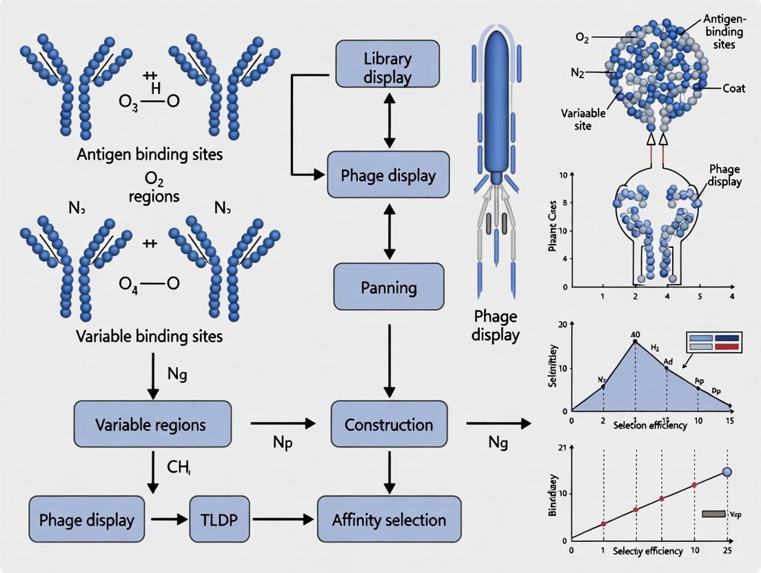
Biopanning is the core selection technique in phage display technology, enabling the isolation of high-affinity antibody fragments from vast combinatorial libraries for affinity maturation research. This process is foundational for developing novel therapeutics, as it mimics natural selection in vitro, allowing researchers to evolve antibodies with progressively higher binding affinities for a specific target antigen over successive rounds. The principle hinges on the physical linkage between the phenotype (the antibody fragment displayed on the phage surface) and the genotype (the DNA encoding that antibody inside the phage particle) [2] [27]. By iteratively selecting for binding to an immobilized target, amplifying the bound phages, and extracting them for the next round, a pool of binders can be enriched from a library of billions to a handful of high-affinity candidates [28].
The M13 Bacteriophage: The Core Vehicle for Display
Central to antibody phage display is the biology of the M13 filamentous bacteriophage. This phage is particularly suited for display because it is non-lytic, establishing a chronic infection in its host E. coli and allowing for continuous phage production without killing the bacterial cell [2]. The phage particle consists of a single-stranded DNA genome encapsulated by several coat proteins. For display applications, antibody fragments (such as scFvs or Fabs) are typically fused to one of the minor coat proteins, most commonly the gene III protein (G3P) [2]. This fusion allows the antibody to be presented on the surface of the phage while its genetic material is housed within, preserving the critical genotype-phenotype link. The M13 phage infects E. coli via the F-pilus, and its assembly is a complex process involving the coordination of multiple proteins to export the single-stranded DNA genome from the cell while simultaneously coating it with the G8P major coat protein and incorporating the minor coat proteins, including the antibody-G3P fusion, at one end [2].
Detailed Biopanning Protocol
The following section provides a detailed, step-by-step protocol for performing a biopanning experiment, from library preparation to the final elution of enriched phage clones.
Pre-Panning: Library and Reagent Preparation
Materials:
- Phage Display Library: A naïve, immunized, or synthetic library displaying antibody fragments (e.g., scFv, sdAb) [28].
- Target Antigen: A purified protein, peptide, or cell-surface receptor.
- Blocking Buffer: 3-5% Bovine Serum Albumin (BSA) or milk protein in a suitable buffer like PBS or TBS.
- Washing Buffer: PBS or TBS, often with a low concentration of a non-ionic detergent (e.g., 0.1% Tween-20).
- Elution Buffer: An acidic solution (e.g., 0.1 M Glycine-HCl, pH 2.2) or a solution containing a competitive ligand to disrupt antibody-antigen binding.
- Neutralization Buffer: 1 M Tris-HCl, pH 9.1.
- E. coli Strain: An F-positive laboratory strain such as TG1 or XL1-Blue.
- Growth Media: Lysogeny Broth (LB) with appropriate antibiotics (e.g., Tetracycline for E. coli maintenance, Ampicillin for phagemid selection).
The Panning Workflow: A Step-by-Step Guide
The biopanning process is an iterative cycle designed to enrich for phage particles that display antibodies binding specifically to a target antigen. The workflow is summarized in the diagram below.
Step 1: Antigen Immobilization and Blocking
- Immobilize the Target Antigen. Coat a solid surface (e.g., an immunotube, a well of a microtiter plate, or magnetic beads) with the target antigen in a suitable buffer (e.g., PBS or carbonate-bicarbonate buffer). Incubate overnight at 4°C or for 1-2 hours at 37°C. The typical antigen concentration ranges from 5-20 µg/mL.
- Block Non-Specific Sites. Remove the antigen solution and wash the surface once with PBS. Add an excess of blocking buffer (e.g., 3-5% BSA) to cover all potential non-specific binding sites on the surface. Incubate for 1-2 hours at room temperature or overnight at 4°C.
- Wash. Perform several washes with a washing buffer (e.g., PBS with 0.1% Tween-20) to remove excess blocking agent.
Step 2: Phage Library Incubation and Binding
- Add the Phage Library. Incubate the pre-blocked phage antibody library (typically 10^10 - 10^13 phage particles in blocking buffer) with the immobilized antigen. To pre-clear non-specific binders, the library can first be incubated with a non-coated, blocked surface.
- Incubate with Agitation. Allow the phage to bind to the antigen by incubating for 30-90 minutes at room temperature with gentle agitation.
Step 3: Washing to Remove Non-Specific Phage
- Stringent Washes. Remove the unbound phage library and perform a series of stringent washes. Start with 10-20 gentle washes using a washing buffer with detergent (e.g., PBS with 0.1% Tween-20) to remove weakly bound phage.
- Increase Stringency. In subsequent panning rounds, increase the stringency by increasing the number of washes (e.g., up to 20 in round 2 and 30 in round 3) or by using buffers with higher detergent concentrations (e.g., 0.5% Tween-20) to select for the highest affinity binders.
Step 4: Elution of Specifically Bound Phage
- Acidic Elution. The most common method is to add an acidic elution buffer (e.g., 0.1 M Glycine-HCl, pH 2.2) to the antigen surface and incubate for 5-15 minutes. This low pH disrupts the antibody-antigen interaction.
- Competitive Elution. Alternatively, a competitive ligand or an excess of soluble antigen can be used to displace the bound phage, which can help select for phages binding to a specific epitope.
- Neutralize. Immediately transfer the eluted phage to a tube containing neutralization buffer (e.g., 1 M Tris-HCl, pH 9.1) to prevent damage to the phage particles. This neutralized eluate contains the enriched pool of antigen-specific phage.
Post-Panning: Amplification and Analysis
Step 5: Amplification of Eluted Phage
- Infect E. coli. Mix the neutralized eluate with a log-phase culture of an F-positive E. coli strain (e.g., TG1). Incubate without antibiotic selection for 30-60 minutes at 37°C to allow for phage infection.
- Plate for Titering. To determine the number of eluted phage (output titer), make a dilution series of the infected cells and plate on LB-agar plates containing the appropriate antibiotic (e.g., Amp for phagemid selection).
- Culture for Phage Production. Inoculate the remaining infected cells into a larger volume of LB broth with antibiotic and a helper phage (if using a phagemid system). Grow overnight at 37°C with shaking. The helper phage provides the necessary proteins for the packaging and assembly of new phage particles displaying the antibody fragments.
- Precipitate Phage. The next day, purify the amplified phage particles from the culture supernatant by precipitation with polyethylene glycol (PEG)/NaCl. Resuspend the PEG-pelleted phage in PBS or a suitable buffer. This amplified phage pool serves as the input for the next round of biopanning.
Step 6: Monitoring Enrichment and Characterization
- Monitor Enrichment. The output titer from each round should be calculated and compared to the input titer. A successful panning campaign will show a significant increase in the output titer over 3-4 rounds, indicating enrichment of antigen-binding clones.
- Characterize Individual Clones. After 3-4 rounds, individual bacterial colonies from the titer plates are picked, and the displayed antibody fragments are produced for characterization. This typically involves:
- ELISA: To confirm specific binding to the target antigen and cross-reactivity against non-target proteins.
- Sequencing: To identify unique antibody clones and group them by sequence families.
- Affinity Measurement: Using techniques like Surface Plasmon Resonance (SPR) or Biolayer Interferometry (BLI) to determine the binding kinetics (KD) of the selected antibodies.
Critical Factors for Successful Biopanning
Quantitative Standards for Key Steps
The following table outlines the critical parameters and their quantitative targets for a successful biopanning campaign.
Table 1: Key Biopanning Parameters and Quantitative Standards
| Parameter | Optimal Value / Range | Purpose and Rationale |
|---|---|---|
| Library Size | >10^9 - 10^11 unique clones [28] | Ensures sufficient diversity for discovering rare, high-affinity binders. |
| Antigen Coating Concentration | 5-20 µg/mL | Provides sufficient antigen density for phage binding without causing steric hindrance. |
| Wash Stringency (Tween-20) | 0.1% (R1) to 0.5% (R3-4) | Removes non-specific and weak binders; increasing stringency selects for higher affinity. |
| Number of Washes | 10 (R1), 20 (R2), 30+ (R3+) | Progressively removes weakly bound phage to enrich for high-affinity clones. |
| Enrichment Factor (EF) | Should increase >10-fold per round [27] | A key metric indicating successful selection of target-specific phage. |
| Target CDR3 Diversity Post-Panning | >50-80% unique sequences (for sdAb) [28] | Indicates a diverse candidate pool, essential for finding optimal leads with different epitopes. |
Library Selection and Bias Mitigation
The choice of antibody library is a fundamental decision. Synthetic libraries, built on a limited number of stable human frameworks with randomized complementarity-determining regions (CDRs), offer the advantage of a fully human origin, bypassing the need for humanization [28]. They are particularly noted for generating high epitope diversity against a target. In contrast, immunized libraries can yield higher initial affinities but may be limited in diversity due to immunological tolerance and a bias towards immunodominant epitopes [28].
A major challenge in biopanning is the enrichment of target-unrelated peptides (TUPs), particularly propagation-related TUPs (Pr-TUPs). These are phage clones that are enriched due to a growth advantage in the E. coli host rather than specific antigen binding [27]. Mitigation strategies include:
- Pre-clearing: Incubating the library with a non-coated, blocked surface before panning.
- Negative Selection: Incubating the amplified output of a round with a non-target surface or a related protein to subtract cross-reactive binders.
- Using Multiple Library Lots: As different lots of the same commercial library can show substantial heterogeneity, using multiple lots can increase the chance of discovering genuine binders [27].
- Next-Generation Sequencing (NGS): Employing NGS to monitor the entire phage pool across rounds allows for the identification of over-represented sequences that may be Pr-TUPs, enabling data cleaning and the focus on truly enriched binders [27].
The Scientist's Toolkit: Essential Reagents and Materials
Table 2: Essential Research Reagent Solutions for Biopanning
| Reagent / Material | Function / Application | Examples / Specifications |
|---|---|---|
| M13 Phage Display Library | Source of antibody diversity for selection. | Synthetic human sdAb library, naïve human scFv library, immunized VH library [28]. |
| F-positive E. coli Strain | Bacterial host for phage infection and amplification. | TG1, XL1-Blue; must express the F-pilus for M13 infection [2]. |
| Helper Phage | Provides structural and assembly proteins in trans for phage production in a phagemid system. | M13K07, VCSM13; often carry a Kanamycin resistance marker. |
| PEG/NaCl Solution | Precipitates and purifies phage particles from bacterial culture supernatants. | Standard solution: 20% PEG-8000, 2.5 M NaCl. |
| Blocking Agent | Reduces non-specific binding of phage to surfaces. | 3-5% BSA (protease-free), Skim milk powder. |
| Tween-20 | Non-ionic detergent used in wash buffers to reduce non-specific binding. | Used at 0.1% - 0.5% (v/v) in PBS or TBS. |
| NGS Services/Kits | For deep sequencing of the phage pool to monitor enrichment and identify biases. | Illumina platform; used for analysis of naive and amplified libraries [27]. |
In the field of therapeutic antibody development, affinity maturation is a critical process for enhancing the binding strength between an antibody and its target antigen. Phage display technology serves as a powerful in vitro platform for this purpose, simulating the natural immune system's evolutionary process [22]. This Application Note details three core diversification techniques—error-prone PCR, chain shuffling, and site-directed mutagenesis—employed during phage display campaigns to generate antibody variants with superior affinity and stability. These methods enable researchers to construct highly diverse mutant libraries from which clones with enhanced biophysical properties can be isolated [29] [30] [31]. The following sections provide comprehensive protocols, quantitative comparisons, and practical guidance for implementing these techniques within the context of antibody affinity maturation research.
The successful application of phage display for affinity maturation relies on the strategic creation of genetic diversity within antibody genes. The three techniques discussed here offer complementary approaches for library generation.
Error-prone PCR introduces random mutations throughout the antibody gene by exploiting the low fidelity of DNA polymerases under optimized PCR conditions [29] [22]. This method is particularly valuable when structural information about the antibody-antigen complex is lacking, as it allows for unexplored regions of sequence space to be sampled. Recent protocols have demonstrated the construction of libraries with theoretical sizes exceeding 10^8 transformants, with functional diversity reaching up to 69% as confirmed by Next Generation Sequencing (NGS) analysis [8].
Chain Shuffling mimics the natural pairing of antibody heavy and light chains by sequentially replacing one chain while keeping the other fixed, effectively accessing novel paratope configurations from pre-existing immune repertoires [30] [32]. This approach was historically proven to achieve substantial affinity improvements, with documented cases of 20-fold enhancement after light chain shuffling followed by an additional 15-fold improvement after heavy chain hypervariable loop shuffling, culminating in antibodies with dissociation constants (Kd) in the nanomolar range [30].
Site-Directed Mutagenesis represents a rational approach that focuses genetic diversity on specific residues within the Complementarity-Determining Regions (CDRs) known to participate in antigen binding [31] [33]. This method requires structural insights from X-ray crystallography or computational modeling but offers higher precision and reduced screening burden. When applied to an anti-polysialic acid antibody, comprehensive mutational scanning of 14 paratope residues led to the discovery of affinity-enhanced variants with up to ~7-fold improved binding in IgG format [31].
Table 1: Comparative Analysis of Diversification Techniques for Antibody Affinity Maturation
| Technique | Mechanism of Diversity | Library Size Typical Range | Affinity Improvement Potential | Structural Information Required | Key Applications |
|---|---|---|---|---|---|
| Error-Prone PCR | Random nucleotide misincorporation during amplification | 10^8 - 10^9 transformants [8] | Varies; can yield functionally diverse libraries (69% unique sequences) [8] | None | Primary library generation, stability maturation [29] |
| Chain Shuffling | Sequential replacement of heavy or light chains with naive repertoires | 10^9 independently transformed clones [32] | ~300-fold combined improvement documented [30] | Limited | Human antibody optimization, mimicking natural affinity maturation [30] [32] |
| Site-Directed Mutagenesis | Targeted substitution of specific CDR residues | ~266 variants for 14 positions (19 substitutions each) [31] | ~7-fold improvement in IgG documented [31] | Extensive (crystal structure or high-quality models) | Fine-tuning of known binders, specificity enhancement [31] [33] |
Table 2: Quantitative Outcomes from Affinity Maturation Studies
| Antibody Target | Diversification Technique | Initial Affinity (Kd) | Evolved Affinity (Kd) | Fold Improvement | Reference |
|---|---|---|---|---|---|
| PhOx hapten | Chain Shuffling | 3.2 × 10^−7 M | 1.1 × 10^−9 M | ~300-fold | [30] |
| PolySia | Site-Directed Mutagenesis | ~4 μM (scFv) | ~0.57 μM (IgG) | ~7-fold | [31] |
| Galectin-3 | Error-Prone PCR + Machine Learning | Not specified | EC50 = 3.46 μM | Not specified | [34] |
| Nucleolin | Random Mutagenesis (in vivo) | Not specified | 50% reduction in sequence diversity post-selection | Enrichment achieved | [8] |
Experimental Protocols
Error-Prone PCR for Random Mutagenesis
Introduction Error-prone PCR enables the introduction of random mutations throughout antibody coding sequences by leveraging low-fidelity polymerization conditions. This protocol is adapted from established methods for antibody affinity and stability maturation [29], incorporating recent advancements in library characterization [8].
Materials
- Template DNA: Plasmid encoding antibody fragment of interest
- Primers: Flanking the variable region with appropriate restriction sites
- PCR reagents: MgCl₂, MnCl₂, unbalanced dNTP concentrations, Taq polymerase
- Mutator strain: E. coli JS200 (alternative to traditional XL1-Red) [8]
Procedure
- Set up error-prone PCR reaction:
- Prepare 100 μL reaction containing: 10-100 ng template DNA, 1× PCR buffer, 0.2 mM each dATP and dGTP, 1 mM each dCTP and dTTP, 0.5 mM MnCl₂, 7 mM MgCl₂, 50 pmol each primer, 5 U Taq DNA polymerase [29].
- Thermal cycling conditions: Initial denaturation at 95°C for 3 min; 25-30 cycles of 95°C for 45 sec, 55°C for 45 sec, 72°C for 1 min/kb; final extension at 72°C for 7 min.
Purify and clone mutated products:
- Purify PCR product using commercial kit to remove enzymes and salts.
- Digest purified product and vector with appropriate restriction enzymes.
- Ligate mutated antibody genes into phage display vector and transform into E. coli.
Iterative mutagenesis (optional):
- For enhanced diversity, subject output from first round to additional error-prone PCR cycles.
- Alternatively, use mutator bacterial strains like JS200 for in vivo diversification over multiple generations [8].
Library validation:
- Determine library size by plating serial dilutions of transformed bacteria.
- Assess diversity by NGS on 10-20 library clones or bulk population [8].
Troubleshooting
- Low mutation rate: Increase MnCl₂ or MgCl₂ concentration; use additional mutagens.
- Excessive stop codons: Reduce cycle number or use mutator strains with lower frameshift rates [8].
- Small library size: Optimize electroporation conditions; use higher quality vector DNA.
Chain Shuffling for Combinatorial Diversity
Introduction Chain shuffling sequentially replaces either heavy or light chains of an existing antibody with naive repertoires, generating combinatorial diversity while preserving some binding characteristics. This protocol is based on the seminal work describing construction of high-affinity human antibodies by chain shuffling [30], with refinements from more recent applications [32].
Materials
- Template: Phagemid encoding parent antibody
- RNA/cDNA: From immunized or naive donors
- PCR reagents: High-fidelity polymerase, primers for antibody variable regions
- Phage display vector: pComb3X or similar with appropriate selection markers
Procedure
- Amplify chain repertoires:
- Isolve total RNA from donor lymphocytes (human/murine).
- Perform RT-PCR using degenerate primers specific for VH, Vκ, and Vλ families.
- Alternatively, use synthetic gene libraries for enhanced diversity control.
First shuffling round (light chain):
- Clone light chain repertoire into vector containing parent heavy chain.
- Transform library and determine size (typically 10^9 clones for comprehensive coverage).
- Select binders through 2-3 rounds of phage panning.
Second shuffling round (heavy chain):
- Isolate improved light chains from first round.
- Clone heavy chain repertoire with selected light chains.
- Repeat selection under increased stringency.
Characterization of shuffled antibodies:
- Express selected clones as soluble Fab or IgG.
- Determine affinity using BLI/SPR; specificity via ELISA/cell binding [32].
Troubleshooting
- Limited diversity: Increase donor number; optimize primer design for comprehensive coverage.
- Loss of binding: Include parent chain as control; use lower stringency in early selection rounds.
- Expression issues: Framework engineering to improve stability of novel chain pairings.
Site-Directed Mutagenesis for Targeted Optimization
Introduction Site-directed mutagenesis focuses diversity on specific CDR residues known to participate in antigen binding, enabling rational affinity enhancement. This protocol draws from successful applications in optimizing anti-glycan antibodies [31] and incorporates contemporary computational approaches [33].
Materials
- Structural data: Crystal structure of antibody-antigen complex or high-quality models
- Template: Phagemid encoding parent antibody
- Primers: Degenerate oligonucleotides covering targeted CDR residues
- PCR reagents: High-fidelity polymerase, DpnI restriction enzyme
Procedure
- Paratope analysis:
- Identify residues contacting antigen from crystal structure if available [31].
- Alternatively, use computational docking and molecular dynamics simulations.
- Select 10-15 key positions for saturation mutagenesis.
Library design and construction:
- Design NNK-degenerate primers for each target position (encodes all 20 amino acids).
- Perform site-saturation mutagenesis individually or in combination.
- Use overlap extension PCR or quick-change mutagenesis protocols.
- Digest template DNA with DpnI to remove methylated parent plasmids.
Library characterization:
- Determine diversity by sequencing 20-30 random clones.
- Assess functional coverage by phage ELISA before selection.
Selection under stringent conditions:
- Apply off-rate selection using acid or competitive elution.
- Gradually decrease antigen concentration over panning rounds.
- Include counter-selection against related antigens to maintain specificity [31].
Troubleshooting
- Low mutation efficiency: Optimize primer design; increase number of PCR cycles.
- Specificity loss: Incorporate negative selection steps during panning.
- Limited improvement: Expand targeted residues to include peripheral contact sites.
The Scientist's Toolkit: Essential Research Reagents
Table 3: Key Research Reagent Solutions for Affinity Maturation
| Reagent/Resource | Function and Application | Examples/Specifications |
|---|---|---|
| Phage Display Vectors | Display antibody fragments on phage surface for selection | pComb3X system [8]; pC3C for Fab display [32] |
| Mutator Bacterial Strains | In vivo random mutagenesis for library generation | E. coli JS200 [8]; XL1-Red for lower mutation frequency |
| NGS Platforms | Library diversity assessment and clone identification | Illumina MiSeq for full-length VHH sequencing (up to 600 bp) [8] |
| Affinity Measurement Instruments | Quantitative assessment of binding improvements | BLI (Octet platform); SPR (Biacore system) [22] |
| Structural Prediction Tools | Antibody-antigen modeling for rational design | AlphaFold, RoseTTAFold, DeepAb [33] |
| Molecular Docking Software | Identification of key residues for mutagenesis | AutoDock, AutoDock Vina, DOCK [22] |
Error-prone PCR, chain shuffling, and site-directed mutagenesis represent powerful and complementary approaches for antibody affinity maturation within phage display research. Error-prone PCR offers the advantage of random, unbiased diversification without requiring structural information. Chain shuffling leverages natural antibody repertoire diversity to generate functional variants through combinatorial pairing. Site-directed mutagenesis provides a rational, focused approach that maximizes the probability of affinity enhancements while minimizing library size. The integration of computational tools and NGS analysis with these traditional methods is revolutionizing the field, enabling more efficient and predictive affinity maturation campaigns [34] [33]. By selecting the appropriate diversification strategy based on available structural information, desired library size, and screening capacity, researchers can systematically engineer antibody variants with dramatically improved binding properties for therapeutic and diagnostic applications.
Affinity maturation is a critical process in antibody development, aimed at increasing the binding affinity and specificity of antibodies for their target antigens. Phage display technology serves as a powerful in vitro counterpart to the natural in vivo process, allowing for the selection of high-affinity antibody variants from large combinatorial libraries. This case study examines two distinct, innovative approaches to antibody affinity maturation: the development of TCR-like antibodies for the recognition of peptide-HLA complexes, and the engineering of anti-nucleolin antibodies for potential therapeutic applications. By comparing these methodologies, we provide a comprehensive framework for researchers undertaking similar antibody engineering projects, highlighting advanced techniques in library construction, selection, and characterization.
TCR-like Antibody Affinity Maturation
Background and Objective
T-cell receptor (TCR)-like antibodies represent a unique class of engineered biologics capable of recognizing peptide-HLA (pHLA) complexes, a ligand traditionally restricted to T-cell recognition. The objective of this case study was to develop HLA-DQ2.5 gluten peptide-specific TCR-like antibodies with very high affinity for application in celiac disease research and potential therapy [35] [36]. The challenge lies in achieving the picomolar affinity required for effective detection and neutralization of specific pHLA complexes.
Experimental Protocol and Workflow
The affinity maturation process followed a sequential development path combining phage display with computational structural modeling.
Library Construction and Selection:
- Initial Lead Identification: A phage-displayed antibody library was panned against the target HLA-DQ2.5 gluten peptide complex.
- CDR Engineering: Complementarity-determining regions (CDRs) were mutated to create a library of variants. In silico structural modeling informed which residues to target for mutagenesis to optimize antigen interactions [35].
- Framework Engineering: Concurrently, framework regions were engineered to improve the overall thermostability of the antibody scaffold, ensuring the protein remained stable throughout the selection process and subsequent applications [35].
Screening and Characterization:
- Phage Display Selection: Multiple rounds of biopanning were performed under conditions of increasing stringency to enrich for clones with the highest affinity.
- Affinity Assessment: Selected clones were expressed as soluble antibodies and their binding affinity was quantified using surface plasmon resonance (SPR) or similar biophysical techniques.
- Structural Validation: The final lead candidates were validated through structural analysis to confirm the predicted binding mode.
The following workflow diagram illustrates this integrated experimental process:
Key Outcomes and Data
The sequential strategy of lead identification, CDR engineering, and framework engineering proved highly successful. The informed approach, guided by structural modeling, allowed for the efficient identification of HLA-DQ2.5 gluten peptide-specific TCR-like antibodies achieving low picomolar affinity [35]. This level of affinity is crucial for targeting the typically low-abundance pHLA complexes on cell surfaces.
Table 1: Key Results from TCR-like Antibody Affinity Maturation
| Parameter | Outcome | Significance |
|---|---|---|
| Final Affinity | Low Picomolar | Essential for sensitive detection of cell-surface pHLA complexes. |
| Engineering Strategy | Combined CDR & Framework Optimization | Improved both binding affinity and molecular stability. |
| Guidance Method | Structural Modeling | Enabled efficient and targeted mutagenesis. |
Anti-Nucleolin Antibody Affinity Maturation
Background and Objective
Nucleolin is a protein overexpressed on the surface of highly proliferating cells within the tumor microenvironment, making it an attractive target for cancer therapy. This case study details a novel protocol for the affinity maturation of an anti-nucleolin VHH (single-domain antibody) previously developed by the research group. The objective was to create an internalizing antibody library and select clones capable of being internalized by target cancer cells, which is a critical property for antibody-drug conjugates [8].
Experimental Protocol and Workflow
This protocol utilized a unique in vivo random mutagenesis approach for library construction, coupled with a cell-based selection strategy.
Library Construction via In Vivo Mutagenesis:
- Plasmid Preparation: The reference anti-nucleolin VHH coding sequence was cloned into a pComb3X vector.
- In Vivo Random Mutagenesis: The constructed plasmid was transformed into an E. coli JS200 mutator strain. This strain harbors a low-fidelity DNA polymerase I that introduces random mutations preferentially in the ColE1 origin-based plasmid, mutating the antibody gene over multiple generations [8].
- Library Characterization: The theoretical library size was 2.19 × 10^8 transformants. Next-Generation Sequencing (NGS) analysis revealed that 78.29% of the sequences were functional (without stop codons), and 69.24% of these were unique sequences, indicating a high-quality and diverse library [8].
Cell-Based Phage Display Selection:
- Selection on Whole Cells: The antibody phage library was panned against nucleolin-overexpressing tumor cells. This approach presents the target in its native conformation and allows for the selection of antibodies that can internalize [8].
- Enrichment Monitoring: A single selection round resulted in an enrichment of the mutated antibody library, evidenced by an almost 50% decrease in sequence diversity in the post-selection pool [8].
The workflow for this method is distinct from the TCR-like approach, as shown below:
Key Outcomes and Data
The use of the JS200 mutator strain provided a simple and effective method for introducing diversity without the need for primer design or subcloning steps typically associated with in vitro mutagenesis. The cell-based selection successfully enriched for antibody clones capable of binding to and internalizing into nucleolin-overexpressing cells in a single round. NGS provided an extensive characterization of the library and post-selection pool, offering valuable data for guiding future selection and analysis [8].
Table 2: Key Results from Anti-Nucleolin Antibody Development
| Parameter | Outcome | Significance |
|---|---|---|
| Mutagenesis Method | In vivo (JS200 E. coli) | Simple method avoiding error-prone PCR. |
| Functional Diversity | 69.24% unique sequences | High-quality library with vast functional repertoire. |
| Selection Method | Whole-cell panning | Ensures target is in native conformation and selects for internalizing clones. |
| Enrichment Efficiency | ~50% diversity drop post-selection | Rapid and efficient enrichment of binders after a single round. |
Comparative Analysis & Technical Considerations
Comparison of Affinity Maturation Strategies
The two case studies demonstrate complementary strategies for antibody affinity maturation. The TCR-like antibody effort employed a rational, structure-guided approach, while the anti-nucleolin project leveraged random mutagenesis coupled with a sophisticated phenotypic screen (internalization).
Table 3: Comparison of the Two Affinity Maturation Protocols
| Aspect | TCR-like Antibody Approach | Anti-Nucleolin Antibody Approach |
|---|---|---|
| Mutagenesis Strategy | Targeted CDR and framework engineering. | In vivo random mutagenesis across the entire gene. |
| Library Size | Not specified, but likely smaller and focused. | 2.19 × 10^8 transformants. |
| Selection Antigen | Defined pHLA complex. | Whole cells (native nucleolin in context). |
| Primary Goal | Maximize affinity (picomolar). | Obtain functional, internalizing antibodies. |
| Key Techniques | Phage display, structural modeling, SPR. | Phage display, NGS, cell-based selection. |
| NGS Role | Implied for clone analysis. | Extensive library and pool characterization. |
Critical Technical Considerations
Successful implementation of these protocols requires attention to several key factors:
Minimizing Bias in Phage Selections: Amplification steps during biopanning can introduce propagation-related target-unrelated peptides (Pr-TUPs), where peptides that confer a growth advantage in the host bacterium become enriched regardless of target binding [27]. Using NGS to monitor the evolution of the phage pool during amplification is crucial for identifying and filtering out these false positives [27].
The Power of NGS: As demonstrated in both case studies, NGS is indispensable for modern antibody engineering. It allows for deep characterization of library diversity, monitoring the enrichment of specific clones during selection, and identifying mutations that contribute to improved affinity and function [27] [8].
Selection Strategy: The choice between a purified antigen and a whole-cell panning has profound implications. Purified antigens allow for stringent selection of high-affinity binders to a specific epitope. In contrast, whole-cell panning selects for antibodies that recognize the target in its native membrane environment, which is critical for developing therapeutic antibodies that must bind to cells in vivo [8].
The Scientist's Toolkit: Key Research Reagents
The following table lists essential materials and reagents used in the featured experiments, which are critical for replicating these affinity maturation workflows.
Table 4: Essential Research Reagents for Phage Display-based Affinity Maturation
| Reagent / Material | Function / Application | Example from Case Studies |
|---|---|---|
| Phagemid Vector | Carries antibody gene and allows display on phage surface. | pComb3X vector [8]. |
| Mutator Bacterial Strain | Introduces random mutations for library generation. | E. coli JS200 strain [8]. |
| Next-Generation Sequencing (NGS) | High-throughput analysis of library diversity and selection outputs. | Illumina MiSeq for VHH library characterization [8]. |
| Target Antigen/Cell Line | The molecule or cell against which selections are performed. | HLA-DQ2.5 gluten complex [35]; nucleolin-overexpressing cancer cells [8]. |
| Structural Modeling Software | Informs rational design of mutagenesis libraries. | Software used for guiding CDR engineering in TCR-like antibodies [35]. |
This case study highlights two successful, yet methodologically distinct, pathways for antibody affinity maturation using phage display. The development of TCR-like antibodies showcases the power of integrating structural modeling with rational library design to achieve extreme, picomolar affinity for a challenging target. Conversely, the anti-nucleolin antibody project demonstrates the efficacy of a random mutagenesis approach combined with a functional, cell-based selection to obtain antibodies with a desired biological property (internalization). Both approaches heavily relied on advanced tools like NGS for deep data analysis. The choice between these strategies depends on the specific project goals, the availability of structural information for the target, and the required functional properties of the final antibody. Together, they provide a robust toolkit for researchers aiming to develop high-affinity antibodies for therapeutic and diagnostic applications.
The pursuit of antibodies against complex antigenic targets, particularly G-protein coupled receptors (GPCRs) and peptide-loaded human leukocyte antigen (pHLA) complexes, represents a frontier in therapeutic drug development. These target classes are critically implicated in a wide array of diseases, including cancer, autoimmune disorders, and metabolic conditions, yet their structural intricacies have historically made them resistant to conventional antibody discovery methods [37]. GPCRs, as seven-transmembrane proteins, present limited extracellular epitope space and require stabilization in a lipid bilayer to maintain native conformation, complicating their use in typical in vitro screening assays [37]. Similarly, pHLA complexes present a challenging target due to their shallow interaction grooves and the subtle molecular differences between closely related allelic variants.
Phage display technology has emerged as a particularly powerful platform for confronting these challenges. Its key advantage lies in the precise control it affords over selection conditions, enabling researchers to bias outcomes toward antibodies that recognize specific conformational states or that bind only in particular microenvironments, such as the acidic conditions of a tumor [11]. Furthermore, the ability to construct and screen exceptionally diverse synthetic libraries—containing over 10^11 unique members—dramatically increases the probability of identifying rare, high-affinity binders to these difficult targets [11] [28]. This application note details successful experimental protocols and strategic insights for leveraging phage display to generate functional antibodies against GPCRs and pHLA complexes, providing a structured framework for researchers engaged in antibody affinity maturation.
Strategic Approaches and Library Design
The foundation of a successful campaign against complex antigens is a well-designed phage display library. Modern synthetic libraries are engineered not just for size, but for the quality of diversity and the inherent developability of the antibodies they encode.
Library Configuration and Diversity Generation
Synthetic libraries can be constructed in various formats, including single-chain variable fragments (scFvs), antigen-binding fragments (Fabs), and single-domain antibodies (sdAbs or VHHs) [28] [38]. The choice of format influences the paratope geometry and screening outcomes. For instance, smaller sdAbs can access cryptic epitopes that might be sterically hindered for larger Fab fragments [28].
A critical design principle is the strategic diversification of Complementarity-Determining Regions (CDRs). Leading libraries, such as the Pioneer library, diversify all six CDRs based on consensus sequences from human rearranged and affinity-matured immunoglobulins [11]. This approach recapitulates natural diversity while being curated to remove sequence liabilities that could lead to poor expression or stability. The following table summarizes the design features of a state-of-the-art synthetic library.
Table 1: Design Features of a Modern Synthetic Antibody Library (Pioneer Library)
| Feature | Description | Rationale |
|---|---|---|
| Library Size | ~2.2 x 10^11 functional members [11] | Maximizes probability of finding rare, high-affinity binders. |
| Germline Genes | IGHV1-69, IGHV3-23, IGKV1-39, IGLV3-21 [11] | Selected for high expression in E. coli, high display rates, and high frequency in clinical-stage antibodies. |
| Diversification | All six CDRs diversified based on natural human antibody sequences [11] | Creates diverse paratope geometries while maintaining favorable biophysical properties. |
| Format | Fab (antigen-binding fragment) [11] | Structurally closest to native immunoglobulin; easy conversion to full-length IgG. |
| Display System | SpyDisplay (utilizes SpyTag-SpyCatcher ligation) [11] | Enables rapid conversion of selected Fabs into various screening and functional formats. |
Specialized Selection Strategies: Moving Beyond In Vitro Panning
Conventional in vitro panning against purified recombinant antigens often fails for GPCRs and pHLA due to difficulties in preserving native conformation. Integrating more physiologically relevant selection strategies is key.
- In vivo Phage Display: This involves administering the phage library directly into an animal model. Phages are subsequently recovered from target tissues. This strategy selects for clones that not only bind the target but also possess the necessary pharmacokinetic properties to reach it in a living organism, including stability in serum and ability to extravasate [38] [39].
- Cell-Based Panning: Using whole cells that overexpress the native target GPCR or pHLA complex for selection helps ensure that identified antibodies recognize the antigen in its natural membrane-embedded context. This can be powerfully combined with negative selection using cells that do not express the target to subtract non-specific binders [37] [40].
- Incorporation of Competitive Elution: To drive selection toward antibodies that bind functionally relevant epitopes (e.g., the ligand-binding pocket of a GPCR), elution can be performed using a known natural ligand or a competing antibody. This enriches for clones with potential antagonistic or agonistic activity [37].
The following workflow diagram integrates these advanced strategies into a cohesive protocol for targeting complex antigens.
Diagram 1: Integrated workflow for phage display selection against complex antigens, combining advanced panning methods with AI-driven analysis.
Application Note 1: GPCR-Targeting Protocol
Background and Objectives
GPCRs form the largest family of cell-surface receptors and are prime therapeutic targets. However, their complex transmembrane structure and limited extracellular domain accessibility have resulted in very few approved antibody therapeutics, with only four FDA-approved GPCR-targeted antibodies as of 2025 [37]. The objective of this protocol is to outline a successful phage display-based strategy for generating high-affinity, functional antibody fragments against a challenging GPCR target.
Detailed Experimental Methodology
1. Antigen Preparation and Library Selection:
- Genetic Immunization: Instead of using purified protein, immunize (for in vivo approaches) or present the target GPCR via plasmid DNA or viral vectors. This ensures the receptor is expressed in its native conformation on the cell surface [37].
- Cell-Based Panning: For in vitro selections, use a stable cell line overexpressing the target GPCR. A critical control is an isogenic cell line lacking the GPCR (e.g., a knockout line) for counter-selection. Incubate the phage library with the GPCR-negative cells first (pre-clearing), then transfer the supernatant to the GPCR-positive cells. After washing, elute specifically bound phages [37] [40].
2. Binding Validation and Screening:
- Primary Screening: Screen monoclonal phage outputs using cell-based ELISA or flow cytometry against the GPCR-expressing cells versus control cells to confirm specific binding to the native target [40].
- Characterization: Convert positive scFv or Fab hits to full-length IgG for downstream functional assays. This is streamlined by systems like SpyDisplay, which allows modular assembly [11].
3. Functional and Developability Assessment:
- Test antibodies for their ability to modulate GPCR signaling (e.g., cAMP accumulation, β-arrestin recruitment) to identify agonists or antagonists.
- Employ AI-driven tools early to predict developability liabilities (e.g., aggregation propensity, poor solubility) from sequence data, allowing prioritization of leads with the highest clinical potential [40].
Key Results and Outcomes
This general approach has been successfully validated against multiple GPCRs. For instance, using a large synthetic Fab library (Pioneer), high-affinity antibodies were directly selected against the GPCRs CXCR4 and C5aR, which are considered challenging targets [11]. These antibodies exhibited excellent affinity, specificity, and potent antagonistic activity in cellular assays, demonstrating parameters comparable to late-stage clinical candidates [11].
Table 2: Summary of Phage Display Successes Against Challenging Target Classes
| Target Class | Specific Target | Library / Platform Used | Key Outcome |
|---|---|---|---|
| GPCR | CXCR4, C5aR [11] | Pioneer Synthetic Fab Library (SpyDisplay) | Antibodies with high affinity and potent antagonistic function selected directly from the library. |
| Immune Checkpoint | TIGIT, TIM3 [40] | Phage Display scFv Library + AI profiling | Identification of leads with defined epitopes and predicted antagonistic function. AI filtered out candidates with poor developability. |
| Drosophila Secreted Proteins | CARPB, Nesfatin-1 [41] | Phage-Displayed Synthetic Nanobody Library | Generated nanobodies suitable for immunostaining and immunoblotting, validating the platform for diverse antigens. |
Application Note 2: pHLA-Targeting Strategy
Background and Objectives
pHLA complexes present tumor-specific or viral peptides to T-cell receptors, making them attractive targets for novel immunotherapies. The challenge lies in generating antibodies that are exquisitely specific for a single pHLA combination, discriminating between the presented peptide and the vast array of other HLAs and peptides. This protocol describes a strategy for isolating such specific antibodies using phage display.
Detailed Experimental Methodology
1. Antigen Design and Production:
- Produce recombinant, biotinylated pHLA monomers. It is crucial to include controls with HLA loaded with irrelevant peptides to enable discrimination of peptide-specific binders.
- Fold and purify stable pHLA complexes, then multimerize them on streptavidin-coated magnetic beads or plates to increase avidity and mimic the natural cell surface presentation.
2. Subtractive Panning Strategy:
- Round 1 (Pre-clearing): Incubate the phage library with beads coupled to HLA molecules loaded with a non-target peptide (or a mixture of common peptides) to deplete phages that bind the HLA framework or common peptides.
- Round 2 (Positive Selection): Incubate the pre-cleared library with beads coupled to the target pHLA complex.
- Round 3 (Negative Selection): To further enhance specificity, perform a subsequent negative selection against the irrelevant pHLA complex. Elute bound phages from the positive selection under stringent conditions (e.g., low pH buffer or protease cleavage).
3. Cross-reactivity Screening:
- Screen outputted clones against a panel of related pHLA complexes (e.g., same HLA with different peptides, or different HLAs with the same peptide) using surface plasmon resonance (SPR) or a multiplexed ELISA. This identifies clones with the desired specificity profile.
Integration with AI and Structural Modeling
The selection process is significantly enhanced by integrating AI tools for epitope mapping and affinity prediction. As demonstrated in the anti-TIM3 and anti-TIGIT campaign, structural models of antibody-target complexes can be generated in silico to predict the epitope and infer function, such as whether the antibody will block the interaction with the natural ligand [40]. This AI-enhanced profiling allows for data-driven candidate selection before costly and time-consuming experimental characterization.
Diagram 2: AI-powered profiling pipeline for characterizing phage-derived antibodies, predicting epitope, function, and developability early in the discovery process.
The Scientist's Toolkit: Essential Research Reagents
Table 3: Key Reagent Solutions for Phage Display Against Complex Antigens
| Research Reagent / Tool | Function in Workflow | Specific Application / Example |
|---|---|---|
| Synthetic Fab Library | Source of diverse, fully human antibody candidates. | Pioneer library (2.2x10^11 members) with pre-optimized developability [11]. |
| SpyTag/SpyCatcher System | Modular conjugation for rapid reformatting of selected Fabs. | SpyDisplay system enables quick conversion of selected Fabs into IgG or other formats for screening [11]. |
| Stable GPCR/pHLA Cell Lines | Provides natively folded antigen for cell-based panning and validation. | Essential for presenting GPCRs in their native conformation; requires proper membrane context [37] [40]. |
| AI-Powered Profiling Software | In silico prediction of epitope, affinity, and developability liabilities. | Tools like those from MAbSilico filter candidates pre-experimentally (e.g., removed 2/5 anti-TIM3 leads for poor developability) [40]. |
| Phagemid Vector System | Genetic backbone for displaying antibody fragments on phage surface. | Vectors based on M13 filamentous phage, typically using pIII or pVIII coat proteins for display [39] [41]. |
Phage display technology, when empowered by sophisticated library design, physiologically relevant selection strategies, and modern computational tools, is capable of overcoming the historic challenges posed by complex antigens like GPCRs and pHLA. The protocols and data outlined herein demonstrate that through integrated workflows—combining in vivo or cell-based panning with AI-driven candidate selection—researchers can directly generate functional, developable lead antibody candidates. This approach significantly de-risks and accelerates the early discovery pipeline, paving the way for a new generation of therapeutics targeting previously intractable disease pathways. As synthetic library diversity continues to grow and in silico prediction tools become more sophisticated, the synergy between phage display and computational biology is poised to unlock an even broader range of therapeutic targets.
Incorporating NGS and Cell-Based Panning for Enhanced Selection
The integration of next-generation sequencing (NGS) with cell-based panning represents a transformative approach in phage display technology, significantly accelerating antibody affinity maturation and the discovery of high-value therapeutic candidates. This application note details standardized protocols for implementing this combined methodology, which enables researchers to quantitatively track enrichment kinetics, identify rare clones, and select for antibodies targeting antigens in their native physiological context. Within the broader scope of phage display for antibody affinity maturation research, this synergistic workflow provides unprecedented depth and efficiency in isolating internalizing antibodies and targeting challenging membrane proteins, such as G-protein coupled receptors (GPCRs).
Phage display has, for decades, provided a robust platform for discovering peptide ligands and antibodies by linking phenotypic expression to genotypic information [42] [43]. The conventional process involves iterative biopanning rounds to enrich phages that bind a target of interest, followed by Sanger sequencing of individual clones. While effective, this approach offers low-throughput monitoring of library diversity and enrichment.
The integration of Next-Generation Sequencing (NGS) revolutionizes this pipeline by enabling the quantitative, high-resolution analysis of millions of phage sequences in parallel [43]. When coupled with cell-based panning—where selections are performed on intact cells to present target antigens in their native conformation and cellular context—this integrated strategy offers a powerful tool for discovering antibodies against complex cell surface targets, such as GPCRs and internalizing receptors for drug delivery applications [11] [8]. This application note provides detailed protocols and data analysis frameworks for implementing this enhanced selection strategy.
Next-Generation Sequencing in Phage Display
The Role of NGS in Affinity Maturation
NGS transforms phage display analysis from a low-throughput screening process to a comprehensive, data-driven discovery engine. Its primary advantages over traditional Sanger sequencing are summarized in Table 1.
Table 1: Comparison of Sanger Sequencing and Next-Generation Sequencing (NGS) in Phage Display
| Feature | Sanger Sequencing | Deep Sequencing (NGS) |
|---|---|---|
| Throughput | 96–384 sequences per run | Millions of reads per run |
| Sensitivity to Minor Clones | Low | High – detects rare binders (<0.01%) |
| Quantitative Clonal Abundance | No | Yes – provides frequency distribution per sequence |
| Round-to-Round Enrichment Tracking | Manual, slow, and limited | Automated, real-time resolution |
| Motif and Family Group Identification | Limited | Robust clustering and alignment possible |
| Time and Cost Efficiency | Expensive per sequence | Low cost per million sequences [43] |
The quantitative data from NGS allows for the high-resolution monitoring of selection dynamics, capturing the entire diversity landscape of a library and tracking the enrichment of specific clones across biopanning rounds [43]. This is particularly valuable in affinity maturation campaigns, where a primary antibody is mutated to create a secondary library, and researchers must identify subtle, beneficial mutations that confer higher affinity [42] [8].
Experimental Protocol: NGS Library Preparation from Phage Pools
This protocol describes how to prepare a phage display library for NGS analysis following a round of biopanning.
Materials & Reagents:
- QIAprep Spin M13 Kit (Qiagen): For high-purity phage DNA extraction.
- Phusion High-Fidelity DNA Polymerase (Thermo Scientific): For PCR amplification with minimal error introduction.
- NGS Indexing Kit (e.g., Illumina Nextera XT): For multiplexing samples.
- Agencourt AMPure XP beads (Beckman Coulter): For PCR purification and size selection.
Procedure:
- Phage DNA Extraction: Isolate phage genomic DNA from an eluted phage pool (approximately 10^12 phage particles) using the QIAprep Spin M13 Kit according to the manufacturer's instructions. Elute DNA in nuclease-free water.
- PCR Amplification of Variable Regions: Set up a PCR reaction to amplify the fragment containing the variable antibody region (e.g., scFv, VHH).
- Template: 10 ng of extracted phage DNA.
- Primers: Design primers flanking the variable region and containing overhangs compatible with your NGS platform.
- Polymerase: Use a high-fidelity polymerase to minimize PCR-introduced errors.
- Cycling Conditions: Follow polymerase manufacturer's guidelines, typically 25-30 cycles.
- Indexing and Library Preparation: Clean the PCR product with AMPure XP beads. In a subsequent limited-cycle PCR, add unique dual-index sequences to each sample to allow for multiplexing.
- Quality Control and Sequencing: Quantify the final library using a fluorometric method (e.g., Qubit). Assess library fragment size distribution using a bioanalyzer (e.g., Agilent 2100). Pool indexed libraries at equimolar concentrations and sequence on an appropriate platform (e.g., Illumina MiSeq for 2x300 bp paired-end reads to cover full CDR regions) [43].
Cell-Based Panning for Native Antigen Selection
Rationale and Applications
Cell-based panning involves using live cells as the selection matrix during biopanning. This presents the target antigen in its native conformation, correct post-translational modifications, and within the context of the cellular membrane and its associated proteins [8]. This method is critical for:
- Targeting Challenging Proteins: Isolating antibodies against complex membrane proteins like GPCRs and ion channels, which are difficult to produce and purify in a functional form [11].
- Selecting Internalizing Antibodies: Identifying antibodies that are not only cell-surface binding but also capable of being internalized. This is a key requirement for developing antibody-drug conjugates (ADCs) that need to deliver their cytotoxic payload inside the cell [8].
- Tissue and Cell-Type Specificity: Selecting for antibodies that bind specifically to a particular cell type (e.g., cancer cells) while minimizing binding to non-target cells.
Experimental Protocol: Cell-Based Panning for Internalizing Antibodies
This protocol is designed for the positive selection of antibodies that bind to and are internalized by a target cell line.
Materials & Reagents:
- Target Cells: A cell line that natively overexpresses the target antigen (e.g., a cancer cell line).
- Control Cells: An isogenic or similar cell line that does not express the target antigen, for counter-selection.
- Dulbecco's Phosphate-Buffered Saline (DPBS): For washing cells.
- Acid Elution Buffer (0.1 M Glycine-HCl, pH 2.2): To elute surface-bound phages.
- Trypsin-EDTA (0.25%): To isolate internalized phages.
- Phage Display Library: A diverse library (e.g., a synthetic human Fab library like Pioneer [11] or a mutated VHH library [8]).
Procedure:
- Pre-clearance: Incubate the phage library with control cells for 1 hour at 4°C on a rotator. Collect the supernatant, which now contains phages depleted of binders to common, non-target cell surface antigens.
- Positive Selection: Incubate the pre-cleared phage library with the target cells for 2 hours at 4°C (to allow binding but inhibit internalization).
- Remove Surface-Bound Phages: Wash cells gently with cold DPBS to remove unbound phages. Add acid elution buffer for 10 minutes on ice to elute phages that are bound to the cell surface but not internalized. Neutralize the eluate immediately.
- Recovery of Internalized Phages: Wash the cells again with DPBS. To recover internalized phages, trypsinize the cells and transfer them to a fresh tube. Lyse the cells using a hypotonic lysis buffer or repeated freeze-thaw cycles.
- Amplification and Iteration: The lysate containing internalized phages is used to infect E. coli for amplification. The amplified output is used as input for 2-4 subsequent rounds of panning, with increasing stringency in washing [8].
The following diagram illustrates the key steps and decision points in this cell-based panning protocol for selecting internalizing antibodies.
Data Integration and Analysis: From NGS Reads to Candidate Hits
The power of integration lies in combining the cellular functional selection of cell-based panning with the deep data analysis provided by NGS.
Bioinformatic Analysis Workflow
The bioinformatic processing of NGS data from phage display experiments follows a structured pipeline to convert raw sequencing reads into a ranked list of candidate antibodies, as visualized below.
- Sequence Processing: Raw NGS reads are quality-filtered, and paired-end reads are merged. Sequences are then translated and filtered to remove those containing stop codons, ensuring the analysis focuses on functional antibody sequences [8].
- Diversity Assessment: The processed sequences are clustered to determine the functional diversity of the library. A successful selection round will show a significant drop in diversity, indicating enrichment. For example, one study reported a decrease in sequence diversity of almost 50% after a single round of cell-based selection [8].
- Enrichment Scoring: The frequency of each unique antibody sequence is tracked across successive rounds of panning. Clones that demonstrate a consistent and significant increase in frequency are prioritized as high-affinity binders.
Case Study: Affinity Maturation of an Anti-Nucleolin VHH
A 2024 study exemplifies this workflow. Researchers constructed a mutagenic library of an anti-nucleolin VHH using an E. coli JS200 mutator strain. They then performed a single round of cell-based panning on nucleolin-overexpressing cancer cells to select for internalizing clones [8].
- NGS Library Characterization: Post-selection NGS of 9,683 sequences revealed 5,249 unique clusters (69% functional diversity), with 92% of clusters consisting of single copies, indicating a highly diverse library.
- Mutation Analysis: NGS coverage of the entire VHH fragment allowed for analysis of mutation frequency per position, showing the highest variability in the CDR3 region, which is critical for antigen binding.
- Outcome: The single selection round successfully enriched the library, reducing its sequence diversity by nearly half and providing a refined pool of candidates for further characterization [8].
Table 2: Key NGS Metrics from an Anti-Nucleolin VHH Library [8]
| Metric | Post-Mutagenesis Library | Post-Cell Panning Pool |
|---|---|---|
| Total Sequences Analyzed | 9,683 | Not Specified |
| Functional Sequences (No Stop Codons) | 7,581 (78.3%) | Not Specified |
| Unique Sequence Clusters | 5,249 | ~50% Reduction (Estimated) |
| Single-Copy Clusters | 92.2% of all clusters | Not Specified |
The Scientist's Toolkit: Essential Research Reagents and Solutions
Successful implementation of this integrated workflow relies on key reagents and platforms. The following table details essential components.
Table 3: Research Reagent Solutions for NGS-Enhanced Cell-Based Panning
| Item Category | Specific Product/System | Function in the Workflow |
|---|---|---|
| Phage Display Library | Pioneer Synthetic Human Fab Library [11] | A highly diverse (2.2x10^11 members), synthetic library designed for high affinity and developability, ideal for discovering leads against diverse antigens like GPCRs. |
| Mutagenesis System | E. coli JS200 Mutator Strain [8] | An in vivo random mutagenesis system for creating diverse secondary libraries from a parent antibody clone during affinity maturation. |
| NGS Platform | Illumina MiSeq/NextSeq [43] | High-throughput sequencing platforms providing the read length (2x300 bp) and depth (millions of reads) required for comprehensive antibody library analysis. |
| Automated Colony Picking | QPix Microbial Colony Picker [44] | High-speed system (3,000 clones/hour) for automating the plating and picking of clones after panning, drastically speeding up the screening process. |
| Cell Culture Reagents | DPBS, Trypsin-EDTA, FBS | Essential reagents for maintaining target and control cell lines used in cell-based panning protocols. |
The confluence of cell-based panning and NGS represents a state-of-the-art methodology in phage display-based antibody discovery and affinity maturation. This synergistic approach allows researchers to move beyond simple binding to select for functionally relevant characteristics—such as internalization and native conformation binding—while using deep sequencing data to make informed, quantitative decisions on candidate selection. By adopting the detailed protocols and analytical frameworks outlined in this application note, research scientists and drug developers can significantly enhance the efficiency and success of their programs to discover novel therapeutic antibodies against even the most challenging targets.
Overcoming Common Challenges and Enhancing Platform Performance
Addressing Expression Bias and Folding Issues in E. coli
Within phage display campaigns for antibody affinity maturation, the efficient production of functional recombinant antibody fragments in Escherichia coli is a critical, yet often limiting, step. Expression bias—where certain sequences are poorly expressed or excluded from libraries—and protein misfolding can severely constrain the diversity and quality of selected candidates. These issues stem from inherent bacterial limitations, including a reducing cytoplasm incompatible with disulfide bond formation, codon usage biases, metabolic burden, and the absence of sophisticated eukaryotic folding machinery. This Application Note details targeted strategies and robust protocols to overcome these challenges, ensuring the generation of high-quality, diverse antibody libraries for therapeutic discovery.
Core Challenges and Strategic Solutions
The following table summarizes the primary bottlenecks in E. coli expression of antibody fragments and the corresponding strategic approaches to mitigate them.
Table 1: Key Challenges and Strategic Solutions for Antibody Expression in E. coli
| Challenge | Impact on Phage Display | Strategic Solution | Key Reagents/Strains |
|---|---|---|---|
| Cytoplasmic Redox Environment | Misfolding and loss of function in antibodies requiring disulfide bonds. | Use of engineered strains with oxidative cytoplasm; periplasmic expression. | SHuffle, Origami strains; strains expressing sulfhydryl oxidase (Erv1p) and isomerase (DsbC) [45]. |
| Metabolic Burden & Expression Bias | Reduced library diversity; overrepresentation of sequences that are easy to express but may have subpar binding characteristics. | Tunable expression systems; antibiotic-free plasmid maintenance; promoter and TIR optimization. | Lemo21(DE3) strain; phosphate-triggered expression systems; infA-based plasmid selection [45] [46]. |
| Translation Inefficiency | Low yields and potential misfolding due to ribosomal stalling at rare codons. | Codon optimization; use of strains carrying rare tRNA genes. | Rosetta and Rosettagami strains [47]. |
| Insufficient Solubility & Aggregation | Formation of inclusion bodies, rendering antibodies non-functional for display. | Fusion with solubility-enhancing tags; co-expression of chaperones; lower temperature induction. | MBP, SUMO, GST tags; CASPON technology [45] [48]. |
Optimizing the Cellular Folding Environment
For antibody fragments, particularly single-domain antibodies (VHH/sdAbs) and Fabs, the formation of correct intra-domain disulfide bonds is essential for stability and function. The standard BL21(DE3) strain has a reducing cytoplasm that inhibits this process.
Engineered Strains for Cytoplasmic Disulfide Bond Formation: Strains like SHuffle and Origami are genetically modified to promote disulfide bond formation in the cytoplasm. SHuffle cells express a mutant disulfide bond isomerase (DsbC) in the cytoplasm, which not only facilitates bond formation but also corrects mis-oxidized proteins [47]. For enhanced production of disulfide-rich proteins like nanobodies, a novel switchable system triggered by phosphate depletion has been developed. This system activates the expression of foldases (e.g., Erv1p and DsbC) at the stationary phase, achieving remarkable yields of up to 800 mg/L in shaker flasks [45].
Periplasmic Expression: Targeting antibody fragments to the periplasm leverages its naturally oxidizing environment and native disulfide bond formation machinery (DsbA, DsbC). This is achieved by cloning the antibody sequence downstream of a pelB or ompA signal peptide. Secretion into the periplasm can also simplify purification and minimize proteolytic degradation [48].
Minimizing Metabolic Burden and Expression Bias
Constitutive high-level expression of recombinant proteins can overwhelm the host cell's metabolism, leading to poor growth and genomic instability, which in turn biases the library representation.
- Tunable Expression Systems: Strains like Lemo21(DE3) allow fine-tuning of expression levels by varying the concentration of rhamnose, which controls the activity of T7 RNA polymerase. This is invaluable for expressing antibodies that are toxic to the host at high levels [47].
- Antibiotic-Free Selection: Traditional antibiotic resistance markers impose a metabolic load. A modern alternative is essential gene complementation, where the host strain is engineered to lack a vital gene (e.g.,
infA), and the expression plasmid provides it. This creates a strong selection pressure for plasmid maintenance without antibiotics, reducing metabolic burden and improving culture stability in high-density fermentations [45]. - Genetic Parts Optimization: The widely used pET vectors have inherent design flaws. Restoring the full consensus sequence of the T7 promoter (T7pCONS) and replacing the suboptimal Translation Initiation Region (TIR) with a synthetically evolved sequence have been shown to increase protein yields by over 100-fold in some cases, ensuring more reliable expression of diverse sequences [46].
Detailed Experimental Protocols
Protocol: High-Yield Production of Disulfide-Bonded Nanobodies Using a Switchable Strain
This protocol leverages a recently developed E. coli strain where the cytoplasmic redox state is switched from reducing to oxidizing at the onset of stationary phase [45].
Workflow Diagram: Switchable Disulfide Bond Formation System
Research Reagent Solutions
| Item | Function | Example |
|---|---|---|
| Switchable E. coli Strain | Engineered host that switches cytoplasm from reducing to oxidizing upon phosphate depletion. | Genetically modified strain with inducible glutaredoxin pathway knockout and foldase expression [45]. |
| Terrific Broth (TB) | High-density growth medium. | Contains glycerol and phosphate buffer; phosphate consumption triggers the system switch. |
| IPTG | Inducer for T7/lac-based expression vectors. | Added at stationary phase to begin recombinant protein production in the new oxidizing environment. |
| Affinity Chromatography Resin | Purification of tagged nanobodies. | Ni-NTA resin for His-tagged proteins; Protein A/G for Fc-fused nanobodies. |
Procedure:
- Day 1: Starter Culture. Inoculate a single colony of the engineered strain harboring your nanobody-pET plasmid into 5 mL of LB medium with appropriate antibiotics. Incubate overnight at 30°C with shaking (220 rpm).
- Day 2: Main Culture. Dilute the overnight culture 1:100 into fresh Terrific Broth (TB) medium. Grow at 37°C with vigorous shaking until the culture reaches the late exponential phase (OD600 ~0.6-0.8).
- Induction. At this point, the culture will naturally enter stationary phase due to phosphate depletion, activating the genetic switch. Immediately add IPTG to a final concentration of 0.5 mM to induce nanobody expression.
- Expression. Continue incubation for 16-20 hours at 30°C. The oxidizing environment and co-expression of foldases will promote the production of soluble, functional nanobodies.
- Day 3: Harvest. Harvest cells by centrifugation (4,000 x g, 20 min, 4°C). The cell pellet can be processed immediately for purification or stored at -80°C.
- Purification. Purify the nanobody using standard immobilized metal affinity chromatography (IMAC) under native, non-denaturing conditions to isolate correctly folded protein.
Protocol: Construction of a Phage Display Library Using a Mutator Strain
This protocol describes an in vivo random mutagenesis approach for affinity maturation using the E. coli JS200 mutator strain [8].
Workflow Diagram: Library Construction via In Vivo Mutagenesis
Research Reagent Solutions
| Item | Function | Example |
|---|---|---|
| JS200 E. coli Strain | In vivo random mutagenesis via low-fidelity DNA polymerase I. | Introduces mutations preferentially near the ColE1 origin of the phagemid [8]. |
| Phagemid Vector | Vector for phage display and mutagenesis. | pComb3X series vectors. |
| ER2738 E. coli Strain | F+ strain for efficient phage propagation and packaging. | Used for the final library expansion and phage rescue. |
| NGS Services | Quality control and diversity assessment of the built library. | Illumina MiSeq for full-length VHH sequencing. |
Procedure:
- Initial Cloning. Clone the gene for your parent antibody (e.g., anti-nucleolin VHH [8]) into a phagemid vector like pComb3X.
- Transformation into Mutator Strain. Transform the constructed plasmid into the JS200 mutator strain. Plate on selective media and incubate at 37°C.
- Iterative Mutagenesis.
- Pick a single colony to inoculate a liquid culture and grow to saturation (approximately 16-24 hours). Each growth cycle allows for the accumulation of random mutations in the plasmid DNA.
- Extract the plasmid pool from the culture.
- Use a portion of the extracted plasmid to re-transform fresh JS200 competent cells to begin the next round. Typically, 3-4 rounds of iterative mutagenesis are performed to achieve sufficient diversity.
- Library Pooling and Transformation. After the final round, extract the plasmid pool. Transform the mutagenized plasmid library into a high-efficiency F+ strain (e.g., ER2738) for phage display. The theoretical library size is determined by the transformation efficiency.
- Quality Control by NGS. Isolate plasmid DNA from a portion of the library and subject the antibody gene region to Next-Generation Sequencing (NGS). This critical step assesses the mutation rate, frequency of stop codons, and functional diversity of the library. A well-constructed library should have a high percentage (>78%) of sequences without stop codons [8].
- Phage Rescue. Rescue the phage library from the remaining cells using a helper phage (e.g., M13KO7) following standard protocols. The resulting phage library is now ready for panning campaigns.
The Scientist's Toolkit
Table 2: Essential Research Reagents for Overcoming Expression and Folding Hurdles
| Category | Item | Specific Function |
|---|---|---|
| Specialized E. coli Strains | SHuffle, Origami | Promotes disulfide bond formation in the cytoplasm. |
| Rosetta, Rosettagami | Supplies tRNAs for rare codons (AGA, AGG, AUA, CUA, etc.). | |
| Lemo21(DE3) | Tunable T7 expression via lysozyme (LysY) control for difficult/toxic proteins. | |
| C41(DE3), C43(DE3) | BL21 derivatives selected for robust expression of membrane and toxic proteins. | |
| Expression Vectors & Systems | pET Series with T7pCONS | High-yield expression vectors with an optimized full-consensus T7 promoter [46]. |
| pBAD, pLemo | Tightly regulated, tunable promoters (arabinose, rhamnose) to minimize metabolic burden. | |
| Fusion Tags & Technologies | CASPON | Fusion tag system for high-yield peptide production with efficient caspase cleavage [45]. |
| His-tag, MBP, SUMO | Affinity purification (His), and significant solubility enhancement (MBP, SUMO) [48]. | |
| Advanced Technologies | SpyTag/SpyCatcher | Modular protein ligation for rapid antibody conjugation and formatting (e.g., in Pioneer library [11]). |
| Cell-Free Protein Synthesis | Bypass cellular viability constraints for high-throughput screening of toxic proteins [49]. |
Success in antibody phage display is contingent upon the ability to generate and present vast libraries of functional, well-folded molecules. By strategically selecting host strains, optimizing genetic parts, and implementing tailored protocols for disulfide bond formation and library diversification, researchers can effectively overcome the classical limitations of the E. coli expression system. The methods outlined herein—from switchable redox strains to in vivo mutagenesis protocols—provide a robust framework for maximizing the quality and diversity of antibody libraries, thereby accelerating the discovery of high-affinity candidates for therapeutic development.
Navigating Library Size Constraints and Amplification Bias
In antibody affinity maturation research, phage display has established itself as a powerful in vitro selection technology. However, two significant technical challenges consistently impact the fidelity and outcome of discovery campaigns: library size constraints and amplification bias. Library size, often limited by bacterial transformation efficiency to approximately 10^7 - 10^10 transformants for phage display, directly restricts the functional diversity of antibody candidates available for selection [50] [1]. Simultaneously, the necessary amplification steps in bacterial hosts introduce systematic biases by preferentially enriching phage clones with superior propagation rates, regardless of their target affinity [27] [51]. These propagation-related target-unrelated peptides (Pr-TUPs) can dominate selection outputs, leading to misinterpretation and costly follow-up on false positives. This application note provides detailed methodologies for quantifying, monitoring, and mitigating these critical constraints within affinity maturation workflows.
Quantitative Assessment of Bias and Diversity
Measuring Library Composition Heterogeneity
Next-Generation Sequencing (NGS) provides the resolution necessary to quantitatively assess library composition and monitor bias introduction during amplification. A comparative analysis of different lots from the same commercial Ph.D.-12 phage display library revealed striking heterogeneity. The percentage of distinct sequences in naïve libraries varied dramatically between lots, from 61.89% in one lot to 96.66% in another, highlighting the inherent variability even in standardized libraries [27].
Table 1: Compositional Differences Between Library Lots During Amplification
| Parameter | Library Lot SA1 | Library Lot SA2 |
|---|---|---|
| Wildtype Clones in Naïve Library | 10.36% | 5.99% |
| Distinct Sequences in Naïve Library | 61.89% | 96.66% |
| Singleton Population in Naïve Library | 45.58% | 93.62% |
| Wildtype Clones After Round 3 Amplification | 90.37% (avg) | 61.81% (avg) |
During serial amplification, the frequency of wildtype clones increases significantly while diversity (number of unique sequences) substantially decreases [27]. This divergence in evolutionary fate between library lots underscores why a single, universal correction factor cannot be applied to biopanning results.
Tracking Amplification-Driven Enrichment
The Enrichment Factor (EF) quantifies how rapidly specific sequences dominate the population during amplification. Research shows that different library lots exhibit not only different starting points but also divergent enrichment patterns during amplification, with substantial differences in EFs for propagation-related sequences [27]. Motif analysis of the most enriched subpopulations in amplified libraries can identify sequence patterns hypothesized to contribute to increased amplification rates, providing signatures for bioinformatic filtering [27].
Experimental Protocols for Bias Evaluation
Protocol: NGS-Based Monitoring of Amplification Bias
This protocol enables researchers to quantify amplification bias in their specific phage display system.
Materials Required:
- Purified phage library (naïve or post-panning)
- Illumina sequencing platform or similar NGS service
- Python and MATLAB scripts for bioinformatic analysis (available in supplementary materials of [27])
- E. coli host strain (e.g., ER2738)
- LB culture medium and PEG/NaCl for phage precipitation
Procedure:
- Serial Amplification: Conduct three rounds of duplicate serial amplification of the phage library. For each round, infect log-phase E. coli culture with phage library, incubate with shaking, precipitate phage particles using PEG/NaCl, and resuspend in appropriate buffer [27].
- DNA Extraction: Purify DNA from all samples (naïve library and each amplification round).
- Library Preparation & Sequencing: Prepare DNA for NGS using an Illumina platform according to manufacturer protocols. Ensure sufficient sequencing depth to capture library diversity.
- Bioinformatic Analysis:
- Process FASTQ files using Python scripts, cleaning reads based on Phred score and peptide insert presence.
- Calculate wildtype percentage, unique sequence counts, and enrichment factors (EFs) for all sequences.
- Perform motif discovery on sequences with EF > 1 to identify propagation-related motifs.
- Data Interpretation: Compare diversity metrics and EF distributions across amplification rounds. Identify sequences with disproportionately high EFs as potential Pr-TUPs for future filtering.
Protocol: Cell-Based Phage Display Selection with Bias Control
This protocol, adapted from affinity maturation work on anti-nucleolin antibodies, integrates methods to minimize bias impact during selection [8].
Materials Required:
- Phage display library (e.g., constructed in pComb3X vector)
- Target cells expressing antigen of interest
- Control cells (antigen-negative)
- ER2738 E. coli strain
- Helper phage (e.g., VCSM13)
- Coating buffers, washing buffers, and elution buffers
Procedure:
- Pre-clearance: Incubate phage library with control cells for 1 hour at 4°C to remove non-specific binders. Collect unbound phage for selection.
- Positive Selection: Incubate pre-cleared library with target cells for 2 hours at 4°C with gentle agitation.
- Stringent Washing: Wash cells 5-10 times with cold PBS containing 0.1% Tween-20 to remove weakly bound phages.
- Internalization Phase (Optional): For internalizing antibodies, incubate cells at 37°C for 30 minutes to allow internalization, then treat with low-pH glycine buffer to remove surface-bound phages.
- Elution: Lyse cells to recover internalized phages or use specific elution buffers for surface-bound phages.
- Amplification & Analysis: Infect E. coli with eluted phage, rescue with helper phage, and precipitate amplified phage for subsequent rounds. After 3-4 rounds, subject output to NGS analysis alongside amplification control data to distinguish true binders from Pr-TUPs.
Visualization of Bias Assessment Workflow
NGS Bias Assessment Workflow - This diagram illustrates the integrated experimental and computational pipeline for systematic identification of amplification bias in phage display libraries.
The Scientist's Toolkit: Essential Research Reagents
Table 2: Key Reagents for Bias-Aware Phage Display
| Reagent / Tool | Function | Application Notes |
|---|---|---|
| pComb3X Phagemid | Phage display vector for antibody fragment expression | Dual-functionality for phage display and soluble antibody production; compatible with helper phage systems [8]. |
| Illumina NGS Platform | High-throughput sequencing of phage pools | Enables diversity assessment and Pr-TUP identification; requires 1.5μg DNA for library characterization [27] [8]. |
| ER2738 E. coli Strain | Host for phage propagation | Standard host for M13 phage; essential for library amplification and production [1]. |
| JS200 E. coli Mutator | In vivo random mutagenesis | Low-fidelity DNA polymerase introduces diversity for affinity maturation; mutation hotspot near ColE1 origin [8]. |
| Helper Phage (VCSM13) | Provides phage structural proteins | Essential for phage particle formation when using phagemid systems; typical multiplicity of infection 20:1 [1]. |
| Python/MATLAB Scripts | Bioinformatic analysis of NGS data | Calculates enrichment factors, diversity metrics, and identifies propagation motifs [27]. |
Mitigation Strategies and Alternative Approaches
Computational Filtering of Pr-TUPs
Bioinformatic analysis enables systematic identification and filtering of propagation-related sequences. Researchers should:
- Establish an amplification control experiment (amplification without selection)
- Calculate enrichment factors for all sequences in amplification control
- Identify sequences with disproportionately high EFs as potential Pr-TUPs
- Cross-reference biopanning outputs against Pr-TUP database before hit selection
- Use motif analysis to identify and filter propagation-related sequence patterns [27] [51]
Alternative Display Technologies
While phage display remains valuable, alternative technologies address some limitations:
CIS Display offers a completely cell-free selection system with library diversity >10^14 variants, bypassing bacterial transformation bottlenecks. Without cellular propagation, it eliminates amplification bias, though it has a less established track record for antibody discovery compared to phage display [50].
Yeast Display provides eukaryotic expression and sorting capabilities, while Mammalian Display offers native post-translational modifications, though with potential inconsistent expression challenges [14].
Practical Guidelines for Library Construction and Handling
- Library Quality Control: Implement NGS characterization of naïve libraries to establish baseline diversity and identify propagation-prone sequences before selection [14].
- Amplification Minimization: Use the minimum number of amplification rounds necessary (typically 3-4) to achieve enrichment [1].
- Library Sourcing: Be aware that different lots of the same commercial library may show significant compositional heterogeneity [27].
- Hybrid Approaches: Consider using phage display for initial selection followed by CIS Display for affinity maturation to leverage the strengths of both platforms [50].
Navigating library size constraints and amplification bias requires integrated experimental and computational strategies. By implementing the protocols and mitigation strategies outlined here, researchers can significantly improve the fidelity of phage display selections for antibody affinity maturation. The combination of systematic bias assessment through NGS, careful experimental design, and bioinformatic filtering of Pr-TUPs provides a robust framework for identifying genuine high-affinity binders amidst the challenges posed by limited library diversity and amplification artifacts.
Affinity maturation is a critical process in therapeutic antibody development, aimed at enhancing antibody binding affinity for improved therapeutic efficacy. This application note provides detailed protocols for implementing two powerful strategies for optimizing selection stringency during phage display-based affinity maturation: thermal challenge and competitive elution. These methodologies enable researchers to impose stringent selection pressures that mimic physiological conditions, facilitating the isolation of high-affinity antibody candidates with superior binding characteristics. By integrating these approaches into standard phage display workflows, developers can efficiently generate antibody variants with picomolar-range affinities essential for next-generation biologics.
In vitro affinity maturation recapitulates the natural immune system's process of generating high-affinity antibodies through iterative rounds of mutation and selection [8]. Phage display technology serves as a powerful platform for this process, allowing presentation and screening of diverse antibody libraries exceeding 10^11 members [42] [52]. The success of affinity maturation hinges on applying appropriate selection stringency to discriminate between clones with modest and superior binding characteristics.
Thermal and competition-based selection strategies introduce controlled stress during biopanning cycles, enabling identification of antibody variants with enhanced structural stability and binding affinity [42] [53]. These approaches are particularly valuable for targeting complex cell surface receptors and disease biomarkers where moderate affinity often limits therapeutic utility [8]. This protocol details the implementation of these stringency branches within standard phage display workflows for antibody affinity maturation.
Key Concepts and Principles
Selection Stringency in Affinity Maturation
Selection stringency refers to controlled experimental conditions that favor the recovery of rare, high-affinity binders from diverse antibody libraries. By manipulating parameters such as temperature, competitor concentration, and washing conditions, researchers can create evolutionary pressure that mimics physiological binding challenges [42].
Theoretical Basis for Stringency Optimization
Thermal stringency leverages the correlation between protein stability and binding affinity. Antibodies with improved structural stability typically demonstrate enhanced resistance to thermal denaturation while maintaining target binding capacity. Introducing controlled heat stress during selection enables enrichment of these thermostable, high-affinity variants [42].
Competition stringency operates on the principle of binding kinetics, where soluble competitors displace weaker binders while high-affinity antibodies remain target-bound. This approach directly selects for variants with slow dissociation rates, a critical parameter for therapeutic efficacy [52].
Table 1: Quantitative Parameters for Selection Stringency
| Stringency Type | Key Parameters | Typical Range | Effect on Selection |
|---|---|---|---|
| Thermal | Incubation Temperature | 37°C - 65°C | Selects for thermostable folds |
| Thermal Challenge Duration | 10 - 60 minutes | Discards unstable variants | |
| Recovery Period | 12 - 24 hours | Enriches functional expressors | |
| Competition | Competitor Concentration | 1 nM - 1 μM | Mimics physiological competition |
| Incubation Time | 30 - 120 minutes | Favors slow off-rates | |
| Target Density | Reduced 10-100 fold | Increases binding competition |
Research Reagent Solutions
Table 2: Essential Research Reagents for Stringency Optimization
| Reagent Category | Specific Examples | Function in Protocol |
|---|---|---|
| Phage Display System | M13 filamentous phage [54], pIII/pVIII display systems [54] | Antibody fragment presentation |
| Antibody Libraries | scFv/Fab libraries [55], Synthetic human antibody libraries [55] | Source of antibody diversity |
| Bacterial Strains | E. coli ER2738 [56], E. coli JS200 [8] | Phage propagation and diversity generation |
| Selection Targets | Bovine insulin [52], Cell surface receptors [53] | Binding affinity assessment |
| Detection Reagents | Anti-M13-biotin conjugate [57], SAPE [57] | Phage binding quantification |
| Competition Agents | Soluble antigen [52], Therapeutic antibodies [42] | Competitive elution |
Protocol: Thermal Stringency Branch
The thermal stringency branch incorporates controlled heat challenges during biopanning to select antibody variants with enhanced structural stability and binding capability under physiological temperatures.
Detailed Experimental Procedures
Library Preparation and Initial Binding
Phage Library Preparation: Amplify naïve scFv phage display library (approximately 10^11 diversity) in E. coli ER2738. Purify phage particles by PEG/NaCl precipitation and resuspend in PBS+ (PBS with 0.5 mM CaCl₂ and 10 mM MgCl₂) [58] [53].
Target Immobilization: Immobilize target antigen (e.g., PD-L1, insulin) on streptavidin-coated plates or magnetic beads using biotinylated antigen at 2.5-5 μg/mL in PBS+. Block remaining binding sites with 3% BSA in PBS for 1 hour at room temperature [58] [57].
Initial Binding Reaction: Incubate phage library (10^10 - 10^11 PFU) with immobilized target in binding buffer (PBS+ with 0.1% BSA) for 2 hours at room temperature with gentle agitation [57].
Thermal Challenge Application
Temperature Gradient Establishment: Prepare parallel samples with identical phage-target complexes. Subject samples to increasing temperature challenges (37°C, 45°C, 55°C, 65°C) for 30 minutes in a calibrated thermal cycler or water bath [42].
Controlled Recovery: Gradually cool samples to room temperature over 30 minutes. Maintain constant agitation to prevent non-specific binding during temperature transitions.
Stringent Washing: Wash samples 5-10 times with PBST (PBS with 0.1% Tween-20) using increasing wash stringency:
- Rounds 1-2: 5 quick washes (30 seconds each)
- Rounds 3-4: 10 extended washes (2 minutes each) [53]
Phage Recovery and Amplification
Acidic Elution: Elute bound phages with 0.1M glycine-HCl (pH 2.2) for 10 minutes with gentle agitation. Neutralize immediately with 1M Tris-HCl (pH 8.0) [56] [53].
Infective Amplification: Mix eluted phages with mid-log phase E. coli ER2738 culture (OD600 = 0.4-0.6). Incubate for 30 minutes at 37°C without shaking, then 30 minutes with shaking at 250 rpm. Plate on LB-tet plates (12 μg/mL tetracycline) for titering or proceed to liquid amplification for subsequent rounds [56].
Phage Precipitation: Amplify eluted phages in 20 mL E. coli ER2738 culture for 4.5 hours at 37°C with vigorous shaking (250 rpm). Precipitate with 1/6 volume PEG/NaCl (20% PEG-8000, 2.5M NaCl) overnight at 4°C. Resuspend pellet in 200 μL TBS for subsequent panning rounds [57].
Data Analysis and Interpretation
Output Titer Monitoring: Calculate phage recovery after each round using titering:
Successful enrichment typically shows 10-1000 fold increase in output titer over 3-4 rounds [53].
Binding Affinity Assessment: Characterize selected clones using Biolayer Interferometry (BLI) to determine kinetic parameters (KD, kon, koff). High-affinity antibodies typically exhibit KD values in low nanomolar to picomolar range after affinity maturation [58] [52].
Protocol: Competition Stringency Branch
The competition stringency branch employs soluble competitors during selection to displace weaker binders and enrich antibody variants with superior binding affinity and slow dissociation kinetics.
Detailed Experimental Procedures
Competition Agent Preparation
Competitor Selection: Choose appropriate competitors based on target:
- Soluble antigen: Same antigen as immobilized target (1-1000 nM)
- Therapeutic antibodies: Known high-affinity antibodies (10-100 μg/mL)
- Natural ligands: Receptor ligands for cell surface targets [52]
Competitor Titration: Prepare serial dilutions of competition agent in binding buffer (PBS+ with 0.1% BSA). Typical concentration range: 1 nM - 1 μM for small molecules, 10-1000 nM for proteins [52].
Competitive Selection Process
Pre-selection Binding: Incubate phage library with immobilized target as described in Section 4.2.1. Allow binding to reach equilibrium (typically 2 hours at room temperature).
Competitor Introduction: Add soluble competitor to binding reaction without disturbing phage-target complexes. Use increasing competitor concentration over selection rounds:
- Round 1: 1-10 nM competitor
- Round 2: 10-100 nM competitor
- Round 3: 100-1000 nM competitor [52]
Competitive Incubation: Incubate with competitor for 60-120 minutes at room temperature with gentle agitation. This extended incubation allows dissociation of weaker binders.
Minimal Washing: Perform 2-3 quick washes with ice-cold PBST to remove displaced phages while maintaining bound populations.
Recovery of High-Affinity Binders
Elution Options:
- Standard elution: 0.1M glycine-HCl (pH 2.2) for 10 minutes
- Proteolytic elution: Trypsin (0.25 mg/mL) for 30 minutes at 37°C for gentle recovery [58]
- Competitive elution: High concentration of soluble antigen (1-10 μM) for 60 minutes
Amplification and Titering: Follow same amplification protocol as thermal branch (Section 4.2.3). Monitor enrichment through increasing output titers despite competitive pressure.
Data Analysis and Interpretation
Competition Resistance Monitoring: Calculate competition resistance index:
Successful enrichment shows increasing CRI over selection rounds.
Next Generation Sequencing Analysis: Extract phage DNA from output populations using Qiagen M13 spin kit. Prepare Illumina sequencing library with two-step PCR using barcoded primers [56]. Analyze CDR3 diversity and mutation frequency to track affinity maturation.
Table 3: Expected Outcomes from Stringency Optimization
| Selection Round | Thermal Stringency | Competition Stringency | KD Range Expected |
|---|---|---|---|
| Initial Library | No thermal resistance | No competition resistance | 100 nM - 1 μM |
| Round 2 | Stable at 37°C | Resistant to 10 nM competitor | 10-50 nM |
| Round 3 | Stable at 45°C | Resistant to 100 nM competitor | 1-10 nM |
| Round 4 | Stable at 55°C | Resistant to 1 μM competitor | 100 pM - 1 nM |
Integrated Application and Validation
Combined Stringency Approaches
For maximum effect, implement thermal and competition branches sequentially or in parallel:
Sequential Application: Perform 2 rounds of thermal stringency followed by 2 rounds of competition stringency to select for both stability and binding affinity.
Simultaneous Application: Incorporate both thermal challenge (45°C) and low-level competition (10 nM) during same selection round for combined selective pressure.
Validation of Affinity-Matured Clones
Binding Kinetics: Characterize purified scFv or IgG antibodies using BLI or SPR. Ribosome-display matured antibodies can achieve KD values below 21 pM after conversion to IgG format [52].
Functional Assessment: Validate using cell-based assays relevant to therapeutic application. For PD-1/PD-L1 inhibitors, perform in vitro T-cell activation assays and in vivo syngeneic mouse models [58].
Structural Analysis: Identify key mutations responsible for affinity enhancement through crystallography or molecular modeling. Common beneficial mutations occur in CDR3 regions and framework regions stabilizing antibody structure [52].
Troubleshooting and Optimization
- Low Phage Recovery: Reduce stringency in early rounds; gradually increase temperature/competition in subsequent rounds
- Limited Diversity: Implement negative selection steps to remove non-specific binders; use larger library sizes (>10^10 diversity)
- Reduced Expression: Screen for improved expression variants; consider framework mutations to improve stability
- Specificity Loss: Incorporate counter-selection with related antigens to maintain binding specificity
By implementing these thermal and competition stringency branches, researchers can significantly enhance the efficiency of antibody affinity maturation, yielding candidates with the high affinity and stability required for therapeutic applications.
Strategies to Minimize Non-Functional Variants and Stop Codons
In antibody affinity maturation using phage display, the quality of the final output is fundamentally constrained by the initial library. A significant challenge in constructing these libraries is the prevalence of non-functional variants and sequences containing premature stop codons, which drastically reduce the pool of potential high-affinity binders. These non-functional sequences consume valuable screening resources and can obscure the identification of genuine hits during the biopanning process. Therefore, implementing robust strategies to minimize their occurrence is paramount to the efficiency and success of any affinity maturation campaign. This application note details practical, evidence-based methodologies to minimize non-functional variants and stop codons, framed within the broader context of optimizing phage display for antibody discovery and development. The protocols herein are designed for researchers, scientists, and drug development professionals engaged in therapeutic antibody engineering.
Strategic Approaches and Underlying Principles
Several strategic approaches can be employed to minimize the generation of non-functional variants, each targeting a different stage of the library construction process. The choice of strategy often depends on the desired diversity, the starting antibody sequence, and available resources. The following diagram illustrates the decision workflow for selecting the appropriate strategy.
Targeted Saturation Mutagenesis Using Degenerate Codons
A primary method for avoiding stop codons involves the strategic use of degenerate codons during oligonucleotide synthesis for library construction. Instead of using the fully degenerate NNK (N = A/T/G/C; K = G/T) codon, which encodes all 20 amino acids and one stop codon, researchers can employ tailored codon mixtures.
- Principle: Certain degenerate codons can provide broad amino acid diversity while completely excluding the introduction of stop (TAA, TAG, TGA) and cysteine codons. The latter is sometimes excluded to prevent the formation of non-native disulfide bonds that can compromise protein stability and folding [6].
- Implementation: In a study optimizing a humanized anti-ErbB2 antibody, a combination of three degenerate codons—NWG, NWC, and NSG (where N = A/T/G/C; W = A/T; S = G/C)—was used for amino acid saturation mutagenesis. This specific mixture encodes for all 20 canonical amino acids without introducing any cysteine or stop residues [6]. This approach allowed for the synthesis of 7,749 degenerate oligonucleotides on a microchip, which were used to construct high-quality single-chain antibody fragment (scFv) gene libraries.
Small Perturbation Mutagenesis (SPM)
For affinity maturation, it is often unnecessary to explore the entire sequence space of a CDR loop. The SPM strategy introduces limited diversity in a targeted manner.
- Principle: SPM focuses on diversifying a small number of candidate positions (e.g., three per CDR) selected through bioinformatic analysis. This minimizes disruptive mutations in framework regions that are critical for structural integrity [6].
- Implementation: Candidate residues for diversification are chosen based on criteria such as:
- Database Analysis: Residues with high observed frequency (Fobs > 90%) in antibody databases are generally conserved and excluded from randomization.
- Deviation from Consensus: If the residue in the parent antibody differs from the most frequent amino acid in the database, it is a candidate for diversification.
- Structural Insights: Residues spatially distant from the binding interface are considered less important and can be excluded [6].
In Vivo Random Mutagenesis with Quality Control
While random mutagenesis can generate unwanted stop codons, it remains a valuable tool when combined with stringent quality control measures.
- Principle: This method uses mutator bacterial strains, such as E. coli JS200, which harbor a low-fidelity DNA polymerase I. This enzyme introduces random mutations preferentially in the plasmid containing the antibody gene [8].
- Quality Assessment: Next-Generation Sequencing (NGS) is critical for characterizing the resulting library. In one study, a VHH library constructed via this method was sequenced, yielding 9,683 merged sequences. After translation, 78.29% (7,581 sequences) were free of stop codons, demonstrating a high functional content. Furthermore, 69.24% of these translated sequences were unique, indicating successful incorporation of functional diversity [8]. This data-driven approach allows researchers to quantify library quality before proceeding to resource-intensive panning.
Machine Learning-Guided Sequence Generation
Emerging approaches leverage NGS data and machine learning to generate novel, high-affinity sequences in silico, bypassing the random introduction of stop codons altogether.
- Principle: An enriched antibody sequence repertoire obtained from NGS of phage display panning outputs is used to train a deep learning model, such as a Long Short-Term Memory (LSTM) network. The trained model learns the sequence patterns associated with binding and can generate new, virtual sequences that mimic these properties [59].
- Implementation: The "likelihood" of a generated sequence, as calculated by the model, correlates with its binding affinity. Researchers can prioritize sequences with high likelihood scores for synthesis and testing. This method has been shown to generate antibody variants with over 1,800-fold higher affinity than the parental clone, all while operating on a curated set of functional sequences derived from experimental data [59].
Table 1: Comparison of Strategic Approaches for Minimizing Non-Functional Variants
| Strategy | Key Principle | Stop Codon Control | Typical Library Size/Functional Diversity | Best Suited For |
|---|---|---|---|---|
| Targeted Saturation Mutagenesis [6] | Uses defined degenerate codons (e.g., NWG, NSG) | Prevents incorporation from the outset | ( 4 \times 10^6 ) DNA sequences [6] | Focused optimization of known binding regions |
| Small Perturbation Mutagenesis (SPM) [6] | Diversifies a limited number of pre-selected residues | Combines controlled codons with rational design | Small, high-quality libraries | Affinity maturation when structural/database info is available |
| In Vivo Random Mutagenesis [8] | Relies on bacterial mutator strains; quality validated post-construction | Does not prevent stop codons; filters them via NGS | Theoretical: ( 2.19 \times 10^8 ) transformants; Functional: ~70% unique functional sequences [8] | Introducing broad diversity without complex oligonucleotide synthesis |
| Machine Learning-Guided Design [59] | Generates sequences in silico from enriched NGS data | Only functional sequences are physically constructed | N/A (Virtual generation) | Maximizing affinity gains from existing selection data |
Detailed Experimental Protocols
Protocol 1: Library Construction Using Targeted Saturation Mutagenesis
This protocol is adapted from a study that achieved a 158-fold affinity increase for an anti-ErbB2 antibody [6].
Research Reagent Solutions
Table 2: Key Reagents for Targeted Saturation Mutagenesis
| Item | Function/Description | Example Product/Catalog Number |
|---|---|---|
| Template scFv Gene | The starting antibody gene for affinity maturation. | e.g., HuA21 scFv in pCANTAB-5E [6] |
| PicoArray Microchips | High-throughput platform for synthesizing thousands of unique degenerate oligonucleotides. | 4k microfluidic PicoArray (LC Sciences) [6] |
| Pfu DNA Polymerase | High-fidelity polymerase for PCR amplification of library fragments. | Pfu polymerase (NEB) [6] |
| Phagemid Vector | Vector for antibody surface display on phage. | pCANTAB-5E (Amersham) [6] |
| Electrocompetent E. coli | High-efficiency bacterial cells for library transformation. | E. coli TG1 cells [6] |
Step-by-Step Procedure
Oligonucleotide Library Design and Synthesis:
- Identify candidate residues for diversification within the CDRs using database analysis (e.g., Absys antibody database) and structural insights [6].
- Design degenerate oligonucleotides that cover the targeted CDR regions. For each position to be randomized, use a mixture of the degenerate codons NWG, NWC, and NSG to saturate amino acid diversity without introducing stop codons or cysteine [6].
- Synthesize the pool of oligonucleotides (e.g., 7,749 oligos) on a programmable microfluidic microchip.
Construction of Mutant scFv Libraries:
- Amplify Diversified CDR Fragments: Perform a primary PCR (15 cycles) using the synthesized OligoMix as a template and specific overlapping primers.
- Amplify Flanking Gene Fragments: Separately amplify the N- and C-terminal gene fragments of the scFv from the wild-type plasmid using frame-specific primers (20 cycles).
- Assemble Full-Length scFv Genes: Use Overlapping Extension (OE)-PCR to fuse the three fragments (CDR, N-terminal, C-terminal) in equal molar amounts. Use vector-cloning primers for 30 cycles to generate the full-length scFv library.
- Clone into Phagemid Vector: Purify the OE-PCR products, digest them with appropriate restriction enzymes (e.g., Sfi I and Not I), and ligate them into the phagemid vector (e.g., pCANTAB-5E).
- Transform and Harvest Library: Transform the ligated product into electrocompetent E. coli TG1 cells. Harvest the plasmid library from the transformed cells for quality control and phage production.
Protocol 2: Quality Control and Candidate Identification via NGS
This protocol is critical for validating any library construction method, especially those involving random mutagenesis [8] [34].
Research Reagent Solutions
Table 3: Key Reagents for NGS-Based Quality Control
| Item | Function/Description | Example Product/Catalog Number |
|---|---|---|
| Phagemid DNA | The genetic material from the constructed library, either pre- or post-selection. | Extracted with QIAprep Miniprep kit (Qiagen) [6] |
| Frame-Specific Primers | PCR primers designed to amplify the diversified CDR regions for sequencing. | Custom designed [6] |
| NGS Sample Prep Kit | Prepares the amplified library for high-throughput sequencing. | TruSeq DNA sample preparation kit v2 (Illumina) [6] |
| NGS Platform | System for deep sequencing the antibody library. | Illumina MiSeq [8] |
Step-by-Step Procedure
Sample Preparation for Sequencing:
- Extract phagemid DNA from the electroporated bacterial cells (for the unselected library) or from phage-infected cells after panning (for the selected library).
- Perform a first PCR to amplify the CDR library fragments using frame-specific primers and high-fidelity Pfu polymerase (20 cycles).
- Purify the PCR products and prepare them for sequencing by adding 3' A-overhangs and ligating Illumina adaptors containing unique index sequences using a commercial kit (e.g., TruSeq DNA kit).
- Perform a second, low-cycle (e.g., 5 cycles) PCR to finalize the sequencing library [6].
Data Analysis and Variant Prioritization:
- Sequence Processing: Quality-filter the raw NGS reads to remove adapters and low-quality sequences. Merge paired-end reads.
- Translate and Filter: Translate the nucleotide sequences into amino acid sequences. Filter out sequences containing premature stop codons and frameshift mutations. Calculate the percentage of functional sequences as a key quality metric [8].
- Identify Enriched Variants: For post-panning analysis, compare the frequency of each unique sequence before and after selection. Variants that show significant enrichment are high-priority candidates for further characterization [6] [34].
- (Optional) Machine Learning: Use the enriched sequence repertoire as training data for an LSTM or other model to generate novel, high-likelihood candidates that may not have been highly abundant in the NGS data but are predicted to have high affinity [59].
The following diagram summarizes the integrated experimental and computational workflow for building and validating a high-quality library.
The strategic minimization of non-functional variants and stop codons is not merely a technical optimization but a foundational requirement for efficient antibody affinity maturation. By adopting methods such as rational degenerate codon design, small perturbation mutagenesis, and rigorous NGS-based quality control, researchers can construct phage display libraries with a high functional content. Furthermore, the integration of machine learning with NGS data presents a powerful, forward-looking paradigm for navigating sequence space intelligently. Together, these strategies ensure that screening efforts are concentrated on a rich pool of viable candidates, significantly accelerating the discovery of high-affinity therapeutic antibodies.
In the pursuit of therapeutic monoclonal antibodies, phage display technology has established itself as a powerful tool for discovering high-affinity binders. However, the isolation of a binding fragment represents merely the initial phase of a much longer developmental pathway. For a candidate to successfully transition into a viable therapeutic, it must possess not only high affinity for its target but also favorable biophysical properties and the capacity to be reformatted into a stable, manufacturable final format. This application note details protocols for assessing the reformattability and developability of antibody fragments—typically single-chain variable fragments (scFvs) or variable domains of heavy-chain-only antibodies (VHHs)—discovered through phage display, within the broader context of antibody affinity maturation research. The primary goal is to provide a structured framework for researchers to identify and advance candidates with the highest probability of clinical success, focusing on properties that minimize immunogenicity and ensure robust production [60].
Background
Therapeutic antibody development has increasingly prioritized fully human sequences to enhance safety and efficacy profiles. The generation of anti-drug antibodies (ADA) in patients can alter pharmacokinetics, reduce efficacy, and pose safety risks. While the aphorism "the more human, the less potential for immunogenic responses" has guided library design, immunogenicity is a multifactorial problem influenced by the antibody sequence, format, formulation, and patient population [60]. Consequently, in vitro selection methods, including phage display, must be designed and executed with these end-goals in mind from the earliest stages.
A critical transition point in the development pipeline is the reformatting of selected antibody fragments (e.g., scFvs, VHHs) into a full-length Immunoglobulin G (IgG) or other bivalent formats. This step is not merely a change in structure; it can profoundly impact the antigen-binding affinity, specificity, and functional activity of the molecule. A binder that performs exceptionally as a scFv may exhibit unforeseen issues—such as aggregation or loss of expression—when reformatted into an IgG [60]. Therefore, a systematic assessment of reformattability is essential.
Parallel to reformattability is the concept of developability, which encompasses the biophysical and biochemical properties that dictate whether an antibody candidate can be developed into a stable, manufacturable, and safe drug product. Key aspects of developability include:
- Structural stability and resistance to aggregation.
- Solubility at high concentrations required for therapeutic dosing.
- Low immunogenicity potential, often assessed through in silico tools.
- Resilience to chemical degradation (e.g., deamidation, oxidation).
The integration of reformattability and developability assessments early in the affinity maturation workflow helps to de-risk downstream development by filtering out problematic candidates and focusing resources on those with the most promising therapeutic profiles [60].
Key Assessment Workflows
The journey from a selected phage display clone to a developable lead candidate involves several critical, interconnected assessment phases. The following workflow diagrams and protocols outline this process.
From Phage Display to Developable Lead
The diagram below illustrates the core pathway for advancing and assessing antibody clones post-selection.
Affinity Maturation Integrating Developability
For affinity maturation campaigns, assessing developability is crucial when working with enriched mutant libraries. The following diagram outlines this integrated process, which can be initiated from a lead candidate requiring optimization.
Experimental Protocols
Protocol 1: Reformatting scFv/VHH to IgG Format
This protocol describes the molecular cloning and initial expression of a full-length IgG from a phage display-derived variable region [60].
- Principle: The genes encoding the variable heavy (VH) and variable light (VL) domains of a selected scFv (or the VHH from a camelid antibody) are cloned into mammalian expression vectors containing the constant regions of human IgG1 and the appropriate light chain (kappa or lambda). The vectors are then co-transfected into a mammalian cell line for expression, allowing for the production of a bivalent, glycosylated antibody.
- Materials:
- Plasmid DNA encoding the scFv/VHH.
- Mammalian expression vectors with human IgG1 constant regions.
- Restriction enzymes and DNA ligase or Gibson Assembly mix.
- Competent E. coli for plasmid amplification.
- HEK293 or CHO cells for transient expression.
- Transfection reagent (e.g., polyethyleneimine).
- Serum-free medium.
- Procedure:
- Amplify and Isolate V-Genes: Amplify the VH and VL genes from the phage display vector using PCR with primers that add appropriate restriction sites or overlap regions for seamless cloning.
- Cloning: Digest both the PCR product and the destination IgG expression vector with the chosen restriction enzymes. Alternatively, use a homology-based assembly method like Gibson Assembly. Ligate the VH fragment into the heavy-chain vector and the VL fragment into the light-chain vector.
- Transform and Verify: Transform the ligation reactions into competent E. coli. Select colonies, grow minicultures, and isolate plasmid DNA. Verify the sequence of the cloned inserts by Sanger sequencing to ensure no mutations were introduced.
- Transient Transfection: Cultivate HEK293 cells in serum-free medium to a density of 1-2 x 10^6 cells/mL. Co-transfect the heavy-chain and light-chain plasmids at a 1:1 mass ratio using a suitable transfection reagent.
- Harvest: Allow the cells to express the antibody for 5-7 days. Collect the culture supernatant by centrifugation to remove cells and debris.
- Analysis: The presence and approximate size of the expressed IgG can be confirmed by reducing and non-reducing SDS-PAGE of the culture supernatant.
Protocol 2: High-Throughput Screening for Aggregation Propensity
This protocol uses Static Light Scattering (SLS) coupled with Size Exclusion Chromatography (SEC) to identify clones with a low propensity for aggregation, a critical developability attribute [60].
- Principle: Size exclusion chromatography separates molecules in solution based on their hydrodynamic radius. By coupling the SEC system to a Multi-Angle Light Scattering (MALS) detector, the absolute molecular weight of the species in each elution peak can be determined. This allows for the precise quantification of monomeric antibody versus higher-order aggregates.
- Materials:
- Purified IgG or Fab fragments.
- SEC column (e.g., Superdex 200 Increase).
- HPLC or FPLC system.
- MALS detector.
- Refractive Index (RI) detector.
- PBS or other suitable formulation buffer.
- Procedure:
- Sample Preparation: Buffer-exchange purified antibodies into the SEC running buffer (e.g., PBS, pH 7.4) using desalting columns or dialysis. Concentrate the sample to 1-2 mg/mL. Centrifuge at high speed (e.g., 14,000 x g) for 10 minutes to remove any pre-formed aggregates or particulates.
- System Equilibration: Equilibrate the SEC-MALS system with the running buffer until a stable baseline is achieved.
- Sample Injection: Inject 50-100 µg of the antibody sample onto the column.
- Data Collection: Run the isocratic method and collect data from the UV, MALS, and RI detectors simultaneously.
- Analysis:
- The primary peak should correspond to the monomeric IgG (molecular weight ~150 kDa).
- The percentage of high molecular weight species (aggregates) is calculated from the integrated peak areas in the light scattering signal.
- Candidates with >95% monomer are generally considered desirable. Clones showing significant aggregation (>5-10%) should be deprioritized.
Protocol 3: Assessment of Poly-specificity by HEp-2 Cell Binding
This protocol assesses non-specific binding, a common liability of antibody candidates that can lead to rapid clearance in vivo [60].
- Principle: The HEp-2 human laryngeal carcinoma cell line expresses a vast array of intracellular antigens. An antibody with poly-specificity will bind to fixed and permeabilized HEp-2 cells in a non-uniform, speckled pattern, which can be visualized using immunofluorescence.
- Materials:
- HEp-2 cells (commercially available slides can also be used).
- Test and control IgG.
- Phosphate Buffered Saline (PBS).
- Blocking buffer (e.g., PBS with 1% BSA).
- Fixation solution (e.g., 4% paraformaldehyde).
- Permeabilization buffer (e.g., PBS with 0.1% Triton X-100).
- Fluorescently-labeled anti-human IgG antibody.
- Fluorescence microscope.
- Procedure:
- Cell Preparation: Culture HEp-2 cells on glass coverslips until ~80% confluent.
- Fixation and Permeabilization: Wash cells with PBS. Fix with 4% PFA for 15 minutes at room temperature. Wash again. Permeabilize with 0.1% Triton X-100 for 10 minutes.
- Blocking: Incubate cells with blocking buffer for 1 hour to prevent non-specific binding.
- Primary Antibody Incubation: Dilute the test and control antibodies in blocking buffer. Apply to the cells and incubate for 1-2 hours.
- Washing: Wash the cells thoroughly 3-5 times with PBS.
- Secondary Antibody Incubation: Apply the fluorescently-labeled anti-human IgG antibody. Incubate for 1 hour in the dark.
- Washing and Imaging: Wash the cells thoroughly. Mount the coverslips and image using a fluorescence microscope.
- Analysis: Compare the staining pattern of the test antibody to a known negative control. A clean, low-background image indicates low poly-specificity. A bright, heterogeneous, or speckled staining pattern indicates high poly-specificity, a negative developability factor.
Data Presentation and Analysis
Quantitative Developability Metrics for Candidate Triage
The following table summarizes key developability assays and target values for lead candidate selection.
Table 1: Key Developability Assays and Target Specifications for Therapeutic Antibody Candidates
| Assay Category | Specific Assay | Target Specification | Rationale |
|---|---|---|---|
| Biophysical Stability | Thermal Melting Point (Tm) | Tm > 65°C | Indicates structural stability; lower Tm may correlate with aggregation. |
| % Monomer (by SEC-MALS) | > 95% | Low aggregation propensity is critical for manufacturability and safety. | |
| Hydrophobic Interaction Chromatography (HIC) Retention Time | Lower retention vs. benchmark | Suggests lower surface hydrophobicity, linked to solubility and low viscosity. | |
| Chemical Stability | Susceptibility to Deamidation (in silico or in vitro) | No hot spots in CDRs | Deamidation can lead to charge variants and potency loss over shelf-life. |
| Non-Specific Binding | HEp-2 Cell Binding Assay | Low/No staining | High poly-specificity correlates with fast clearance in vivo. |
| Self-Interaction Nanoparticle Spectroscopy (SINS) | Low kD value | Quantifies self-interaction, a key predictor of solution behavior. |
The Scientist's Toolkit: Essential Research Reagents
The table below lists key reagents and solutions essential for performing the reformatting and developability assessments described in this note.
Table 2: Research Reagent Solutions for Reformattability and Developability Assessment
| Reagent / Solution | Function / Application | Key Considerations |
|---|---|---|
| Mammalian Expression Vectors | Reformatting scFv/VHH to full-length IgG for functional testing. | Vectors must contain the appropriate constant regions and selection markers (e.g., ampicillin, neomycin). |
| HEK293 or CHO Cell Lines | Transient or stable expression of reformatted IgG. | HEK293 cells are common for transient, high-yield expression; CHO cells are the industry standard for stable production. |
| Protein A Resin | Affinity purification of IgG from culture supernatant. | Ensures high-purity samples crucial for accurate biophysical characterization. |
| SEC-MALS System | Quantifying monomeric purity and aggregate content of purified antibodies. | The MALS detector provides absolute molecular weight, eliminating the need for column calibration. |
| Biacore or BLI System | Validating binding affinity (KD) and kinetics (ka, kd) post-reformatting. | Confirms that the binding characteristics of the original fragment are retained in the IgG format. |
| Differential Scanning Calorimetry (DSC) | Measuring thermal stability (Tm) of the antibody's Fab and Fc domains. | A single, high-Tm transition for the Fab domain indicates high conformational stability. |
| HEp-2 Cell Line | Screening for polyspecificity (non-specific, off-target binding). | A high-content, functional assay for a key pharmacokinetic risk factor. |
Integrating rigorous, early-stage reformattability and developability assessments into the phage display workflow is no longer optional but a necessity for efficient antibody drug discovery. The protocols and metrics outlined herein provide a roadmap for researchers to transition from simple binding fragments to robust, therapeutic-grade lead candidates. By systematically evaluating stability, specificity, and aggregation propensity—particularly after the critical step of reformatting to IgG—teams can significantly de-risk their development pipelines. This focused approach ensures that resources are channeled towards candidates with the greatest potential to become safe, effective, and manufacturable biologic medicines, ultimately accelerating the journey from the laboratory to the clinic.
Assessing Affinity, Specificity, and Platform Selection
In the field of antibody discovery and optimization, particularly within phage display research, the accurate determination of binding affinity is a critical step for success. Affinity maturation, the process of improving antibody binding strength, relies on robust analytical techniques to identify and characterize superior clones [22] [25]. Among the available methods, Surface Plasmon Resonance (SPR), Bio-Layer Interferometry (BLI), and Enzyme-Linked Immunosorbent Assay (ELISA) have emerged as foundational tools. Each technique offers distinct advantages and limitations, making them suitable for different stages of the drug development pipeline. This application note provides a detailed comparison of these gold-standard methods, framed within the context of phage display for antibody affinity maturation, to guide researchers and drug development professionals in selecting and implementing the most appropriate assay for their needs.
The following table summarizes the core characteristics of SPR, BLI, and ELISA, providing a high-level comparison to guide initial technology selection.
Table 1: Core Characteristics of SPR, BLI, and ELISA
| Feature | SPR (Surface Plasmon Resonance) | BLI (Bio-Layer Interferometry) | ELISA (Enzyme-Linked Immunosorbent Assay) |
|---|---|---|---|
| Detection Principle | Label-free; measures refractive index change on a sensor surface [61] [62] | Label-free; measures interference pattern shift on a biosensor tip [63] | End-point; relies on enzyme-mediated colorimetric, fluorescent, or chemiluminescent signal [61] [22] |
| Data Output | Real-time kinetics (kon, koff) and affinity (KD) [64] | Real-time kinetics (kon, koff) and affinity (KD) | End-point; semi-quantitative affinity [61] [22] |
| Throughput | Moderate to High (Modern systems) [63] | High ("dip-and-read" format) [63] | High (96- or 384-well plate format) [22] |
| Sample Requirement | Purified samples typically required [63] | Tolerates unpurified samples (e.g., crude supernatants, lysates) [63] | Purified samples recommended to avoid background interference |
| Assay Time | Minutes to hours [62] | Minutes to hours | Hours to days [61] [62] |
| Labeling | Not required | Not required | Required (enzyme-conjugated antibodies) [61] |
| Key Advantage | High sensitivity & robust kinetics; considered a gold standard [22] | Throughput & ease of use; minimal maintenance [63] | Low cost, accessibility, and high throughput [61] [22] |
For researchers engaged in phage display, the choice of technique often depends on the stage of the project. ELISA is exceptionally valuable for the initial high-throughput screening of thousands of clones due to its low cost and scalability [22]. Following initial screening, SPR and BLI are indispensable for the detailed characterization of lead candidates, providing critical kinetic data that ELISA cannot [61] [63]. The real-time, label-free nature of SPR and BLI allows for a more accurate representation of the binding event, which is crucial for predicting in vivo performance.
Detailed Methodologies and Protocols
Surface Plasmon Resonance (SPR) Protocol
SPR is a powerful technique for obtaining detailed kinetic parameters of antibody-antigen interactions. The following workflow and protocol are based on Biacore systems, a leading SPR technology [64].
Diagram 1: SPR Experimental Workflow
Protocol Steps:
- Sensor Chip Preparation: Select an appropriate sensor chip (e.g., CM5 for amine coupling). The chip is activated using a mixture of N-ethyl-N'-(3-dimethylaminopropyl)carbodiimide hydrochloride (EDC) and N-hydroxysuccinimide (NHS) [64].
- Ligand Immobilization: The antigen (ligand) is diluted in a suitable low-salt buffer (e.g., sodium acetate, pH 4.0-5.5) and injected over the activated chip surface, resulting in covalent immobilization. Remaining activated groups are deactivated with ethanolamine [64].
- Analyte Binding Kinetics:
- The antibody (analyte) is serially diluted in running buffer (e.g., HBS-EP).
- Each dilution is injected over the ligand and reference surfaces at a constant flow rate (e.g., 30 μL/min).
- The association phase is monitored during analyte injection.
- The dissociation phase is monitored by switching back to running buffer.
- Surface Regeneration: After each cycle, the sensor surface is regenerated to remove all bound analyte without damaging the immobilized ligand. This is typically achieved with a short injection of a regeneration solution (e.g., 10 mM Glycine, pH 2.0-3.0) [64].
- Data Analysis: The resulting sensorgrams (response units vs. time) are processed by double-referencing (subtracting both the reference surface and a buffer blank). The data is then fit to a suitable binding model (e.g., 1:1 Langmuir) to calculate the association rate (kon), dissociation rate (koff), and the equilibrium dissociation constant (KD = koff/kon) [64].
Bio-Layer Interferometry (BLI) Protocol
BLI offers kinetic analysis in a flexible "dip-and-read" format, making it ideal for faster screening and analysis of crude samples [63].
Diagram 2: BLI Experimental Workflow
Protocol Steps:
- Biosensor Selection and Hydration: Choose a biosensor functionalized with the appropriate chemistry (e.g., Anti-Human Fc Capture for antibodies, Ni-NTA for his-tagged proteins). Hydrate all sensors in buffer for at least 10 minutes before use.
- Baseline (60 sec): Immerse the biosensor in a well containing only kinetics buffer to establish an optical baseline.
- Loading (180-300 sec): Immerse the biosensor in a well containing the ligand solution to capture the ligand onto the biosensor surface. The goal is to achieve an adequate loading level for detection.
- Baseline (60 sec): Return the biosensor to a kinetics buffer well to wash away unbound ligand and stabilize the signal.
- Association (180-300 sec): Immerse the biosensor in a well containing the analyte solution. Binding of the analyte to the immobilized ligand causes a wavelength shift, which is recorded in real-time.
- Dissociation (180-300 sec): Return the biosensor to a kinetics buffer well to monitor the dissociation of the analyte from the ligand.
- Data Analysis: The interference pattern data is processed and fit to a binding model using the instrument's software (e.g., Octet Data Analysis HT) to extract kon, koff, and KD values [63].
Enzyme-Linked Immunosorbent Assay (ELISA) Protocol
ELISA remains a workhorse for semi-quantitative affinity assessment and high-throughput screening due to its simplicity and low cost [61] [22].
Protocol Steps (Sandwich ELISA for detection):
- Coating (Overnight): Dilute the antigen in a coating buffer (e.g., carbonate-bicarbonate buffer, pH 9.6). Add 100 μL/well to a 96-well microplate and incubate overnight at 4°C.
- Washing and Blocking (1-2 hours): Empty the plate and wash 3 times with a washing buffer (e.g., PBS containing 0.05% Tween-20, PBST). Add 200 μL/well of a blocking buffer (e.g., 1-5% BSA or non-fat dry milk in PBS) to prevent non-specific binding. Incubate for 1-2 hours at room temperature.
- Primary Antibody Incubation (2 hours): Wash the plate 3 times. Add serially diluted primary antibodies (e.g., phage display outputs) in blocking buffer to the wells. Incubate for 2 hours at room temperature.
- Washing (5-10 min): Wash the plate 3-5 times to remove unbound antibodies.
- Secondary Antibody Incubation (1 hour): Add an enzyme-conjugated secondary antibody (e.g., HRP-anti-M13 for phage, or HRP-anti-human Fc for IgG) diluted in blocking buffer. Incubate for 1 hour at room temperature, protected from light.
- Washing (5-10 min): Wash the plate 3-5 times thoroughly to remove unbound secondary antibody.
- Signal Detection (10-30 min): Add a substrate solution appropriate for the enzyme (e.g., TMB for HRP). Incubate for 5-30 minutes until color develops.
- Stop and Read: Stop the reaction by adding a stop solution (e.g., 1M H2SO4 for TMB). Immediately measure the absorbance of each well using a microplate reader [61] [22].
Quantitative Data Comparison
The quantitative performance of these techniques varies significantly, influencing their application in affinity maturation workflows. The following table provides a detailed comparison based on key performance metrics.
Table 2: Quantitative Performance and Application Comparison
| Parameter | SPR | BLI | ELISA |
|---|---|---|---|
| Affinity Range | Broad (pM - μM) [64] | Broad (pM - μM) | Best for high-affinity (nM - pM) [61] |
| Kinetics Measurement | Direct (kon, koff) [61] [64] | Direct (kon, koff) [63] | Not direct; inferred from endpoint [61] |
| Sensitivity | High (picomolar range) [64] | High (picomolar range) | High (picomolar range) [61] |
| Low-Affinity Interaction Detection | Excellent (real-time monitoring prevents wash-off) [61] [62] | Good | Poor (multiple washes remove low-affinity binders) [61] [62] |
| Sample Consumption | Low (tens of microliters) [61] | Low (hundreds of microliters) | Moderate (milliliters for full plate) |
| Typical Assay Duration (Kinetics) | 30 min - 2 hours per cycle [62] | 15 - 90 min per cycle | 1 - 2 days (including overnight steps) [61] |
| Throughput (Samples/Day) | Moderate to High (96-768 with automation) [63] | High (96-384) [63] | Very High (96-1536) |
| Cost per Sample (Consumables) | High (sensor chips) | Moderate (biosensor tips) | Low (plates and reagents) [61] |
A critical advantage of label-free techniques like SPR in affinity maturation is their superior ability to detect low-affinity interactions. One study demonstrated that SPR identified 4.1% of patients as positive for a low-affinity anti-drug antibody response, compared to only 0.3% detected by ELISA. This is because the stringent washing steps in ELISA can remove low-affinity binders, leading to false negatives, whereas SPR monitors binding in real-time without such disruptions [61] [62].
The Scientist's Toolkit: Key Reagents and Materials
Successful implementation of these affinity measurement techniques requires specific reagents and instruments. The following table lists essential materials for setting up these assays.
Table 3: Essential Research Reagents and Solutions
| Item | Function/Description | Key Considerations |
|---|---|---|
| SPR Sensor Chips (e.g., CM5, NTA, SA) | Solid support with a gold film for ligand immobilization via various chemistries (amine coupling, streptavidin-biotin, His-capture) [64]. | Choice depends on ligand properties and desired immobilization strategy. |
| BLI Biosensors (e.g., Anti-Human Fc, Ni-NHT, Streptavidin) | Disposable fiber-optic tips functionalized for ligand capture [63]. | Match the capture molecule to the tag on your ligand (e.g., use Anti-Human Fc for human IgG). |
| HRP-Conjugated Antibodies | Enzyme-linked secondary antibodies for signal generation in ELISA (e.g., anti-M13 for phage, anti-human IgG for antibodies) [61]. | Must be specific for the host species of the primary antibody. |
| ELISA Plates | 96- or 384-well polystyrene plates with high protein-binding capacity. | Ensure plate material is compatible with the detection reader. |
| Running Buffer (e.g., HBS-EP) | Standard buffer for SPR/BLI; maintains pH and ionic strength, and contains a surfactant to minimize non-specific binding [64]. | Must be analyte-free and compatible with the interaction. |
| Regeneration Solution (e.g., Glycine pH 2.0) | Solution used in SPR to break the antibody-antigen interaction without damaging the immobilized ligand, allowing for chip re-use [64]. | Must be optimized for each specific interaction to be effective yet gentle. |
| Chromogenic Substrate (e.g., TMB) | Colorless substrate converted to a colored product by HRP in ELISA; signal intensity correlates with bound antibody [61]. | Stop solution is required to halt the reaction for measurement. |
SPR, BLI, and ELISA each hold a vital place in the antibody affinity maturation workflow. ELISA provides an unmatched combination of throughput and cost-efficiency for initial clone screening. In contrast, SPR and BLI deliver the detailed kinetic profiles necessary for lead optimization and critical quality control. The choice between SPR and BLI often involves a trade-off between the high sensitivity and robust fluidics of SPR and the operational simplicity, throughput, and tolerance for crude samples offered by BLI [63]. For a comprehensive and orthogonal strategy, many leading laboratories now employ BLI for high-throughput screening and SPR for definitive characterization, leveraging the strengths of both label-free technologies to efficiently advance the best candidates from phage display campaigns into development [63].
Within antibody discovery pipelines, functional validation is a critical stage that bridges the identification of candidate molecules and their progression as therapeutic leads. For antibodies derived from phage display libraries, this process confirms that affinity-matured variants not only bind their target but also elicit the desired biological response in a relevant physiological context [42]. Cell-based assays provide this context, measuring biological activity—such as receptor blockade, agonist activity, or cell killing—rather than mere binding affinity [65]. This Application Note details standardized protocols for the functional validation of phage-derived antibodies, framing them within the broader scope of affinity maturation research. The methodologies described herein are designed to provide researchers with robust, reproducible workflows to quantify the in vitro potency of lead candidates, a key determinant of their therapeutic potential.
Key Cell-Based Assay Platforms
Functional validation leverages several cell-based platforms to interrogate the biological activity of antibodies from different angles. The table below summarizes the core assays used in potency testing.
Table 1: Core Cell-Based Assay Platforms for Functional Validation
| Assay Platform | Measured Parameter | Information Output | Typical Workflow |
|---|---|---|---|
| Flow Cytometry | Antigen binding to live cells; Surface receptor occupancy [65] | Binding specificity; Cross-reactivity; Apparent affinity on native receptors | Incubation of antibodies with live cells, washing, detection with fluorescent secondary reagent |
| Live-Cell Staining & Microscopy | Spatial localization of antigen-antibody interaction; Cell morphology changes [65] | Target expression; Antibody internalization; Cytopathic effects | Antibody incubation, fixation (optional), imaging via fluorescent or confocal microscopy |
| Signaling/Reporter Assays | Activation or inhibition of intracellular signaling pathways [11] | Functional potency (IC50, EC50); Signal transduction modulation | Engineering of reporter cell lines (e.g., luciferase), antibody treatment, luminescence/fluorescence readout |
| Cell Proliferation/Viability Assays | Metabolic activity or cell count [11] | Antagonistic or agonistic biological function; Cytotoxicity | Co-culture of cells with antibodies, followed by metabolic (MTT) or ATP-based (CellTiter-Glo) quantitation |
Flow Cytometry for Surface Antigen Binding
This protocol quantifies the binding of purified antibodies to target antigens expressed in their native conformation on the surface of live cells [65].
Materials:
- Cells: Target cell line endogenously or recombinantly expressing the antigen of interest; Antigen-negative cell line for control.
- Antibodies: Purified phage-derived antibody (e.g., Fab, scFv, IgG) and an isotype-matched control antibody.
- Buffer: Flow Cytometry Staining Buffer (e.g., PBS + 1% BSA).
- Detection Antibody: Fluorescently-labeled secondary antibody specific to the human Fc region or the antibody format used.
- Equipment: Flow cytometer.
Procedure:
- Cell Preparation: Harvest and count cells. Wash cells twice with ice-cold staining buffer. Aliquot (1 \times 10^5) to (5 \times 10^5) cells per tube or well of a U-bottom plate.
- Antibody Staining: Resuspend cells in 100 µL of staining buffer containing the primary antibody (phage-derived or control) at a range of concentrations (e.g., 10 nM to 0.1 nM) for generating a saturation binding curve. Incubate for 60 minutes on ice.
- Washing: Pellet cells and wash twice with 200 µL of ice-cold staining buffer to remove unbound antibody.
- Secondary Detection: Resuspend cell pellets in 100 µL of staining buffer containing the appropriate fluorescently-labeled secondary antibody at the manufacturer's recommended dilution. Incubate for 30-45 minutes on ice, protected from light.
- Final Wash and Acquisition: Wash cells twice with staining buffer. Resuspend the final pellet in 200-300 µL of staining buffer or PBS. Analyze cells immediately on a flow cytometer, collecting data from at least 10,000 viable cell events.
- Data Analysis: Plot the geometric mean fluorescence intensity (MFI) against the antibody concentration. Fit the data with a non-linear regression (saturation binding) model to determine the apparent equilibrium dissociation constant (KD, app) on cells.
Functional Potency via Reporter Gene Assay
This protocol measures a functional outcome—modulation of a signaling pathway—to determine the half-maximal inhibitory/effective concentration (IC50/EC50) of an antibody, a key potency metric [11].
Materials:
- Cells: Reporter cell line (e.g., HEK293 or CHO) engineered with a responsive element (e.g., NF-κB, STAT, CRE) driving the expression of a luciferase or GFP gene.
- Stimulus: Recombinant ligand (e.g., cytokine, chemokine) that activates the pathway under investigation.
- Antibodies: Purified phage-derived antibodies and relevant controls.
- Buffer: Appropriate cell culture medium.
- Reagent: Luciferase assay substrate or other detection reagent.
- Equipment: Luminometer or plate reader, tissue culture incubator, multi-well plates.
Procedure:
- Cell Plating: Harvest and count reporter cells. Seed cells into a white-walled, clear-bottom 96-well plate at a density of (2 \times 10^4) to (5 \times 10^4) cells per well in complete medium. Incubate for 16-24 hours at 37°C, 5% CO2.
- Antibody Pre-treatment: Prepare serial dilutions of the test and control antibodies in medium. Remove the old medium from the plated cells and add the antibody dilutions. Incubate for 30-60 minutes.
- Pathway Stimulation: Add the pathway-specific ligand at a pre-determined EC80-EC90 concentration to all wells except the background control wells (medium only) and the unstimulated control wells.
- Incubation: Incubate the plate for 4-6 hours (or as optimized for the specific pathway) at 37°C, 5% CO2.
- Signal Measurement: Equilibrate the plate to room temperature. Add luciferase substrate according to the manufacturer's instructions. Measure luminescence immediately on a plate-reading luminometer.
- Data Analysis: Normalize luminescence readings: 0% activity = unstimulated control, 100% activity = stimulated control (ligand alone). Plot normalized response against the log of antibody concentration and fit the data with a four-parameter logistic (4PL) curve to determine the IC50/EC50 value.
Experimental Workflow & Logical Pathway Diagrams
Integrated Validation Workflow
The following diagram outlines the logical progression from post-panning candidate pools to fully validated leads, integrating the protocols described in this note.
Signaling Pathway Assay Logic
For targets like GPCRs or cytokine receptors, reporter assays are a mainstay of potency testing. The diagram below generalizes the logical components of such an assay for an antagonistic antibody.
The Scientist's Toolkit: Research Reagent Solutions
The table below lists essential materials and their functions for establishing the protocols described in this note.
Table 2: Essential Research Reagents for Cell-Based Functional Validation
| Reagent/Material | Function in Assay | Example Application |
|---|---|---|
| Live Cells Expressing Native Antigen | Provides the physiological target in its native conformation and membrane context for binding and functional assays [65]. | Binding validation via flow cytometry; Potency assessment in reporter assays. |
| Isogenic Knockout Cell Lines | Critical negative controls to confirm antibody specificity by demonstrating lack of binding or function in absence of the target antigen [65]. | Specificity control in flow cytometry; Confirmation of on-target activity in functional assays. |
| Fluorescently-Labeled Secondary Antibodies | Detection of unlabeled primary antibodies that bind to cell surface targets. Enables quantification via flow cytometry or microscopy [65]. | Detection of human IgG, Fab, or scFv antibodies in live-cell staining protocols. |
| Engineered Reporter Cell Lines | Cells containing a construct with an inducible promoter element driving a reporter gene. Convert pathway activation into a quantifiable signal [11]. | Measuring antibody-mediated agonism or antagonism of receptor signaling (e.g., GPCRs, cytokine receptors). |
| Cell Viability/Proliferation Assay Kits | Quantify metabolic activity (e.g., MTT) or ATP content (e.g., CellTiter-Glo) as a surrogate for cell health and proliferation [11]. | Assessing cytotoxic or proliferative effects of antibody candidates. |
| PURExpress In Vitro Translation System | A reconstituted, ribosome-based translation system. Used in advanced display technologies for in situ expression from mRNA templates [66]. | Ribosome display; Deep screening methodologies on next-generation sequencing platforms. |
Phage Display vs. Yeast, Ribosome, and Mammalian Cell Display
In vitro display technologies represent a cornerstone of modern therapeutic antibody development, enabling the high-throughput screening and engineering of antibodies by physically linking the protein (phenotype) to its genetic information (genotype) [67]. For researchers focused on antibody affinity maturation—the process of enhancing binding strength to a target antigen—selecting the appropriate display platform is a critical strategic decision. This article provides a detailed comparative analysis of four key technologies: Phage Display, Yeast Display, Ribosome Display, and Mammalian Cell Display, with a specific focus on their application in affinity maturation protocols. Each platform offers a unique combination of library diversity, selection methodology, and expression environment, factors that directly influence the efficiency and outcome of affinity maturation campaigns [68] [67]. The following sections and tables provide a quantitative framework to guide this selection, followed by detailed experimental protocols essential for implementation.
Technology Comparison Tables
Table 1: Core Characteristics of Display Technologies
| Feature | Phage Display | Yeast Display | Ribosome Display | Mammalian Cell Display |
|---|---|---|---|---|
| Library Size | Up to 10¹¹ variants [68] [67] | 10⁷–10⁹ variants [68] [67] | >10¹² variants [50] | ~10⁵–10⁶ variants [69] |
| Expression System | Prokaryotic (E. coli) [68] | Eukaryotic (S. cerevisiae) [68] | Cell-free [70] [26] | Eukaryotic (e.g., HEK-293T) [71] [67] |
| Selection Method | Biopanning (immobilized target) [68] | FACS (quantitative sorting) [68] | Cell-free selection (immobilized target) [70] | FACS (quantitative sorting) [71] [69] |
| Typical Antibody Format | scFv, Fab, sdAb [67] | scFv, Fab [68] | scFv, Fab [70] [26] | scFv, full-length IgG [71] [67] |
| Key Advantage | Largest library diversity; established platform [68] [67] | Quantitative screening; eukaryotic folding [68] | Largest theoretical library; no transformation bias [26] [50] | Native IgG context; human glycosylation [67] [69] |
Table 2: Performance in Affinity Maturation Context
| Aspect | Phage Display | Yeast Display | Ribosome Display | Mammalian Cell Display |
|---|---|---|---|---|
| Affinity Resolution | Coarse; qualitative [68] [72] | Precise; quantitative (FACS) [68] [72] | Fine; enables off-rate selection [50] | Precise; quantitative (FACS) [69] |
| Reported Affinity Gains | 3,500-fold (70 nM to 20 pM) [12] | Low picomolar range achievable [68] | >40-fold; 152 pM Kd from 5.8 nM parent [70] [26] | Effective for nanomolar to picomolar maturation [69] |
| Throughput | High [68] | Medium to High [68] | High [50] | Medium [69] |
| Mutagenesis & Library Construction | Error-prone PCR, site-saturation [12] [8] | Homologous recombination in yeast [68] | PCR-based; no transformation [70] [26] | CRISPR/HDR with ssODN donors [69] |
Detailed Experimental Protocols
Phage Display for TCR-like Antibody Affinity Maturation
This protocol outlines a structure-informed affinity maturation campaign, as used to develop low picomolar TCR-like antibodies, and can be adapted for other targets [12].
Key Research Reagent Solutions
- Phagemid Vector (e.g., pComb3X): For scFv or Fab expression fused to phage coat protein pIII [8] [67].
- E. coli SS320 and ER2738 Strains: For high-efficiency library transformation and phage propagation [12] [8].
- Helper Phage (e.g., M13K07, Hyperphage): Provides wild-type coat proteins for phage assembly; Hyperphage (no gene 3) enables higher display valency [67].
- Degenerate Oligonucleotides (NNK codons): For targeted mutagenesis of specific CDR loops [12] [26].
Workflow Diagram:
Procedure:
- Library Design and Construction:
- Targeted CDR Libraries: Design degenerate oligonucleotides to randomize specific CDR loops (e.g., H1 and H3) identified by structural modeling as suboptimal for binding. Use NNK codons for full amino acid coverage. For critical residues, consider partial randomization (e.g., 50% wild-type, 50% mutated) [12].
- Random Library: Use error-prone PCR with dNTP analogues to introduce random mutations across the entire scFv gene with a controlled mutation rate (e.g., 1-4 amino acid substitutions per gene) [12].
- Perform separate PCR amplifications for each library. Clone the PCR products into a phagemid vector via restriction digestion (e.g., SfiI/SacII) and ligation. Electroporate the ligated DNA into electrocompetent E. coli SS320 cells to achieve a high diversity of primary transformants (>10⁹ for targeted libraries) [12].
- Phage Production and Selection:
- Rescue the library by superinfection with helper phage (e.g., M13K07) to produce phage particles displaying the antibody variants. Purify phage particles from the culture supernatant via PEG/NaCl precipitation [12] [67].
- For the first selection round (R1), use low-stringency conditions: incubate the phage library with immobilized antigen (e.g., on a plate or magnetic beads), wash with a mild buffer (e.g., PBS with 0.1% Tween-20), and elute bound phage with acid (e.g., 100 mM Triethylamine) or trypsin if a trypsin-sensitive tag is used [12].
- Diversified Selection Campaign:
- In subsequent rounds, split the selection into parallel branches to apply different pressures [12]:
- Thermostability Branch: Challenge the phage library by incubating at a temperature that unfolds the parent clone (e.g., 65°C for 15 min) before the selection step to remove unstable variants.
- Competition Branch: Include a high concentration of soluble parent IgG (e.g., 1 µM) during the antigen incubation step to favor phage with faster association rates or higher affinity that can outcompete the parent.
- Increase stringency in R2 by displaying scFvs at low valence (using a helper phage with wild-type pIII) and reducing antigen concentration. In R3, increase antigen concentration to recover and amplify binders [12].
- In subsequent rounds, split the selection into parallel branches to apply different pressures [12]:
- Output Analysis and Validation:
- Monitor enrichment through polyclonal phage ELISA after each round [12].
- Pick single clones from enriched outputs for sequencing.
- Express soluble scFv or Fab fragments in E. coli for quantitative analysis using surface plasmon resonance (SPR) or bio-layer interferometry (BLI) to determine binding kinetics (KD, kon, koff) [12].
Ribosome Display for Affinity Maturation of Phage-Derived Populations
This cell-free protocol is highly effective for further maturing antibody populations initially isolated by phage display, as it allows for larger library sizes and different mutation spectra [70] [26].
Key Research Reagent Solutions
- DNA Template from Phage Output: PCR-amplified scFv genes (lacking stop codon) from a phage display selection [26].
- In Vitro Transcription/Translation Kit: E. coli S30 or T7-based systems for coupled or separate transcription and translation [70].
- Selection Antigen: Biotinylated antigen for capture on streptavidin-coated magnetic beads.
Workflow Diagram:
Procedure:
- Library Diversification and Template Preparation:
- Use error-prone PCR or targeted mutagenesis of CDR regions (e.g., VH and VL CDR3 using NNS randomization) on the scFv gene pool from phage output to create diversity [26].
- Perform a PCR to generate the DNA template for ribosome display. This PCR must add crucial regulatory elements: a T7 promoter, a ribosome binding site (RBS), and a spacer sequence at the 3' end. Critically, the template must lack a stop codon to prevent ribosome release and maintain the genotype-phenotype link [70].
- In Vitro Transcription and Translation:
- Transcribe the PCR template DNA into mRNA using a T7 RNA polymerase.
- Purify the mRNA and use it in a cell-free translation system (e.g., E. coli S30 extract) to synthesize the scFv protein. The ribosome will stall at the end of the mRNA, forming a stable ternary complex of mRNA, ribosome, and nascent protein [70].
- Selection and Maturation:
- Dilute the translation mixture in an appropriate binding buffer and incubate with biotinylated antigen. To favor off-rate selection, add a large excess of non-biotinylated antigen partway through the binding incubation and continue incubation (e.g., for hours to days) to allow dissociation of weaker binders [70].
- Capture the antigen-antibody-ribosome-mRNA complexes using streptavidin-coated magnetic beads.
- Wash the beads extensively with high-salt buffers (e.g., containing 500 mM KCl) and detergents to remove non-specific binders and ribosome complexes that have dissociated.
- Elution and mRNA Recovery:
- Elute the bound ribosome complexes by disrupting the ribosome with EDTA, which chelates Mg²⁺ ions essential for ribosome stability.
- Recover the mRNA from the eluate and purify it.
- Use reverse transcription-PCR (RT-PCR) to generate a new DNA template for the next round of selection or for cloning and analysis [70] [26].
- Validation:
Mammalian Cell Display for Affinity Maturation in IgG Format
This protocol utilizes CRISPR/Cas9 and Homology-Directed Mutagenesis (HDM) to generate diverse libraries directly in a mammalian host, enabling screening of full-length, glycosylated antibodies [69].
Key Research Reagent Solutions
- PnP Hybridoma or HEK-293T Cells: Mammalian cell lines for display. HEK-293T is suited for transient display, while engineered PnP hybridomas allow targeted genomic integration [71] [69].
- CRISPR/Cas9 Components: Cas9 protein/gRNA RNP complexes for efficient DNA cleavage [69].
- ssODN Donor Templates: Single-stranded oligodeoxynucleotides containing degenerate codons (NNK, NNB) flanked by homology arms for HDR [69].
- Flow Cytometry Reagents: Fluorescently labeled antigen and detection antibodies for FACS.
Workflow Diagram:
Procedure:
- Library Design and ssODN Preparation:
- Design a single-guide RNA (gRNA) targeting the genomic region of the antibody variable gene (e.g., within the CDRH3) in the display cell line [69].
- Design and synthesize a library of single-stranded oligonucleotide (ssODN) donor templates. These should contain the desired degenerate codons (e.g., NNK for full randomization) flanked by homology arms (typically 30-90 nt) complementary to the sequences surrounding the Cas9 cut site [69].
- Homology-Directed Mutagenesis (HDM) in Mammalian Cells:
- Form ribonucleoprotein (RNP) complexes by pre-complexing Alt-R Cas9 protein with the synthesized gRNA.
- Harvest display cells (e.g., PnP hybridomas) and electroporate them with the RNP complex and the ssODN donor library. This co-transfection facilitates Cas9 cleavage and subsequent repair via HDR, incorporating the mutated sequence from the ssODN [69].
- Culture the transfected cells to allow recovery and expression of the surface-displayed IgG library.
- Screening by Fluorescence-Activated Cell Sorting (FACS):
- Detach the displayed cell library and stain with a concentration of fluorescently labeled antigen that is limiting (e.g., near or below the KD of the parent antibody) to favor the selection of high-affinity binders.
- Include a detection antibody against a display tag (e.g., anti-c-myc) to gate for cells with high expression levels, ensuring selected clones are both high-affinity and well-expressed [71].
- Use a FACS sorter to isolate the top 0.1-1% of cells displaying the highest antigen binding relative to expression.
- Sort the selected cells directly into culture medium for expansion.
- Analysis and Validation:
- After sorting, cells can be expanded, and the process of staining and sorting can be repeated for additional rounds to further enrich for the highest affinity clones.
- Extract genomic DNA from the final sorted population and amplify the integrated antibody variable regions by PCR for next-generation sequencing (NGS) to identify enriched mutations and sequence families [69].
- Clone identified lead sequences into an IgG expression vector for transient production in HEK-293 cells. Purify the antibodies and characterize their affinity and biological activity in functional assays [69].
Within the context of antibody affinity maturation research using phage display, the selection of high-affinity binders is only the first step. A critical subsequent phase is developability assessment—the evaluation of key biophysical and biological properties that determine whether a candidate antibody can be successfully developed into a stable, safe, and efficacious therapeutic. Antibodies with poor developability are associated with high failure rates in later development stages due to issues such as aggregation, low solubility, high viscosity, and unwanted immunogenicity. This application note details standardized protocols for evaluating three cornerstone developability properties: stability, specificity, and immunogenicity risk. By integrating these assessments early in the phage display screening pipeline, researchers can de-prioritize problematic clones and focus resources on leads with the highest clinical success potential.
Quantitative Developability Assessment
A comprehensive developability profile is built by quantifying critical attributes. The following parameters should be measured and tracked for candidate antibodies emerging from phage display campaigns.
Table 1: Key Metrics for Developability Assessment
| Property | Target Metric | High-Risk Indicator | Common Assessment Method |
|---|---|---|---|
| Stability | Melting Temperature (Tm) > 65°C | Tm < 60°C | Differential Scanning Fluorimetry (DSF) |
| Aggregation Propensity < 5% | Aggregation > 10% | Size-Exclusion Chromatography (SEC) | |
| Specificity | High affinity to target antigen (KD in nM-pM range) | Significant off-target binding | Surface Plasmon Resonance (SPR) or BLI |
| Cross-reactivity with related proteins absent | Binds to non-target homologs | Protein Microarray or ELISA | |
| Immunogenicity Risk | Low polyspecificity (low signal in PSR assay) | High polyspecificity | Polyspecificity Reagent (PSR) ELISA |
| No predicted T-cell epitopes | Presence of multiple predicted T-cell epitopes | In silico T-cell epitope mapping |
Data-driven decisions are crucial. For instance, one study evaluating machine learning models for antibody design used experimental binding affinity data from over 184,500 antibody mutants across 9 antigens to benchmark predictions, underscoring the importance of robust, quantitative datasets for functional validation [73].
Experimental Protocols
Protocol 1: Assessing Stability via Thermal Melting
This protocol determines the conformational stability of an antibody by measuring its thermal melting temperature (Tm) using Differential Scanning Fluorimetry (DSF).
Materials:
- Purified monoclonal antibody (≥ 0.2 mg/mL)
- DSF-compatible fluorescent dye (e.g., SYPRO Orange)
- Real-time PCR instrument or dedicated thermal shift instrument
- Microplate (96-well or 384-well, PCR-compatible)
Procedure:
- Sample Preparation: Dilute the purified antibody to a final concentration of 0.1 - 0.5 mg/mL in a suitable buffer (e.g., PBS). Avoid buffers with strong absorbance in the UV range.
- Dye Addition: Combine the antibody solution with the fluorescent dye according to the manufacturer's recommendations. A typical final dye dilution is 1:1000 to 1:5000 from the stock solution.
- Loading: Dispense 20-50 µL of the antibody-dye mixture into a minimum of three replicate wells of a microplate. Seal the plate to prevent evaporation.
- Run Method: Program the real-time PCR instrument with a thermal ramp, for example, from 25°C to 95°C with a gradual increase of 0.5°C to 1°C per minute, with fluorescence data collection at each interval.
- Data Analysis: Plot the fluorescence signal as a function of temperature. The Tm is defined as the temperature at the midpoint of the protein unfolding transition, which corresponds to the peak of the first derivative of the fluorescence curve.
Interpretation: A higher Tm generally indicates a more conformationally stable antibody. Candidates with a Tm greater than 65°C are typically preferred. A low Tm suggests a risk of instability during manufacturing and storage.
Protocol 2: Evaluating Specificity and Cross-Reactivity
This protocol uses an enzyme-linked immunosorbent assay (ELISA) to screen for off-target binding, a key risk for therapeutic safety and efficacy.
Materials:
- Purified monoclonal antibody
- Target antigen and a panel of related/non-related proteins (e.g., homologs, serum proteins)
- ELISA plates (96-well)
- Coating buffer (e.g., carbonate-bicarbonate buffer, pH 9.6)
- Blocking buffer (e.g., PBS with 1-5% BSA or non-fat dry milk)
- Washing buffer (e.g., PBS with 0.05% Tween 20, PBST)
- Detection antibody (e.g., HRP-conjugated anti-human Fc)
- HRP substrate (e.g., TMB)
- Stop solution (e.g., 1M H2SO4)
- Plate reader
Procedure:
- Plate Coating: Coat individual wells of the ELISA plate with 100 µL of the target antigen and each off-target protein in the panel (2-10 µg/mL in coating buffer). Incubate overnight at 4°C.
- Washing: Empty the plate and wash three times with washing buffer.
- Blocking: Add 200 µL of blocking buffer to each well. Incubate for 1-2 hours at room temperature. Wash three times.
- Primary Antibody Incubation: Add the purified monoclonal antibody, diluted in blocking buffer, to wells coated with the target and off-target proteins. Include a negative control well (blocking buffer only). Incubate for 1-2 hours at room temperature. Wash three times.
- Detection Antibody Incubation: Add the HRP-conjugated detection antibody, diluted in blocking buffer, to all wells. Incubate for 1 hour at room temperature. Wash three times.
- Signal Development: Add HRP substrate to all wells and incubate until color develops. Stop the reaction with an equal volume of stop solution.
- Readout: Measure the absorbance at the appropriate wavelength (e.g., 450 nm for TMB) using a plate reader.
Interpretation: A specific antibody will show strong signal only for the target antigen and minimal signal (comparable to the negative control) for off-target proteins. Significant binding to off-targets indicates a high risk of cross-reactivity in vivo.
Protocol 3: Profiling Immunogenicity Risk via Polyspecificity
This protocol assesses polyspecificity, a property where an antibody non-specifically binds to many diverse antigens, which is a major red flag for immunogenicity and rapid clearance.
Materials:
- Purified monoclonal antibody
- Polyspecificity Reagent (PSR): A preparation of membrane proteins from human tissues (e.g., HEK293 cell lysate) or a defined mixture of unrelated proteins [73].
- ELISA plates (96-well)
- Control antibodies: A known polyspecific antibody (positive control) and a known non-polyspecific therapeutic antibody (negative control).
- Standard ELISA reagents (as listed in Protocol 3.2)
Procedure:
- Plate Coating: Coat the ELISA plate with the Polyspecificity Reagent (e.g., 2-5 µg/mL) and, in separate wells, with your target antigen as a positive binding control. Incubate overnight at 4°C.
- Washing and Blocking: Wash the plate and block as described in Protocol 3.2.
- Antibody Incubation: Add the test antibody, positive control, and negative control to both the PSR-coated and target antigen-coated wells. A dilution series is recommended for a more quantitative assessment.
- Detection and Readout: Follow steps 5-7 from Protocol 3.2 for detection antibody incubation, signal development, and readout.
Interpretation: The signal from the PSR-coated well is compared to that from the target antigen-coated well and to the controls. A high PSR signal relative to the negative control indicates high polyspecificity. Such candidates carry a high immunogenicity risk and should be deprioritized.
Workflow Visualization
The following diagram illustrates the integrated workflow for evaluating antibody developability within a phage display pipeline.
Antibody Developability Assessment Workflow
The Scientist's Toolkit
Table 2: Essential Research Reagent Solutions for Developability Assessment
| Reagent / Material | Function in Developability Assessment | Example Use Case |
|---|---|---|
| Polyspecificity Reagent (PSR) | A complex mixture of antigens used to measure an antibody's non-specific binding, a key immunogenicity risk factor. | Polyspecificity ELISA (Protocol 3.3) to identify clones with promiscuous binding. |
| Differential Scanning Fluorimetry (DSF) Dye | A fluorescent dye (e.g., SYPRO Orange) that binds to hydrophobic regions of proteins exposed upon thermal unfolding. | Thermal shift assay (Protocol 3.1) to determine melting temperature (Tm) and conformational stability. |
| Protein A/G Magnetic Beads | Used for efficient immunoprecipitation of antibody-phage complexes during selection and validation steps. | PHIP-Seq workflow [74]; can be adapted for pull-down assays to check target specificity. |
| Phage Display Peptide Library | A diverse collection of phage particles, each displaying a unique peptide or protein variant on its surface. | The starting point for affinity maturation [75]; library quality is critical for success. |
| Biosensor Chips (e.g., for SPR/BLI) | Sensor surfaces used in label-free techniques to quantify binding kinetics (kon, koff) and affinity (KD) in real-time. | Confirmatory affinity measurement and kinetic profiling of leads after initial screening [73]. |
Integrating these developability assessments is paramount for modern antibody discovery. As the field advances, benchmarks like AbBiBench are highlighting the importance of evaluating the antibody-antigen complex as a functional unit, moving beyond sequence-level analysis alone [73]. By adopting a rigorous and early-stage developability screening strategy, researchers can significantly de-risk the development pipeline and accelerate the delivery of safer, more effective biologic therapeutics.
The integration of artificial intelligence (AI), machine learning (ML), and computational design is revolutionizing the field of therapeutic antibody discovery. These technologies are transforming traditional experimental paradigms, enabling the in-silico prediction and design of high-affinity antibody candidates with precision and speed previously unattainable [76] [77]. Within the context of phage display research for antibody affinity maturation, these tools are overcoming inherent limitations of conventional methods, such as the labor-intensive screening process and the vastness of the possible antibody sequence space [34] [73]. This document details specific application notes and experimental protocols for leveraging these advanced computational methods to enhance and accelerate phage display campaigns.
AI and ML Applications in Antibody Discovery
Key Computational Models and Their Functions
Advanced computational models are now capable of tackling various aspects of antibody design, from sequence generation to affinity prediction. The table below summarizes the key models and their primary applications in antibody research.
Table 1: Key Computational Models for Antibody Design and Affinity Prediction
| Model Name | Model Type | Primary Application in Antibody Research | Notable Performance |
|---|---|---|---|
| ProteinMPNN [77] | Inverse Folding / Sequence Optimization | Generates sequences that fold into a given protein backbone structure. | 53% sequence recovery rate; improves stability & solubility [77]. |
| RFDiffusion [77] | Diffusion-based Generative Model | De novo design of novel protein backbones and binders constrained to a target. | Enables design of de novo binders with higher success rates than previous methods [77]. |
| ESM-IF [77] | Inverse Folding Model | Optimizes protein sequences for a given structural scaffold. | Achieves 51% sequence recovery rate [77]. |
| AntiBERTy [73] | Masked Language Model | Models antibody-specific sequence likelihood; used for assessing structural integrity. | Provides perplexity scores as a proxy for binding affinity [73]. |
| ESM (Evolutionary Scale Modeling) [77] | Protein Language Model | Learns from evolutionary sequences to generate new, functional antibody sequences. | Used to model the distribution of natural sequences for generative design [77]. |
Protocol: Recovering Functional Variants from Unfavorable Phage Selections Using Deep Sequencing and ML
This protocol is adapted from Kawada et al. and addresses a common challenge in phage display: the undesirable enrichment of target-unrelated binders, which can obscure the discovery of true hits [34].
Application Note: This pipeline is particularly valuable when polyclonal phage ELISA suggests successful enrichment, but subsequent monoclonal screening fails to yield high-affinity binders, indicating potential amplification bias [34].
Materials:
- Phage Pool: An unfavorably enriched phage display library from a previous biopanning campaign (e.g., after 3-4 rounds of selection).
- Antigen: The target of interest (e.g., galectin-3 [34]).
- Software: Access to deep sequencing services and machine learning tools (e.g., Python with scikit-learn).
Procedure:
- Deep Sequencing of Biopanned Pools: Extract and prepare DNA from the phage pool of each round of biopanning for deep sequencing. This provides a comprehensive profile of all enriched variants, not just those identified by limited Sanger sequencing [34].
- Sequence Similarity Analysis: Cluster the deep sequencing data based on sequence similarity. This helps mitigate the effects of amplification bias by grouping related sequences and identifying families of variants that may have been underrepresented in conventional analysis [34].
- Machine Learning-Guided Mutation: a. Training Data Preparation: Use the deep sequencing data (variant sequences and their frequencies) as input features. The frequency can serve as an indicator of binding affinity to prepare training data [34]. b. Model Training and Prediction: Train a machine learning model (e.g., regression model) to predict affinity based on sequence. The model can then propose effective mutations for increasing affinity [34]. c. Variant Synthesis and Testing: Synthesize the top ML-proposed variants and characterize their binding affinity (e.g., via ELISA). This step led to the identification of a variant with an EC50 of 3.46 µM in the referenced study [34].
Research Reagent Solutions
Table 2: Essential Research Reagents for AI-Enhanced Phage Display
| Reagent / Resource | Function and Description | Example |
|---|---|---|
| Synthetic Phage Display Library | Provides a highly diverse, fully human source of antibody fragments for selection. Designed for good expressibility and developability. | Pioneer library (~2.2 x 10^11 members); uses SpyTag/SpyCatcher for modular reformatting [11]. |
| Benchmarking Dataset | Standardized datasets for training and evaluating computational models on antibody-antigen binding affinity. | AbBiBench (184,500+ experimental measurements across 14 antibodies and 9 antigens) [73]. |
| Structure Prediction Tool | Generates high-quality 3D models of antibodies and antibody-antigen complexes for structure-based design. | AlphaFold2 and AlphaFold-Multimer [77]. |
| Protein Design Software Suite | A comprehensive platform for molecular modeling and design, using energy functions for sequence and structure optimization. | Rosetta [77]. |
Computational Design and Benchmarking
Application Note: Antigen-Conditioned Antibody Design
A paradigm shift in computational antibody design is the move from evaluating antibodies in isolation to designing them within the context of the antibody-antigen (Ab-Ag) complex. AI-driven generative methods have been successfully applied to antigen-conditioned antibody design, where the antigen's structure is used as an input to generate novel, specific binders de novo. Experimental studies have confirmed the binding of these computationally designed antibodies to their intended targets, validating the approach [76].
Protocol: Benchmarking Protein Models for Affinity Maturation Using AbBiBench
AbBiBench provides a framework to objectively evaluate and select the best computational model for an affinity maturation project before committing to costly experimental work [73].
Application Note: This benchmark is crucial because standard metrics like amino acid recovery rate are poor indicators of success for antibodies, as the natural antibody repertoire is extremely diverse. AbBiBench instead evaluates a model's ability to score the structural integrity and binding potential of the full Ab-Ag complex [73].
Materials:
- AbBiBench Dataset: The publicly available dataset containing over 184,500 experimental measurements of antibody mutants [73].
- Protein Models: The set of models to be evaluated (e.g., inverse folding models, language models, diffusion models).
Procedure:
- Model Selection: Choose a set of protein models to benchmark (see Table 1 for examples).
- Zero-Shot Correlation Analysis: a. For each antibody mutant in the benchmark dataset, compute the model's likelihood (or a related score like perplexity) for the sequence or structure. b. For each model, calculate the correlation (e.g., Spearman's rank) between its computed likelihoods and the experimental binding affinity measurements from the dataset. c. Rank the models based on the strength of this correlation. Structure-conditioned inverse folding models have been shown to outperform others in this task [73].
- Generative Design and Validation: a. Sampling: Use the top-performing model(s) to generate new antibody variant sequences. b. In-silico Ranking: Rank the generated variants using the model's own scores or by assessing the structural integrity (e.g., interface pLDDT) of their predicted Ab-Ag complex. c. Experimental Testing: Synthesize the top-ranked designs and validate their binding affinity in vitro (e.g., using ELISA or surface plasmon resonance). This step confirmed the successful introduction of binding to influenza H1N1 for antibody F045-092 in the benchmark study [73].
Workflow Visualization
The following diagram illustrates the integrated experimental-computational workflow for AI-enhanced antibody affinity maturation.
Diagram 1: AI-enhanced antibody affinity maturation workflow.
Diagram 2: Computational *de novo antibody design and benchmark.*
Conclusion
Phage display remains a powerful and versatile platform for antibody affinity maturation, proven by its ability to generate high-affinity leads against even the most challenging targets, including GPCRs. Success hinges on a deep understanding of foundational principles, the strategic application of diversification methods, and the proactive management of technical limitations such as expression bias and library constraints. The integration of novel approaches—including NGS, cell-based panning, and computational design—is progressively enhancing the efficiency and output quality of the platform. Future developments will likely focus on creating even more intelligent libraries, improving in vitro to in vivo translatability, and deepening the integration of AI to predict and design antibodies with optimal affinity and developability profiles, thereby accelerating the development of next-generation biologic therapeutics.
References
- 1. Phage Display: Definition, Protocol, Library, and Applications [https://www.sinobiological.com/resource/antibody-technical/phage-display-antibody]
- 2. Basics of Antibody Phage Display Technology - PMC [https://pmc.ncbi.nlm.nih.gov/articles/PMC6024766/]
- 3. Antibody Phage Display: Technique and Applications - PMC [https://pmc.ncbi.nlm.nih.gov/articles/PMC3951127/]
- 4. Overview of Phage Display Technology [https://www.creative-biolabs.com/overview-of-phage-display-technology.html]
- 5. A Primer on Phage Display Libraries [https://bitesizebio.com/35807/phage-display-libraries/]
- 6. Effective Optimization of Antibody Affinity by Phage Display ... [https://pmc.ncbi.nlm.nih.gov/articles/PMC4457833/]
- 7. Affinity maturation by phage display [https://pubmed.ncbi.nlm.nih.gov/19252854/]
- 8. Development of a new affinity maturation protocol for the ... [https://www.nature.com/articles/s41598-024-61230-z]
- 9. Protocol for High Throughput Screening of Antibody Phage ... [https://pmc.ncbi.nlm.nih.gov/articles/PMC9257838/]
- 10. Somatic hypermutation generates antibody specificities beyond the primary repertoire - PubMed [https://pubmed.ncbi.nlm.nih.gov/40339575/]
- 11. Pioneer: a synthetic human antibody phage display library for rapid therapeutic lead generation - PMC [https://pmc.ncbi.nlm.nih.gov/articles/PMC12355719/]
- 12. Affinity maturation of TCR-like antibodies using phage ... [https://pmc.ncbi.nlm.nih.gov/articles/PMC9536190/]
- 13. Phage-displayed synthetic library and screening platform ... [https://elifesciences.org/reviewed-preprints/105887]
- 14. Guidelines in the Preparation of Fully Synthetic, Human ... [https://www.mdpi.com/2073-4468/14/3/71]
- 15. by Affinity | SpringerLink Maturation Phage Display [https://link.springer.com/protocol/10.1007/978-1-59745-554-1_16]
- 16. Next-Generation Phage Display: Integrating and Comparing ... [https://journals.plos.org/plosone/article?id=10.1371/journal.pone.0008338]
- 17. design using LSTM based deep generative model from... Antibody [https://www.nature.com/articles/s41598-021-85274-7?error=cookies_not_supported&code=8ad416f4-de93-4431-aee9-344908087fa2]
- 18. Effective Optimization of Antibody by Affinity ... Phage Display [https://journals.plos.org/plosone/article?id=10.1371/journal.pone.0129125]
- 19. Progress in phage display: evolution of the technique and ... [https://pmc.ncbi.nlm.nih.gov/articles/PMC11115567/]
- 20. Generation of a helper phage for the fluorescent detection ... [https://www.nature.com/articles/s41598-023-45087-2]
- 21. Helper Phages: Small Particles with Big Impact - StackWave [https://www.stackwave.com/antibody-discovery-faqs/helper-phages-small-particles-with-big-impact]
- 22. Antibody Affinity Maturation: Review, Strategies, and Process [https://www.sinobiological.com/resource/antibody-technical/antibody-affinity-maturation]
- 23. Accelerating antibody discovery and optimization with high ... [https://pmc.ncbi.nlm.nih.gov/articles/PMC12063268/]
- 24. Phage Display Libraries for Antibody Therapeutic ... [https://pmc.ncbi.nlm.nih.gov/articles/PMC6784186/]
- 25. Affinity maturation: highlights in the application of in vitro ... [https://pmc.ncbi.nlm.nih.gov/articles/PMC8726058/]
- 26. VH and VL recombination using Antibody and ribosome... phage [https://pmc.ncbi.nlm.nih.gov/articles/PMC3929446/]
- 27. A comparative analysis of sequence composition in different ... [https://pmc.ncbi.nlm.nih.gov/articles/PMC11786364/]
- 28. Guidelines in the Preparation of Fully Synthetic, Human Single ... [https://pmc.ncbi.nlm.nih.gov/articles/PMC12371903/]
- 29. Antibody Affinity and Stability Maturation by Error-Prone PCR [https://pubmed.ncbi.nlm.nih.gov/37679631/]
- 30. Building High Affinity Human Antibodies by Chain Shuffling [https://www.nature.com/articles/nbt0792-779]
- 31. Engineering affinity-matured variants of an anti-polysialic ... [https://pmc.ncbi.nlm.nih.gov/articles/PMC11870402/]
- 32. Generation, affinity maturation, and characterization of a ... [https://pmc.ncbi.nlm.nih.gov/articles/PMC2659651/]
- 33. Affinity maturation of antibody fragments: A review ... [https://www.sciencedirect.com/science/article/pii/S0141813023026272]
- 34. Discovery and affinity maturation of antibody fragments ... [https://www.sciencedirect.com/science/article/abs/pii/S1389172325001094]
- 35. Affinity maturation of TCR-like antibodies using phage display ... [https://pubmed.ncbi.nlm.nih.gov/35871543/]
- 36. Affinity maturation of TCR-like antibodies using phage ... [https://pure.johnshopkins.edu/en/publications/affinity-maturation-of-tcr-like-antibodies-using-phage-display-gu]
- 37. Overcoming Antibody Discovery Challenges Against ... [https://alloytx.com/overcoming-antibody-discovery-challenges-against-gpcr-targets/]
- 38. In vivo Phage Display: A promising selection strategy for ... [https://www.frontiersin.org/journals/microbiology/articles/10.3389/fmicb.2022.962124/full]
- 39. Improving Pharmacokinetics of Peptides Using Phage ... [https://www.mdpi.com/1999-4915/16/4/570]
- 40. AI-enhanced profiling of phage-display-identified anti-TIM3 ... [https://www.frontiersin.org/journals/immunology/articles/10.3389/fimmu.2025.1499810/full]
- 41. Phage-displayed synthetic library and screening platform ... [https://elifesciences.org/articles/105887]
- 42. A Beautiful Bind: Phage Display and the Search for Cell ... [https://pmc.ncbi.nlm.nih.gov/articles/PMC12298957/]
- 43. How Next-Gen Sequencing Enhances Phage Display for ... [https://www.creative-biolabs.com/phage-display-sequencing.html]
- 44. Phage Display Technology for Antibodies Discovery [https://www.moleculardevices.com/applications/monoclonal-antibodies-mabs/phage-display]
- 45. Recent advances in recombinant production of soluble ... [https://pmc.ncbi.nlm.nih.gov/articles/PMC11736966/]
- 46. Improved designs for pET expression plasmids increase ... [https://www.nature.com/articles/s42003-020-0939-8]
- 47. Protein Expression in E. coli: A Powerful System ... [https://www.sinobiological.com/resource/protein-review/ecoli-protein-expression-system]
- 48. Advances in Escherichia coli-Based Therapeutic Protein ... [https://www.mdpi.com/2673-8449/3/4/21]
- 49. Accelerating antibody discovery and optimization with high ... [https://jbiomedsci.biomedcentral.com/articles/10.1186/s12929-025-01141-x]
- 50. CIS Display™ vs. Phage Display technology [https://isogenica.com/cis-display-vs-phage-display/]
- 51. On the origin of non-specific binders isolated in ... [https://www.frontiersin.org/journals/microbiology/articles/10.3389/fmicb.2025.1571679/full]
- 52. Affinity maturation of phage display antibody populations ... [https://www.sciencedirect.com/science/article/abs/pii/S0022175906001049]
- 53. Biopanning of Phage Displayed Peptide Libraries for the ... [https://pmc.ncbi.nlm.nih.gov/articles/PMC4053471/]
- 54. The Overview of Phage display [https://www.cusabio.com/c-20680.html?srsltid=AfmBOop2f8rfqbYY9vHix-3WUMFQTfWH8OCkdHqQsG5WcaWHNLUFUVT8]
- 55. Unveiling the Potential of Phage Display Screening [https://www.creative-biolabs.com/unveiling-the-potential-of-phage-display-screening-techniques-applications-and-challenges.html]
- 56. Phage display protocol and materials - Harms Lab [https://harmslab.uoregon.edu/wiki/phage-display-protocol-and-materials/]
- 57. Identification of novel human IgE-binding peptides from a ... [https://www.nature.com/articles/s41598-025-12574-7]
- 58. BLI-Based Functional Assay in Phage Display Benefits the ... [https://pmc.ncbi.nlm.nih.gov/articles/PMC7354572/]
- 59. Antibody design using LSTM based deep generative model from phage display library for affinity maturation | Scientific Reports [https://www.nature.com/articles/s41598-021-85274-7]
- 60. High Affinity, Developability and Functional Size: The Holy ... [https://www.mdpi.com/1420-3049/16/5/3675]
- 61. SPR vs ELISA | Comparing techniques for biomolecular ... [https://nicoyalife.com/blog/spr-vs-elisa-comparing-techniques-for-biomolecular-detection/]
- 62. Advantages of SPR over ELISA [https://www.affiniteinstruments.com/post/advantages-of-spr-over-elisa]
- 63. BLI vs. SPR: Choosing the Ideal Method for Analyzing ... [https://www.sartorius.hr/en/news/blog/bli-vs-spr-choosing-the-ideal-method-for-analyzing-biomolecular-interactions/]
- 64. Biacore SPR for Antibody Affinity Measurement [https://www.creative-proteomics.com/resource/biacore-spr-for-antibody-affinity-measurement.htm]
- 65. Bacterial cell surface characterization by phage display ... [https://www.nature.com/articles/s41467-024-51912-7]
- 66. Rapid discovery of high-affinity antibodies via massively ... [https://www.nature.com/articles/s41551-023-01093-3]
- 67. Advancements in mammalian display technology for ... [https://www.frontiersin.org/journals/immunology/articles/10.3389/fimmu.2024.1469329/full]
- 68. Phage Display vs. Yeast Display [https://www.creative-biolabs.com/phage-display-vs-yeast-display.html]
- 69. High-throughput antibody engineering in mammalian cells ... [https://pmc.ncbi.nlm.nih.gov/articles/PMC6101513/]
- 70. Affinity maturation of phage display antibody populations ... [https://pubmed.ncbi.nlm.nih.gov/16730741/]
- 71. Mammalian Cell Display for Antibody Engineering - PMC [https://pmc.ncbi.nlm.nih.gov/articles/PMC3475965/]
- 72. Phage Display vs Yeast Display: Choosing the Right Method [https://www.linkedin.com/posts/leo-wan_when-it-comes-to-selecting-the-right-antibody-activity-7344024857426214913-fZ59]
- 73. AbBiBench: A Benchmark for Antibody Binding Affinity ... [https://arxiv.org/html/2506.04235v2]
- 74. Guide to PHIP-Seq: Experimental Design, Workflow, and ... [https://www.creative-proteomics.com/proteinseq/resource/phip-seq-guide-design-workflow.htm]
- 75. The Ultimate Guide to Phage Display Technology [https://www.proteogenix.science/scientific-corner/antibody-production/phage-display-technology-guide/]
- 76. Artificial intelligence-driven computational methods for ... [https://pubmed.ncbi.nlm.nih.gov/40677216/]
- 77. Applying computational protein design to therapeutic ... [https://www.frontiersin.org/journals/immunology/articles/10.3389/fimmu.2025.1571371/full]