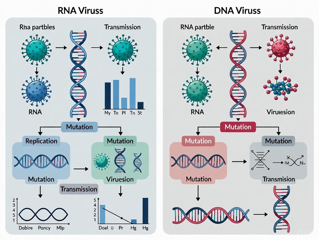
RNA vs DNA Virus Mutation Rates: Mechanisms, Measurement, and Therapeutic Exploitation
This article provides a comprehensive analysis of the fundamental differences in mutation rates between RNA and DNA viruses, a critical parameter shaping viral evolution, pathogenesis, and therapeutic design.
RNA vs DNA Virus Mutation Rates: Mechanisms, Measurement, and Therapeutic Exploitation
Abstract
This article provides a comprehensive analysis of the fundamental differences in mutation rates between RNA and DNA viruses, a critical parameter shaping viral evolution, pathogenesis, and therapeutic design. We explore the biochemical and structural basis for the 100 to 1,000,000-fold higher mutation rates in RNA viruses, dominated by error-prone RNA-dependent RNA polymerases (RdRps) lacking proofreading. The scope extends to advanced methodologies for quantifying mutational landscapes, the clinical implications of high mutation rates including drug resistance and immune evasion, and the emerging therapeutic strategy of lethal mutagenesis. A comparative framework validates these concepts against real-world challenges like SARS-CoV-2 variant emergence, offering virologists and drug developers a synthesized perspective on leveraging viral mutation rates for next-generation antiviral interventions.
The Genetic Instability Spectrum: Unpacking Core Mechanisms of Viral Mutation
In viral evolution, the terms "mutation rate" and "mutation frequency" represent fundamentally distinct concepts that are often incorrectly used interchangeably. Mutation rate refers to the probability of genetic changes occurring per nucleotide per replication cycle, representing a biochemical reality of the replication process. In contrast, mutation frequency measures the observed proportion of mutations in a viral population at a specific time, representing a snapshot of genetic variation shaped by both replication and evolutionary forces. This technical guide examines the distinction between these concepts within the broader context of RNA versus DNA virus research, providing experimental methodologies, quantitative comparisons, and practical frameworks for researchers and drug development professionals working in antiviral therapeutic development.
Conceptual Foundations: Rate Versus Frequency
Biochemical Reality Versus Population Observation
The distinction between mutation rate and frequency is fundamental to understanding viral evolution. Mutation rate is a biochemical parameter that quantifies the number of mutations introduced during a single replication cycle, expressed as substitutions per nucleotide per cell infection (s/n/c). This parameter reflects the inherent fidelity of the viral replication machinery and remains largely constant for a given virus-replication system [1].
In contrast, mutation frequency represents a population-level snapshot of existing genetic variation at a specific point in time, calculated as the proportion of mutated sequences in a population. Unlike rate, frequency is highly dynamic and influenced by multiple post-replication processes including natural selection, genetic drift, population bottlenecks, and selective sweeps [1].
Implications for Viral Evolution and Therapeutic Design
The relationship between mutation rate and frequency has profound implications for viral pathogenesis and control strategies. While mutation rate determines the raw material for evolution, mutation frequency reflects the outcome of evolutionary processes acting upon this variation. RNA viruses typically exhibit mutation rates between 10⁻⁶ to 10⁻⁴ s/n/c, approximately 100-1000 times higher than DNA viruses (10⁻⁸ to 10⁻⁶ s/n/c) [1]. This elevated rate generates extensive mutant spectra (quasispecies) that facilitate rapid adaptation to environmental challenges, including antiviral drugs and host immune responses [2].
Table 1: Key Conceptual Distinctions Between Mutation Rate and Frequency
| Parameter | Mutation Rate | Mutation Frequency |
|---|---|---|
| Definition | Probability of mutation per nucleotide per replication cycle | Observed proportion of mutations in a population at a specific time |
| Timeframe | Per generation (replication cycle) | Single time point measurement |
| Primary determinants | Polymerase fidelity, proofreading activity, replication mechanisms | Mutation rate plus selection, genetic drift, population history |
| Stability | Relatively constant for a virus-replication system | Highly dynamic over time |
| Therapeutic relevance | Target for lethal mutagenesis (e.g., nucleoside analogs) | Measure of standing genetic variation available for adaptation |
Quantitative Landscape of Viral Mutation Rates
Comparative Analysis Across Viral Families
Viral mutation rates span approximately five orders of magnitude, with nucleic acid type being a primary determinant. RNA viruses and single-stranded DNA (ssDNA) viruses occupy the higher ranges of this spectrum, while double-stranded DNA (dsDNA) viruses generally exhibit lower mutation rates. This relationship, however, is not exclusively determined by genome composition alone, as genomic architecture, replication speed, and access to repair mechanisms also contribute significantly to observed rates [3].
The higher mutation rates in RNA viruses stem primarily from their RNA-dependent RNA polymerases (RdRps), which typically lack proofreading activity. An important exception exists in coronaviruses, which encode a proofreading 3' exonuclease that substantially reduces their mutation rate compared to other RNA viruses [1]. This exception demonstrates how evolutionary innovations can modulate fundamental biochemical constraints.
Table 2: Mutation Rates Across Major Virus Classes
| Virus Class | Representative Viruses | Mutation Rate (s/n/c) | Key Influencing Factors |
|---|---|---|---|
| ss(+)RNA | Poliovirus, Hepatitis C virus | 10⁻⁵–10⁻⁴ | RdRp fidelity, template structure, replication complex |
| ss(-)RNA | Influenza A virus, Measles virus | 10⁻⁵–10⁻⁴ | RdRp fidelity, replication speed |
| dsRNA | Bacteriophage Φ6 | ~10⁻⁶ | RNA duplex stability, replication machinery |
| Retroviruses | HIV-1, Murine leukemia virus | 10⁻⁵–10⁻⁴ | Reverse transcriptase fidelity, host factors |
| ssDNA | Parvoviruses, φX174 | 10⁻⁶–10⁻⁵ | Host polymerase errors, replication mechanism |
| dsDNA | Papillomaviruses, Herpesviruses | 10⁻⁸–10⁻⁶ | Proofreading, post-replicative repair, polymerase fidelity |
SARS-CoV-2: A Case Study in RNA Virus Mutation Parameters
Recent research utilizing circular RNA consensus sequencing (CirSeq) has precisely quantified the mutation rate of SARS-CoV-2 at approximately 1.5 × 10⁻⁶ mutations per nucleotide per viral passage [4] [5]. This places it at the lower end of the RNA virus spectrum, consistent with its coronavirus-specific proofreading mechanism.
The mutation spectrum of SARS-CoV-2 is dominated by C→U transitions, which occur approximately four times more frequently than any other substitution type [5]. This biased spectrum likely results from frequent cytidine deamination by host apolipoprotein B mRNA-editing enzymes (APOBECs) or other RNA editing mechanisms [4]. The mutation rate is significantly reduced in genomic regions that form stable secondary structures, as mutations disrupting these essential structures are strongly selected against, highlighting the complex interplay between biochemical constraints and evolutionary selection [4] [5].
Methodological Approaches: Measuring Mutation Rate Versus Frequency
Experimental Workflow for Mutation Rate Determination
Accurately determining mutation rates requires specialized approaches that account for the rarity of replication errors and the confounding effects of natural selection. The following workflow illustrates the precise experimental methodology used in contemporary viral mutation rate studies:
Virus Culture & Passage Conditions: Mutation rate studies require carefully controlled passage conditions. For SARS-CoV-2, researchers typically use susceptible cell lines (e.g., VeroE6, Calu-3, or primary human nasal epithelial cells) with low multiplicity of infection (MOI = 0.1) to minimize co-infection and complementation effects that could rescue defective genomes [4] [5]. Serial passages are performed to distinguish newly generated mutations from pre-existing variants.
CirSeq Methodology: Circular RNA consensus sequencing (CirSeq) provides the sensitivity required for accurate mutation rate determination. This ultra-sensitive approach involves: (1) RNA fragmentation and circularization of short RNA fragments; (2) Rolling-circle reverse transcription to generate cDNA molecules containing tandem repeats of the original template; (3) High-throughput sequencing to read these tandem repeats; and (4) Consensus sequence generation by comparing tandem repeats to eliminate sequencing and reverse transcription errors [4] [5]. This method enables detection of mutations at frequencies as low as 10⁻⁶, far below conventional sequencing approaches.
Mutation Rate Calculation: The mutation rate is calculated specifically from lethal or highly detrimental mutations (e.g., premature stop codons in essential genes like RNA-dependent RNA polymerase) that cannot be carried between passages and must be generated anew each generation [5]. This approach ensures that the measured frequency reflects the true biochemical error rate rather than selectively neutral or beneficial mutations that may accumulate over time.
Mutation Frequency Assessment Methods
In contrast to rate measurements, mutation frequency analysis employs different methodological approaches focused on capturing standing genetic variation:
Population Sequencing: Bulk RNA sequencing of viral populations without consensus refinement provides a direct measurement of mutation frequency. The key limitation is the inability to distinguish between true replication errors and sequencing artifacts at low frequencies.
Clone Sequencing: Sanger sequencing of individual molecular clones can provide accurate frequency measurements but is limited by throughput constraints and may miss low-frequency variants.
Bioinformatic Filtering: Analysis of large sequence databases (e.g., GISAID for SARS-CoV-2) can identify mutations present in consensus sequences, but these represent only the successfully fixed variants that have reached high frequency in populations [5].
Research Toolkit: Essential Reagents and Methods
Table 3: Essential Research Reagents for Viral Mutation Studies
| Reagent/Method | Function | Application Context |
|---|---|---|
| CirSeq Protocol | Ultra-sensitive mutation detection | Gold-standard for mutation rate determination in RNA viruses |
| VeroE6 Cells | Permissive cell line for viral replication | Supports high genetic diversity; useful for evolution studies |
| Calu-3 Cells | Human lung epithelial cell line | Models human respiratory infection more physiologically |
| Primary HNEC (ALI culture) | Human nasal epithelial cells at air-liquid interface | Mimics natural infection conditions in human upper airway |
| RdRp Inhibitors | Suppress viral replication | Controls replication cycles in passage experiments |
| Lethal Mutagenesis Agents | Nucleoside analogs (e.g., ribavirin) | Experimental elevation of mutation rates to probe error thresholds |
| UShER/Ensembl Pipelines | Phylogenetic placement of mutations | Identifies mutations absent from global databases (indicates detrimental effects) |
Evolutionary Implications and Therapeutic Applications
Mutation Rate and Frequency in Viral Adaptation
The relationship between mutation rate and frequency creates distinct evolutionary dynamics across virus classes. RNA viruses maintain high mutation rates that generate extensive mutant spectra, providing substrates for rapid adaptation to changing environments [2]. This adaptive capacity comes with a cost—excessive mutation loads can push viral populations toward error catastrophe, where the accumulation of deleterious mutations causes population collapse [6].
The concept of error threshold has significant therapeutic implications. Mutagenic nucleoside analogs that increase viral mutation rates beyond sustainable levels can drive populations to extinction—an approach termed lethal mutagenesis [6]. This strategy has demonstrated efficacy against several RNA viruses, including poliovirus and influenza [6].
Structural Constraints on Mutational Landscapes
Recent research reveals that RNA secondary structures in viral genomes create heterogeneous mutation landscapes. In SARS-CoV-2, genomic regions involved in base-pairing interactions show significantly reduced mutation rates, as mutations disrupting these essential structures are strongly selected against [4] [5]. This finding demonstrates how natural selection shapes not only mutation frequencies but also exerts upstream influence on the effective mutation rate across different genomic contexts.
The following diagram illustrates the complex relationship between mutation processes and evolutionary outcomes in viral populations:
The distinction between mutation rate and frequency provides a critical conceptual framework for understanding viral evolution and developing effective antiviral strategies. Mutation rate represents a biochemical reality of replication fidelity, while mutation frequency reflects the complex interplay of replication errors and evolutionary forces. For RNA viruses, high mutation rates generate diverse mutant spectra that facilitate rapid adaptation but also create vulnerabilities to lethal mutagenesis. Emerging methodologies like CirSeq now enable precise measurement of these parameters, revealing how structural constraints and host factors shape mutational landscapes. These insights provide foundations for predicting viral evolution trajectories and designing therapeutic interventions that exploit the fundamental constraints of viral replication.
The replication of viral genomes is a critical process governed by polymerase enzymes, whose fidelity—or accuracy—varies tremendously between DNA and RNA viruses. This disparity creates a fundamental "fidelity divide" with profound implications for viral evolution, pathogenesis, and therapeutic development. DNA viruses typically replicate with relatively high fidelity using DNA-dependent DNA polymerases, many of which incorporate proofreading mechanisms. In stark contrast, RNA viruses rely on RNA-dependent RNA polymerases (RdRps) that lack robust proofreading capabilities, resulting in error-prone replication and high mutation rates [7] [8]. This biochemical distinction explains why RNA viruses generally exhibit mutation rates approximately 100 to 10,000 times higher than their DNA counterparts, with significant consequences for their evolutionary dynamics and the challenges they pose for drug and vaccine development [9].
The high mutation rates of RNA viruses are credited with facilitating their rapid adaptation to new hosts, immune evasion, and evolution of drug resistance. However, emerging evidence suggests these extreme mutation rates may not be exclusively adaptive but rather a byproduct of selection for rapid genomic replication, where a trade-off exists between speed and accuracy [10] [11]. This review examines the molecular basis of the polymerase fidelity divide, its quantitative dimensions, exceptional cases that challenge this dichotomy, experimental approaches for its study, and its implications for antiviral therapeutic strategies.
Molecular Mechanisms Underlying the Fidelity Divide
Error-Prone Replication in RNA Viruses
RNA virus replication is characterized by high error frequencies resulting from several biochemical limitations. The intrinsic selectivity of viral RdRps toward correct nucleotides is typically on the order of 10⁴-10⁵, similar to DNA polymerases; however, most RdRps lack associated 3′→5′ exonuclease activity that would allow for proofreading [7] [8]. Without this critical proofreading function, misincorporated nucleotides remain in the nascent RNA strand, resulting in established mutations. Additionally, RNA viruses do not benefit from post-replicative repair systems that correct errors in cellular DNA genomes [7]. The one notable exception to this rule exists within the nidovirus family (including coronaviruses), which encodes a proofreading exoribonuclease within non-structural protein 14 (nsp14) [12].
Biochemical studies indicate that RdRp fidelity is governed by multiple checkpoints mediated by amino acids both proximal and distal to the enzyme's active site [8]. The architecture of RdRps resembles a cupped "right hand" with fingers, palm, and thumb domains, similar to other polymerase classes, though with distinct structural features that influence their function [13]. Factors beyond intrinsic polymerase selectivity further contribute to error-prone replication, including sequence context, divalent cation concentrations, relative abundance of nucleoside triphosphates, and RNA secondary structure [7].
Proofreading and Repair in DNA Viruses
DNA viruses exhibit more diverse replication strategies with generally higher fidelity. Many larger DNA viruses encode their own DNA polymerases that include 3′→5′ exonuclease proofreading domains, analogous to cellular replicative DNA polymerases [7]. This proofreading capability allows for the detection and removal of misincorporated nucleotides before chain elongation continues. For instance, bacteriophage T4 possesses a DNA polymerase with 3′ exonuclease activity, and amino acid replacements that inactivate this domain produce a strong mutator phenotype [7].
Some DNA viruses have evolved mechanisms to manipulate host DNA repair systems. Small DNA viruses like polyomaviruses can encode proteins that inactivate the 3′ exonuclease proofreading domain of host DNA polymerases, potentially increasing mutation rates [7]. Others, like bacteriophage ΦX174, avoid post-replicative repair entirely—its genome is devoid of GATC motifs that would be recognized by the host's methyl-directed mismatch repair system [7]. Interestingly, some large DNA viruses such as African swine fever virus encode their own DNA repair systems, including an error-prone repair polymerase (pol X) that may contribute to genetic diversity [7].
Table 1: Molecular Mechanisms Creating Genetic Diversity in Different Virus Types
| Mechanism | dsDNA Viruses | ssDNA Viruses | RNA Viruses |
|---|---|---|---|
| Lack of 3′ exonuclease proofreading | − | +/− | + |
| Avoidance of post-replicative repair | − | +/− | + |
| Use of error-prone repair polymerases | +/− | +/− | − |
| Diversity-generating retro-elements | +/− | − | − |
| APOBEC hypermutation | +/− | +/− | + |
| ADAR hypermutation | − | − | +/− |
| Template switching/recombination | − | − | + |
Source: Adapted from [7]. Key: + = generally present; +/− = present in some cases; − = not shown or infrequent
Quantitative Comparison of Viral Mutation Rates
Accurate estimates of viral mutation rates reveal the dramatic consequences of the polymerase fidelity divide. Comprehensive analyses indicate that mutation rates for DNA viruses range from 10⁻⁸ to 10⁻⁶ substitutions per nucleotide per cell infection (s/n/c), while RNA viruses exhibit markedly higher rates from 10⁻⁶ to 10⁻⁴ s/n/c [9]. This difference spans two to four orders of magnitude, establishing fundamentally distinct evolutionary dynamics between these viral classes.
The measurement of viral mutation rates presents significant methodological challenges. Estimates must account for different replication modes—"stamping machine" replication (where multiple copies are made sequentially from the same template) versus binary replication (where progeny strands immediately become templates)—which affect the relationship between mutations per strand copying and mutations per cell infection [9]. Additionally, selection bias must be corrected since deleterious mutations are eliminated and underrepresented in frequency measurements. Advanced statistical methods have been developed to account for these factors, providing more accurate comparisons across virus families [9].
Table 2: Comparison of Mutation Rates and Genomic Properties Across Virus Types
| Virus Category | Mutation Rate (substitutions/nucleotide/cell infection) | Typical Genome Size | Proofreading Activity |
|---|---|---|---|
| DNA Viruses | 10⁻⁸ to 10⁻⁶ | 5-300 kb | Present in many |
| RNA Viruses | 10⁻⁶ to 10⁻⁴ | 3-32 kb | Generally absent |
| Retroviruses | ~10⁻⁵ | 7-12 kb | Absent in RT |
| Coronaviruses | ~10⁻⁶ | 26-32 kb | Present (ExoN) |
Source: Compiled from [7] [9] [12]
Beyond nucleotide substitutions, insertions and deletions (indels) represent another mutation category, though they occur approximately four times less frequently than substitutions in viral genomes [9]. The inverse correlation observed between mutation rate and genome size among RNA viruses suggests a "error threshold" that constrains genomic complexity—excessively high mutation rates prevent maintenance of genetic information in larger genomes [9].
The Coronavirus Exception: An RNA Virus with Proofreading
Coronaviruses represent a remarkable exception to the typical error-prone nature of RNA viruses. As members of the order Nidovirales, coronaviruses possess genomes ranging from 26-32 kb—the largest among RNA viruses—which would be unsustainable with typical RNA virus mutation rates [12]. This genomic stability is enabled by a unique proofreading system encoded within the viral replication complex.
The coronavirus proofreading machinery centers on non-structural protein 14 (nsp14), which contains an N-terminal 3′→5′ exoribonuclease (ExoN) domain and a C-terminal N7-methyltransferase domain [12]. The ExoN activity requires interaction with nsp10 as a cofactor and demonstrates proofreading capability by removing misincorporated nucleotides during replication. Experimental studies with SARS-CoV lacking ExoN activity demonstrate a significantly increased sensitivity to mutagens like 5-fluorouracil, with ExoN-deficient viruses accumulating 14-fold more mutations compared to wild-type viruses when exposed to the mutagen [14]. This proofreading system reduces the coronavirus mutation rate to approximately 10⁻⁶ s/n/c, intermediate between typical RNA viruses and DNA viruses [12].
The coronavirus proofreading complex represents a sophisticated multi-enzyme apparatus. The RNA-dependent RNA polymerase (nsp12) first misincorporates a nucleotide, creating an RNA duplex with a mismatch. This aberrant product is then recognized by the nsp14-nsp10 complex, which excises the misincorporated nucleotide. Following excision, replication resumes with the correct nucleotide incorporation [12]. This process enhances replication fidelity while still permitting sufficient genetic diversity for adaptation.
Figure 1: Coronavirus Proofreading Mechanism. The ExoN complex (nsp14-nsp10) recognizes and excises misincorporated nucleotides, enabling correct nucleotide incorporation by the RdRP (nsp12).
Experimental Approaches for Studying Polymerase Fidelity
Isolation and Characterization of Fidelity Variants
A key methodology for studying viral polymerase fidelity involves isolating and characterizing fidelity variants through selective pressure with mutagenic agents. The general protocol begins with determining the maximum concentration of mutagens (such as ribavirin, 5-fluorouracil, or 5-azacytidine) that can be applied to host cells without causing excessive cytotoxicity [15]. Viruses are then passaged repeatedly under sublethal mutagenic pressure, which selects for variants with altered fidelity—typically higher-fidelity "antimutator" strains that better resist the mutagenic effects [15].
Following selection, the mutagen-resistant viral population is sequenced to identify mutations in the polymerase or associated replication proteins. Candidate mutations are regenerated in infectious clones or isolated via plaque purification, and their resistance phenotypes are confirmed by testing against multiple mutagens with different structures [15]. True fidelity variants typically demonstrate broad resistance across multiple mutagen classes rather than specific resistance to a single compound.
To confirm that identified polymerase changes alter replication fidelity, mutation frequencies must be quantitatively measured. This involves extracting viral RNA, reverse-transcriptase PCR amplification of specific genomic regions (typically 800-1200 nucleotides), molecular cloning of the amplified products, and sequencing of multiple clones (often 96 or more per population) [15]. Mutation frequencies are calculated by dividing the total number of single nucleotide polymorphisms by the total nucleotides sequenced, expressed as mutations per 10,000 nucleotides sequenced. This comprehensive approach allows researchers to distinguish genuine fidelity variants from mutants with other resistance mechanisms.
The Scientist's Toolkit: Essential Research Reagents
Table 3: Key Research Reagents for Viral Fidelity Studies
| Reagent/Condition | Function in Fidelity Research | Example Applications |
|---|---|---|
| Ribavirin | RNA mutagen; base analog that promotes transition mutations | Selection of fidelity variants; lethal mutagenesis studies |
| 5-Fluorouracil | Pyrimidine analog mutagen | Proofreading studies; coronavirus ExoN validation |
| Manganese Chloride | Divalent cation that decreases polymerase fidelity | Fidelity modulation; biochemical assays |
| Plasmid-based Infectious Clones | Recovery of specific fidelity mutants | Structure-function studies |
| TOPOTA Cloning Kit | Molecular cloning of RT-PCR products | Mutation frequency measurements |
| Next-generation Sequencing | Deep sequencing of viral populations | Comprehensive diversity analysis |
| Cell Viability Assays | Assessment of mutagen cytotoxicity | Determination of selective conditions |
Figure 2: Workflow for Isolation of Viral Fidelity Variants. The process involves selective pressure with mutagens, identification of resistance mutations, and comprehensive characterization of fidelity changes.
Evolutionary Implications and Therapeutic Applications
The Speed-Fidelity Trade-off in Viral Evolution
The conventional view that RNA viruses maintain high mutation rates primarily for adaptive benefit has been challenged by recent research suggesting that extreme mutation rates may be a byproduct of selection for rapid replication. This "speed-fidelity trade-off" hypothesis proposes that viral polymerases face biochemical constraints that force a compromise between replication speed and accuracy [10] [11]. Studies with poliovirus fidelity variants provide compelling evidence for this model. The well-characterized 3DG64S antimutator variant of poliovirus demonstrates significantly reduced replication rates alongside its approximately 3-fold increase in fidelity [10]. Experimental evolution of this variant under selection for replicative speed led to compensatory mutations that restored replication kinetics without necessarily affecting the fidelity phenotype, suggesting that speed is more critical than accuracy for within-host spread and virulence [10] [11].
The kinetic proofreading model for biosynthetic reactions provides a theoretical framework for understanding this trade-off. According to this model, higher fidelity requires additional time for substrate verification and error correction, inevitably slowing the catalytic cycle [10]. For viruses competing within hosts, rapid replication and dissemination may provide greater selective advantages than genetic diversity per se, particularly when considering that most mutations are deleterious rather than beneficial [11]. This perspective helps explain why RNA viruses tolerate mutation rates perilously close to the "error threshold" beyond which genetic information cannot be maintained.
Therapeutic Targeting of Viral Polymerase Fidelity
The fidelity divide between DNA and RNA viruses presents distinctive opportunities for therapeutic intervention. For RNA viruses, lethal mutagenesis represents a promising strategy that exploits their high mutation rates. This approach involves administration of nucleoside analogs that increase viral mutation frequencies beyond sustainable levels, driving populations to extinction through accumulation of deleterious mutations [9] [11]. Ribavirin, used against several RNA viruses including hepatitis C virus, may exert part of its antiviral effect through this mechanism, particularly when combined with interferon [9].
The coronavirus proofreading system presents both a challenge and opportunity for antiviral development. The ExoN activity protects against nucleoside analogs, complicating drug development [12]. However, combination therapies targeting both the polymerase and proofreading functions show promise. One proposed strategy involves administering nucleoside analogs alongside compounds that inhibit the proofreading complex, potentially overcoming the viral defense mechanism [12]. Alternatively, antisense oligonucleotides (ASOs) might be designed to exploit the proofreading system, potentially tricking it into damaging the viral genome [12].
For DNA viruses, traditional nucleoside analogs like acyclovir continue to be mainstays of treatment, often exploiting differences between viral and cellular polymerases for selectivity. The generally lower mutation rates of DNA viruses reduce the likelihood of drug resistance emergence compared to RNA viruses, though resistance remains a significant clinical concern for many DNA viral infections.
The fundamental divide in polymerase fidelity between DNA and RNA viruses represents a cornerstone of virology with far-reaching implications. The presence of proofreading mechanisms in many DNA viruses and their general absence in RNA viruses creates dramatically different evolutionary landscapes for these pathogen classes. While the high mutation rates of RNA viruses facilitate rapid adaptation, emerging evidence suggests this may be a tolerated byproduct of selection for replication speed rather than a directly optimized trait. The exceptional proofreading capability of coronaviruses demonstrates that evolutionary solutions exist to overcome the constraints typically faced by RNA viruses. Understanding these fundamental mechanisms continues to inform therapeutic strategies, from lethal mutagenesis for RNA viruses to proofreading disruption for coronaviruses, highlighting the importance of basic virology research for addressing emergent viral threats.
The mutation rate is a critical biological parameter that profoundly influences viral evolution, pathogenesis, and the development of control strategies. Research has consistently demonstrated a fundamental divide in the genetic stability of viruses, with mutation rates spanning approximately four orders of magnitude from 10⁻⁸ to 10⁻⁴ substitutions per nucleotide per cell infection (s/n/c) [9] [16]. This variation is not random but fundamentally correlates with viral genome composition and structure. DNA viruses typically exhibit mutation rates clustered at the lower end of this spectrum (10⁻⁸ to 10⁻⁶ s/n/c), while RNA viruses occupy the higher range (10⁻⁶ to 10⁻⁴ s/n/c) [9] [16]. This disparity arises primarily from differences in replication machinery; RNA-dependent RNA polymerases (RdRps) and reverse transcriptases (RTs) generally lack the proofreading activity inherent to many DNA-dependent DNA polymerases [17] [18]. This technical guide explores the quantitative landscape of viral mutation rates, details the experimental methodologies for their determination, and discusses the implications of this fidelity gap for viral evolution and therapeutic intervention, providing a resource for researchers and drug development professionals.
Quantitative Landscape of Viral Mutation Rates
Comprehensive Mutation Rate Spectrum
The mutation rates of viruses have been systematically characterized across diverse families, revealing a consistent pattern based on genomic material and replication strategy. The table below summarizes the documented ranges for different virus types.
Table 1: Ranges of Viral Mutation Rates
| Virus Type | Mutation Rate Range (substitutions/nucleotide/cell infection) | Primary Polymerase Type | Proofreading Activity |
|---|---|---|---|
| DNA Viruses | 10⁻⁸ – 10⁻⁶ [9] [16] | DNA-dependent DNA polymerase | Often present [18] |
| RNA Viruses | 10⁻⁶ – 10⁻⁴ [9] [16] | RNA-dependent RNA polymerase | Generally absent [17] [19] |
| Retroviruses | ~10⁻⁵ (within RNA virus range) [9] [16] | Reverse Transcriptase (RT) | Generally absent [18] |
It is crucial to distinguish between two common units of measurement: the rate per strand copying (s/n/r) and the rate per cell infection (s/n/c). The latter accounts for the total number of replication cycles within an infected cell and is therefore typically higher, as some viruses, particularly double-stranded DNA viruses, undergo several rounds of genomic copying per cell infection [9]. Furthermore, across all virus types, nucleotide substitutions are approximately four times more common than insertions or deletions (indels) [9].
Representative Mutation Rates in Selected Viruses
Specific estimates for model viruses illustrate the practical implications of these ranges. For instance, the vesicular stomatitis virus (VSV), an RNA virus, has a mutation rate measured at approximately 1.64 × 10⁻⁵ per round of copying for a specific phenotype, translating to a per-nucleotide rate of about 6.15 × 10⁻⁶ s/n/r [20]. This high rate is a hallmark of RNA virus replication. In contrast, some large RNA viruses, such as coronaviruses, have evolved a degree of replication fidelity through an exonucleolytic proofreading-repair activity (3′ to 5′ exonuclease) that can decrease their error rate [17] [21]. This exception highlights that mutation rates are themselves evolvable traits.
Experimental Methodologies for Mutation Rate Quantification
Accurately measuring viral mutation rates is methodologically challenging due to the rarity of the event and confounding factors like selection. The following section details two cornerstone experimental approaches.
The Luria-Delbrück Fluctuation Test
This classic genetic method is used to determine the rate at which a specific phenotypic mutation arises.
- Objective: To calculate the mutation rate to a defined phenotype (e.g., drug resistance, antibody escape) per round of genomic replication.
- Workflow: The experimental protocol involves multiple parallel cultures, each initiated from a small number of viral particles to ensure the mutation of interest is not pre-existing. The cultures are expanded independently, and the number of cultures without mutants (the "null class") is used to calculate the rate, as this metric is robust to the effects of natural selection during the experiment [9].
- Calculation: The mutation rate to a specific phenotype (m) is derived from the proportion of cultures showing no mutants (P₀) and the final population size (N), using the formula m = -ln(P₀) / N [9] [20]. This phenotypic rate can be converted to a per-nucleotide substitution rate (μ) if the mutational target size (T) is known: μ = m / (3T), where 3 represents the three possible nucleotide substitutions at a given site.
Diagram: Luria-Delbrück Fluctuation Test Workflow
Molecular Clone Sequencing
This direct sequencing approach provides a genome-wide view of accumulated mutations.
- Objective: To directly observe the frequency of mutations across a genomic region after a controlled number of replication cycles.
- Workflow: Cells are infected at a low multiplicity of infection (MOI) to ensure infection by a single, genotypically defined viral genome. The resulting progeny virions are harvested, and their RNA is reverse-transcribed, PCR-amplified, and molecularly cloned. Multiple clones are then sequenced and compared to the original inoculum sequence to identify new mutations [9] [20].
- Calculation: The observed mutation frequency (f) is the number of mutations divided by the total number of nucleotides sequenced. To convert this frequency to a mutation rate (μ), the number of viral generations (c) and a statistical correction factor for selection bias (α) must be applied: μ = f / (T * c * α) [9]. The selection correction is necessary because many deleterious mutations are lost from the population before they can be sampled, and this bias can be accounted for using empirically derived distributions of mutational fitness effects [9].
Diagram: Molecular Clone Sequencing Workflow
Evolutionary and Therapeutic Context
Evolutionary Trade-Offs and the "Error Threshold"
The high mutation rates of RNA viruses are a double-edged sword. While they generate the genetic diversity necessary for rapid adaptation to new hosts, immune evasion, and drug resistance, most mutations are deleterious [11] [19]. This creates a fundamental trade-off. The prevailing hypothesis has been that RNA virus mutation rates are optimized by natural selection to be as high as possible without exceeding the error threshold—the point where the accumulation of deleterious mutations leads to population collapse, a phenomenon known as lethal mutagenesis [11] [19].
However, an alternative explanation posits that high mutation rates may be a byproduct of selection for rapid genomic replication [11]. There appears to be a trade-off between speed and fidelity; faster polymerases tend to make more mistakes. Since rapid replication is a key fitness advantage for viruses, selection may favor faster but less accurate polymerases, tolerating the consequent high mutation rate as a cost of doing business [11] [19].
Implications for Drug Development
The high mutation rate of RNA viruses has direct consequences for therapeutic strategies:
- Antiviral Resistance: The high error rate means that pre-existing variants resistant to a single drug are likely present in a population. This explains the rapid emergence of resistance and validates the use of combination antiviral therapies, as demonstrated for HIV-1 [9].
- Lethal Mutagenesis: The proximity of RNA viruses to the error threshold is exploitable. The administration of mutagenic nucleoside analogues (e.g., ribavirin) can increase the mutation rate beyond the sustainable threshold, pushing the viral population toward extinction [9] [11]. This approach has shown efficacy against several RNA viruses in model systems [9].
- Vaccine Design: The malleability of viral antigens necessitates vaccines that elicit broad immune responses. For live-attenuated vaccines, understanding mutation rates is critical for assessing and minimizing the risk of reversion to virulence, a known issue with the Sabin poliovirus vaccine [17] [21].
The Scientist's Toolkit: Essential Research Reagents
Table 2: Key Reagents for Viral Mutation Rate Studies
| Research Reagent / Method | Function in Mutation Rate Studies |
|---|---|
| Monoclonal Antibodies | Used in fluctuation tests as a selective agent to isolate and quantify antibody-escape mutants [20]. |
| Nucleoside Analogues | Serve as chemical mutagens to experimentally induce lethal mutagenesis and study error thresholds [9] [11]. |
| Fidelity Mutants (e.g., 3D:G64S) | Engineered viral polymerases with altered fidelity (higher or lower) used to dissect the relationship between mutation rate, replication speed, and fitness [11]. |
| APOBEC3/ADAR proteins | Host factors that actively edit viral genomes, representing a host-driven source of mutations that must be accounted for in certain systems [20]. |
| Luria-Delbrück Fluctuation Analysis | A statistical framework and experimental design used to calculate mutation rates from phenotypic data while accounting for random mutation events [9] [20]. |
| Next-Generation Sequencing (NGS) | Enables deep sampling of the mutant spectrum within a population, allowing for direct estimation of mutation frequencies and spectra [18]. |
The quantification of viral mutation rates from 10⁻⁸ to 10⁻⁴ s/n/c reveals a fundamental principle of virology: genome composition dictates replicative fidelity, which in turn shapes evolutionary potential and pathogenic strategy. The divide between DNA and RNA viruses underscores the different evolutionary constraints they face. For researchers and drug developers, a precise understanding of these rates and the methods used to measure them is indispensable. It informs the battle against antiviral resistance, validates novel strategies like lethal mutagenesis, and guides the design of robust vaccines. Future research will continue to refine these measurements and explore the intricate balance between the adaptive benefits and the destructive costs of the error-prone replication that defines the RNA viral world.
RNA viruses have historically been characterized by high mutation rates due to the error-prone nature of their RNA-dependent RNA polymerases (RdRp), which lack proofreading capabilities. This evolutionary strategy generates diverse quasispecies populations that facilitate rapid adaptation but also constrains genome size, with most RNA viruses maintaining genomes under 15 kilobases (kb). Coronaviruses, with their exceptionally large 26-32 kb RNA genomes, represent a striking exception to this rule. This anomaly is explained by the presence of a unique exoribonuclease domain within nonstructural protein 14 (nsp14) that provides proofreading functionality—a feature exceptionally rare in RNA viruses [22] [23]. The bifunctional nsp14 protein, containing both 3'-to-5' exoribonuclease (ExoN) and N7-methyltransferase (N7-MTase) activities, enables coronaviruses to maintain genome integrity while operating with an expanded genetic code [22] [23]. This review examines the molecular mechanisms of coronavirus proofreading, its role in viral replication and evolution, and the surprising exceptions that challenge our understanding of this sophisticated RNA surveillance system.
Molecular Architecture and Mechanism of the nsp14 Proofreading Complex
Structural Organization of Bifunctional nsp14
The coronavirus nsp14 is a 60 kDa bifunctional enzyme that plays a pivotal role in replication fidelity. Its N-terminal domain harbors the ExoN activity, while the C-terminal domain possesses N7-MTase activity involved in mRNA capping [22] [23]. SARS-CoV-2 and SARS-CoV nsp14 share more than 95% amino acid sequence similarity, underscoring the evolutionary conservation of this critical protein [22]. The ExoN domain belongs to the DEDD exonuclease superfamily, which includes proofreading domains of many DNA polymerases and various eukaryotic and prokaryotic exonucleases [24] [23]. This evolutionary relationship to DNA proofreading systems highlights the unique position of coronaviruses in the RNA viral world.
The ExoN active site contains five conserved residues distributed across three canonical motifs: Motif I (D90/E92), Motif II (E191), and Motif III (H268/D273) [24] [23]. These residues coordinate two divalent metal ions (preferentially Mg²⁺) and a reactive water molecule to catalyze nucleoside monophosphate excision in the 3'-to-5' direction [22] [23]. The nsp14 structure also incorporates three zinc finger motifs (ZF1, ZF2, ZF3) that contribute to structural stability and catalytic function [24]. The C-terminal N7-MTase domain contains a conserved DxG S-adenosyl-L-methionine (SAM)-binding motif essential for its methyltransferase activity [22].
Allosteric Activation by nsp10 Cofactor
The exonuclease activity of nsp14 is functionally dependent on interaction with nsp10, a small cofactor protein that enhances ExoN activity up to 35-fold [25] [23]. Structural analyses reveal that nsp10 binding induces significant conformational changes in nsp14, particularly refolding of a "lid" subdomain that releases exonuclease activity [25]. This allosteric regulation ensures that proofreading occurs specifically within the context of the viral replication-transcription complex (RTC), where nsp10 is present to activate nsp14. The nsp10/nsp14 complex subsequently interacts with other RTC components, including the nsp12 RdRp and nsp13 helicase, forming a sophisticated multi-enzyme machine capable of both RNA synthesis and error correction [25].
Table 1: Key Functional Domains and Motifs of Coronavirus nsp14
| Domain/Motif | Location | Key Residues | Function |
|---|---|---|---|
| ExoN Domain | N-terminal (1-290) | D90, E92, E191, H268, D273 | 3'-to-5' exoribonuclease activity; proofreading |
| Zinc Finger 1 (ZF1) | ExoN domain | C207, C210, C226, H229 | Structural stability and catalytic function |
| Zinc Finger 2 (ZF2) | ExoN domain | H257, C261, H264, C279 | Structural stability and catalytic function |
| Zinc Finger 3 (ZF3) | C-terminal | C452, C473, C484, C487 | Structural stability |
| N7-MTase Domain | C-terminal (291-527) | D331, G333, P335, A/G337 | mRNA capping; SAM binding |
| nsp10 Binding Site | Multiple interfaces | Various hydrophobic and polar residues | Allosteric activation of ExoN |
Figure 1: nsp14 Proofreading Complex Architecture and Activation Mechanism. The bifunctional nsp14 protein contains distinct ExoN and N7-MTase domains, with allosteric activation by nsp10 cofactor enhancing ExoN activity 35-fold.
Experimental Evidence for Proofreading Function
Reverse Genetics and Mutator Phenotypes
The proofreading function of nsp14 was conclusively demonstrated through reverse genetics approaches where ExoN active-site residues were mutated. Initial studies with murine hepatitis virus (MHV) and SARS-CoV showed that ExoN knockout mutants were viable but exhibited 15-21-fold increases in mutation frequency during replication [26] [23]. Complete genome sequencing of SARS-CoV ExoN mutant viruses revealed unique mutation sets in every genome examined, with 100 unique mutations distributed across the genome, demonstrating dramatically increased mutational load [26]. These mutants also showed increased sensitivity to mutagenic agents like 5-fluorouracil, to which wild-type coronaviruses are relatively resistant [23].
Unexpectedly, the same ExoN knockout approaches yielded different results across coronavirus genera. While alphacoronaviruses (HCoV-229E) and gammacoronaviruses failed to produce viable ExoN knockout mutants, most betacoronaviruses (MHV, SARS-CoV) yielded viable mutants with hypermutation phenotypes [23]. Surprisingly, despite 95% amino acid identity with SARS-CoV nsp14, SARS-CoV-2 ExoN knockout mutants were nonviable, as were equivalent mutants of MERS-CoV [23]. This stark contrast between closely related viruses suggests that nsp14 ExoN has additional critical functions beyond proofreading that vary in their essentiality across coronaviruses.
In Vitro Biochemical Assays
Biochemical characterization of recombinant nsp14 has provided detailed insights into its enzymatic mechanism. Nsp14 hydrolyzes both single-stranded and double-stranded RNA, processing them to final products of 8-12 nucleotides and 5-7 nucleotides, respectively [27]. The exonuclease activity is metal ion-dependent, with preference for Mg²⁺ over Mn²⁺, Co²⁺, and Zn²⁺, while Ca²⁺, Ni²⁺, and Cu²⁺ do not support catalysis [22]. The ExoN domain specifically removes mismatched nucleotides from the 3' end of RNA strands, efficiently excising RdRp misincorporation products [24] [23]. This activity is particularly important for maintaining the integrity of the large coronavirus genome, as the error rate of the RdRp alone would otherwise lead to unacceptably high mutational loads.
Table 2: Experimental Evidence for nsp14 Proofreading Function Across Coronaviruses
| Virus | Genus | ExoN Knockout Viability | Mutation Rate Increase | Key Observations |
|---|---|---|---|---|
| MHVA | Betacoronavirus | Viable | 15-fold | Increased sensitivity to mutagens |
| SARS-CoV | Betacoronavirus | Viable | 21-fold | 100+ unique mutations per genome |
| SARS-CoV-2 | Betacoronavirus | Nonviable | N/A | Essential function beyond proofreading |
| MERS-CoV | Betacoronavirus | Nonviable | N/A | Occasional reversion to wild-type |
| HCoV-229E | Alphacoronavirus | Nonviable | N/A | Lethal despite RNA synthesis competence |
| TGEV | Alphacoronavirus | Conditionally viable | Variable | ZF-C mutant with reduced antiviral response |
Quantitative Analysis of Mutation Rates and Spectra
Advanced sequencing technologies have enabled precise measurement of coronavirus mutation rates. Circular RNA consensus sequencing (CirSeq), an ultra-sensitive method that eliminates sequencing and reverse-transcription errors, revealed that SARS-CoV-2 mutates at a rate of approximately 1.5 × 10⁻⁶ per base per viral passage [4]. This rate is significantly lower than that of most RNA viruses, which typically exhibit mutation rates of 10⁻³ to 10⁻⁵ per base per replication cycle, positioning coronaviruses closer to DNA viruses in terms of replication fidelity.
The mutation spectrum of SARS-CoV-2 is dominated by C→U transitions, consistent with cytidine deamination as a major mutagenic process [4]. Notably, mutation rates are significantly reduced in regions with RNA secondary structure, and mutations that disrupt these structures are particularly harmful to viral fitness [4]. This relationship between RNA structure, mutation rate, and fitness highlights the complex evolutionary constraints acting on the coronavirus genome.
Analysis of naturally occurring nsp14 variants has identified specific mutations that alter viral evolvability. The P203L substitution in nsp14, not found in other coronaviruses but observed in SARS-CoV-2, is associated with significantly higher evolutionary rates [24]. Recombinant SARS-CoV-2 carrying the P203L mutation acquired more diverse genomic mutations than wild-type virus during replication in hamsters, suggesting that such substitutions can accelerate genomic diversity and potentially drive variant emergence [24]. Epidemiological studies further support this concept, demonstrating that SARS-CoV-2 isolates with nsp14 mutations show the strongest association with increased genome-wide mutation load compared to mutations in other components of the RNA synthesis complex [28].
Table 3: Mutation Rates and Spectra Across RNA Viruses With and Without Proofreading
| Virus Family | Genome Size (kb) | Proofreading Mechanism | Mutation Rate (per base per replication) | Dominant Mutation Type |
|---|---|---|---|---|
| Coronaviridae | 26-32 | nsp14 ExoN | ~1.5 × 10⁻⁶ | C→U transitions |
| Picornaviridae | 7-9 | None | 10⁻³ to 10⁻⁵ | Various |
| Flaviviridae | 9-12 | None | 10⁻⁴ to 10⁻⁶ | Various |
| Orthomyxoviridae | 13-15 | None | ~3 × 10⁻⁶ | Various |
| Arenaviridae | 10-14 | ExoN (NP domain) | ~2 × 10⁻⁶ | Various |
Research Reagents and Methodologies for nsp14 Studies
Essential Research Tools
The investigation of nsp14 proofreading mechanisms relies on specialized reagents and methodologies. Reverse genetics systems have been developed for multiple coronaviruses, allowing introduction of specific mutations into nsp14 and recovery of recombinant viruses [26] [23]. These systems typically employ bacterial artificial chromosomes or vaccinia virus vectors to maintain the large coronavirus genome. For biochemical characterization, recombinant nsp14 and nsp10 proteins are expressed in Escherichia coli or insect cell systems and purified using affinity chromatography tags [25] [23].
Cell culture models form the foundation of coronavirus replication studies. VeroE6 cells (African green monkey kidney cells) are particularly susceptible to SARS-CoV-2 infection and support efficient viral replication, though they may permit accumulation of higher genetic diversity than other cell lines [4]. For more physiologically relevant models, Calu-3 (human lung adenocarcinoma) cells and primary human nasal epithelial cells (HNEC) cultured at air-liquid interface (ALI) provide human respiratory system context [4].
Advanced sequencing methodologies are crucial for detecting the relatively rare mutations that escape proofreading. Circular RNA consensus sequencing (CirSeq) provides exceptional accuracy by circularizing short RNA fragments to generate tandem cDNA repeats, enabling distinction between true mutations and technical artifacts [4]. This approach has been successfully applied to multiple SARS-CoV-2 variants, including USA-WA1/2020, Alpha, Beta, Gamma, Delta, and Omicron strains [4].
Experimental Protocols
Protocol 1: Reverse Genetics for ExoN Mutant Generation
- Introduce desired mutations into nsp14 ExoN domain (e.g., active site residues D90A/E92A) via site-directed mutagenesis of full-length cDNA clone
- Generate infectious RNA through in vitro transcription
- Transfect RNA into permissive cells (e.g., VeroE6, BHK-21)
- Recover viral progeny and sequence entire genome to confirm introduced mutations
- Passage virus to assess stability of ExoN mutations and potential reversion events [26] [23]
Protocol 2: In Vitro ExoN Activity Assay
- Express and purify recombinant nsp14 and nsp10 proteins with affinity tags
- Synthesize RNA substrates (typically 20-40 nt) with fluorescent labels or radiolabels
- Prepare reaction mixture containing: 50 mM HEPES (pH 8.0), 5 mM MgCl₂, 1 mM DTT, 0.1 mg/mL BSA, nsp14 (0.1-1 µM), nsp10 (1-5 µM), and RNA substrate (0.1-1 µM)
- Incubate at 30-37°C for 15-60 minutes
- Terminate reactions with EDTA or formamide loading buffer
- Analyze products by denaturing polyacrylamide gel electrophoresis or capillary electrophoresis [23]
Protocol 3: Mutation Rate Measurement Using CirSeq
- Extract viral RNA from culture supernatants or infected cells
- Fragment RNA and circularize fragments using RNA ligase
- Perform reverse transcription to generate tandem repeat cDNAs
- Prepare sequencing library and sequence on high-throughput platform
- Analyze data to distinguish true mutations from technical errors by comparing repeats within each cDNA molecule
- Calculate mutation frequencies by dividing observed mutations by total bases sequenced [4]
Figure 2: Experimental Workflows for nsp14 Proofreading Research. Three complementary approaches—reverse genetics, biochemical analysis, and mutation detection—provide comprehensive understanding of ExoN function.
The Scientist's Toolkit: Essential Research Reagents
Table 4: Key Research Reagents for nsp14 and Proofreading Studies
| Reagent/Cell Line | Specifications | Research Application | Key Features |
|---|---|---|---|
| VeroE6 Cells | African green monkey kidney cells | Viral propagation and evolution studies | High susceptibility to SARS-CoV-2; permits accumulation of genetic diversity |
| Calu-3 Cells | Human lung adenocarcinoma cells | Physiologically relevant infection models | Human respiratory origin; more representative of human infection |
| Primary HNEC-ALI | Human nasal epithelial cells, air-liquid interface | Most physiologically relevant model | Maintains cellular differentiation and mucociliary function |
| Reverse Genetics System | Infectious cDNA clones | Generation of engineered viruses | Enables introduction of specific mutations into nsp14 |
| Recombinant nsp14/nsp10 | E. coli or insect cell expression | Biochemical characterization | Enables in vitro study of ExoN and MTase activities |
| CirSeq Methodology | Circular RNA consensus sequencing | Mutation rate quantification | Ultra-high accuracy; distinguishes true mutations from artifacts |
Discussion: Implications for Antiviral Development and Viral Evolution
The exceptional proofreading capability of coronaviruses presents both challenges and opportunities for therapeutic intervention. The ExoN activity represents a formidable barrier to nucleoside analog therapies, as it can efficiently excise incorporated mutagenic nucleotides before they can cause lethal mutagenesis [22] [23]. This explains the relative resistance of coronaviruses to many nucleoside analogs that are effective against other RNA viruses. However, combination therapies targeting both the RdRp and ExoN activities may overcome this barrier by simultaneously introducing mutations and inhibiting their repair [22].
The variability in essentiality of ExoN activity across coronaviruses reveals important nuances in nsp14 function. While proofreading represents a conserved activity, nsp14 appears to have additional roles in primary viral RNA synthesis that are essential in some coronaviruses (SARS-CoV-2, MERS-CoV) but not others (SARS-CoV, MHV) [23]. This suggests that nsp14 may participate in other aspects of RNA metabolism beyond proofreading, possibly including RNA recombination or the regulation of innate immune recognition [27]. The zinc finger motifs, particularly ZF1, appear to modulate the antiviral response, with specific mutations reducing dsRNA accumulation and subsequent interferon signaling [27].
From an evolutionary perspective, the coronavirus proofreading system represents a remarkable adaptation that permits expansion of genome size while maintaining sequence integrity. This innovation may have enabled the acquisition of additional genes and regulatory elements that enhance viral fitness and host adaptability. The emergence of variants with altered proofreading efficiency, such as the nsp14-P203L mutant, demonstrates that coronaviruses can dynamically regulate their evolutionary rate in response to selective pressures [24]. This plasticity in mutation rate represents an additional layer of evolutionary strategy not available to most RNA viruses.
Future research should focus on elucidating the structural basis of nsp10-mediated nsp14 activation, developing specific ExoN inhibitors, and understanding how proofreading efficiency correlates with viral transmission and pathogenicity across different coronavirus species. The exquisite balance between replication fidelity and evolutionary flexibility makes the nsp14 system a fascinating example of viral adaptation and a promising target for therapeutic intervention against current and future coronavirus threats.
Host-factor mediated mutagenesis represents a fundamental interface between innate immunity and viral evolution. This whitepaper provides a comprehensive technical examination of how host enzymes, particularly APOBEC cytidine deaminases, actively shape viral mutation landscapes. Within the context of RNA versus DNA virus research, we delineate the molecular mechanisms, quantitative mutation profiles, and experimental methodologies essential for investigating these processes. The content specifically addresses the differential susceptibility of viral genetic material to host-mediated editing, with particular emphasis on the implications for antiviral drug development and therapeutic target identification. Structured data presentation and detailed protocols aim to equip researchers with the practical tools necessary to advance this critical field of study.
The evolutionary arms race between viruses and their hosts has driven the development of sophisticated host immune mechanisms that extend beyond conventional pathways. Among these, host-factor mediated mutagenesis represents a paradigm-shifting concept where cellular enzymes, primarily intended for host defense, directly alter viral genetic material. The apolipoprotein B mRNA-editing enzyme catalytic polypeptide (APOBEC) family of cytidine deaminases stands as a prime exemplar of this mechanism, demonstrating potent antiviral activity through cytosine deamination in single-stranded DNA or RNA substrates [29] [30]. These enzymes initiate a mutational cascade by catalyzing the hydrolytic deamination of cytidine to uridine, thereby introducing permanent genetic alterations that can cripple viral functionality [30].
Understanding these processes is crucial within the broader framework of mutation rate disparities between RNA and DNA viruses. RNA viruses traditionally exhibit higher mutation rates due to error-prone replication machinery; however, host-mediated mutagenesis introduces an additional layer of complexity that impacts both RNA and DNA viruses differently. The differential susceptibility stems from the nature of the viral genetic material, its exposure in single-stranded form during replication, and the specific tropism of host deaminases [31] [32]. This review systematically dissects the APOBEC-mediated mutagenesis pathway, provides quantitative comparisons of resulting mutational signatures, details experimental methodologies for its investigation, and frames these findings within the overarching thesis of viral mutation rate determinism.
The APOBEC Enzyme Family: Structure and Function
The APOBEC family comprises eleven primary members in humans: APOBEC1, Activation-Induced Deaminase (AID), APOBEC2, APOBEC3 (A–H), and APOBEC4 [29]. These enzymes share a conserved catalytic domain characterized by a zinc-coordination motif (H-X-E-X23–28-P-C-X-C) essential for cytidine deamination activity [30]. Despite structural similarities, family members demonstrate distinct functions, substrate preferences, and tissue expression patterns. AID, expressed in activated B cells, facilitates antibody diversification through somatic hypermutation of immunoglobulin genes. APOBEC1, primarily expressed in the small intestine, edits apolipoprotein B mRNA to generate tissue-specific protein isoforms. The APOBEC3 subfamily (A3A-A3H), widely expressed across human tissues, constitutes the primary defense against viral pathogens and retrotransposons [29] [30].
Structurally, several APOBEC3 enzymes (A3G, A3F, A3B, A3DE) contain two catalytic domains, while others (A3A, A3C) possess a single domain [30]. The N-terminal domains of A3G and A3F are enzymatically inactive but crucial for RNA binding, virion incorporation, and oligomerization, whereas their C-terminal domains contain the active deamination site. In contrast, both domains of APOBEC3B remain catalytically active [30]. This structural modularity enables functional specialization, with different domains contributing to nucleic acid binding, subcellular localization, and pathogen restriction through both deamination-dependent and independent mechanisms.
Table 1: APOBEC Family Members and Primary Functions
| Enzyme | Primary Function | Substrate Preference | Biological Role |
|---|---|---|---|
| AID | Somatic hypermutation; Class switch recombination | ssDNA (WRCY motifs) | Adaptive immunity in B cells [30] |
| APOBEC1 | mRNA editing | RNA (apoB mRNA) | Lipid metabolism [30] |
| APOBEC3A | Viral genome restriction | ssDNA | Innate immunity against viruses [29] |
| APOBEC3B | Viral genome restriction | ssDNA | Innate immunity; often overexpressed in cancers [29] |
| APOBEC3G | Viral genome restriction | ssDNA | Innate immunity; potent HIV-1 restriction [30] |
| APOBEC4 | Unknown | Unknown | Unknown function [30] |
Molecular Mechanisms of APOBEC-Mediated Mutagenesis
APOBEC enzymes function by deaminating cytidine to uridine within single-stranded DNA or RNA substrates. This conversion initiates a molecular cascade that ultimately generates stable mutations. The mechanism proceeds through several well-defined stages:
Substrate Access and Deamination
During viral replication, transient single-stranded DNA (ssDNA) regions become accessible to APOBEC enzymes. APOBEC3s target these substrates with distinct sequence preferences: APOBEC3A and APOBEC3B primarily deaminate cytidine in TpC dinucleotide contexts, with APOBEC3A favoring pyrimidines preceding TpC and APOBEC3B preferring purines [29]. APOBEC3G demonstrates preference for CCC motifs and other trinucleotide contexts [29]. The deamination reaction itself involves zinc-mediated hydrolytic deamination that converts cytidine to uridine, creating a uracil lesion within the viral genome [30].
Mutation Fixation Pathways
The uracil lesion created by APOBEC activity can be processed through multiple cellular pathways, leading to different mutational outcomes:
C-to-T Transition: During subsequent replication cycles, DNA polymerases misread the uracil as thymine, resulting in C-to-T transitions. This represents the most common mutation outcome and corresponds to COSMIC Signature 2 [29].
C-to-G Transversion: Alternatively, uracil DNA glycosylase can recognize and excise the uracil base, creating an abasic site. Error-prone translation synthesis past this abasic site can generate C-to-G transversions, corresponding to COSMIC Signature 13 [29].
Cluster Mutagenesis: APOBEC activity can cause localized hypermutation termed "kataegis," with over 75% of such clustered mutations in cancer genomes attributed to APOBEC3 activity [29].
Figure 1: Molecular Pathway of APOBEC-Mediated Mutagenesis. APOBEC enzymes deaminate cytosine in single-stranded DNA to uracil, which is then processed through replication or repair pathways to generate characteristic mutation signatures.
The resulting mutational patterns are ubiquitous in cancer genomes, with APOBEC3-induced mutations constituting up to 68% of the tumor mutation burden in some cancers and being found in over half of all tumors [29]. This demonstrates the potent mutagenic capacity of these enzymes when improperly regulated.
Mutation Landscapes in RNA versus DNA Viruses
The differential impact of host-mediated mutagenesis on RNA versus DNA viruses reflects fundamental distinctions in their replication strategies and genetic material composition. RNA viruses, particularly +ssRNA viruses like SARS-CoV-2 and Zika virus, demonstrate distinctive vulnerability and evolutionary responses to host editing enzymes.
Quantitative Mutation Profiles
Advanced sequencing methodologies have enabled precise quantification of viral mutation rates and spectra. Circular RNA Consensus Sequencing (CirSeq) studies of SARS-CoV-2 reveal a mutation rate of approximately 1.5 × 10⁻⁶ mutations per base per viral passage, with a spectrum dominated by C→U transitions [4]. This signature is consistent with APOBEC-mediated cytidine deamination and represents the most frequent substitution type observed during SARS-CoV-2 evolution. Notably, mutation rates are significantly reduced in genomic regions with stable secondary structures, indicating that RNA structural elements provide protection against host editing enzymes [4].
Table 2: Mutation Profiles of Representative Viruses
| Virus | Virus Type | Mutation Rate | Dominant Substitution | Associated Host Factor |
|---|---|---|---|---|
| SARS-CoV-2 | +ssRNA | ~1.5 × 10⁻⁶/base/passage [4] | C→U transitions [4] | APOBEC3A, APOBEC1 [32] |
| HIV-1 | ssRNA-RT | Not quantified in results | G→A hypermutation [30] | APOBEC3G [30] |
| HBV | dsDNA-RT | Not quantified in results | C→T transitions [30] | APOBEC3G, A3F, A3B, A3C [30] |
| HPV | dsDNA | Not quantified in results | C→T transitions [30] | APOBEC3A, A3C, A3H [30] |
Mechanistic Divergence in Antiviral Defense
DNA and RNA viruses encounter different selective pressures from host mutagenic factors, leading to distinct evolutionary adaptations:
RNA Virus Interactions: +ssRNA viruses like Enterovirus 71 (EV71) and Hepatitis C Virus (HCV) are primarily targeted by APOBEC3G through deamination-independent mechanisms. For EV71, APOBEC3G inhibits replication by competitively binding to the 5'UTR region, interacting with viral RNA-dependent RNA polymerase, and incorporating into progeny virions—all without requiring catalytic activity [32]. Similarly, HCV replication is inhibited by APOBEC3G without significant hypermutation of the viral genome [32].
DNA Virus Interactions: DNA viruses, particularly those undergoing reverse transcription (e.g., HIV-1, HBV) or replicating as single-stranded DNA, are vulnerable to enzymatic deamination by multiple APOBEC3 enzymes. HIV-1 exemplifies this interaction, where APOBEC3G incorporates into virions, deaminates minus-strand cDNA during reverse transcription, and induces G→A hypermutation that inactivates the provirus [30]. The HIV-1 Vif protein counteracts this defense by targeting APOBEC3G for proteasomal degradation, highlighting the intense co-evolutionary arms race [30].
These differential interactions underscore a fundamental principle: the mutational burden imposed by host factors is heavily influenced by viral replication strategy and the nature of the viral genome, with significant implications for viral evolution and therapeutic targeting.
Experimental Protocols and Methodologies
Circular RNA Consensus Sequencing (CirSeq) for Viral Mutation Detection
CirSeq represents an ultra-sensitive approach for precisely determining viral mutation rates and spectra, having been successfully applied to SARS-CoV-2, polio virus, Ebola virus, and other RNA viruses [4]. The protocol proceeds as follows:
RNA Fragmentation and Circularization: Viral RNA is purified and fragmented into short segments (~200-400 nt). These fragments are circularized using RNA ligase, creating templates for rolling-circle amplification [4].
cDNA Synthesis and Amplification: Circular RNA templates undergo reverse transcription with rolling-circle amplification, generating long cDNA molecules containing tandem repeats of the original sequence. This amplification enables error correction through consensus generation [4].
Library Preparation and Sequencing: The cDNA is fragmented, and standard sequencing libraries are prepared. High-throughput sequencing generates reads covering each original RNA molecule multiple times [4].
Consensus Generation and Mutation Calling: Bioinformatic pipelines generate consensus sequences for each original RNA molecule by comparing multiple reads from the same template. This approach eliminates sequencing and reverse transcription errors, allowing detection of authentic mutations at frequencies as low as 10⁻⁶ [4].
Mutation Rate Calculation: Lethal or highly detrimental mutations (e.g., premature stop codons in essential genes like RNA-dependent RNA polymerase) are used to calculate baseline mutation rates, as they cannot be carried over between passages and must arise anew each generation [4].
Figure 2: CirSeq Workflow for Viral Mutation Detection. This ultra-sensitive sequencing approach uses circularization and consensus generation to accurately identify authentic mutations while filtering technical errors.
Gene-Trap Insertional Mutagenesis for Host Factor Identification
Gene-trap insertional mutagenesis is a high-throughput forward genetics approach to identify host genes essential for viral replication [33]:
Library Generation: A murine leukemia virus (MLV)-based shuttle vector containing a promoterless neomycin-resistance gene randomly integrates into host cell genomes, disrupting gene function ("trapping") when inserted between a promoter and early exon [33].
Selection and Viral Challenge: Gene-trap library cells are selected with neomycin, then challenged with a lytic virus. Disruption of host genes essential for viral replication but not cell survival confers resistance [33].
Clone Isolation and Validation: Surviving clones are isolated and resistance is confirmed through challenge with higher viral doses. Genomic DNA is digested to liberate shuttle vectors, which are self-ligated, transformed into bacteria, and sequenced to identify the trapped host genes [33].
Systems Biology Analysis: Identified host factors are analyzed through protein-protein interaction networks, evolutionary conservation profiling, and disease association mapping to identify central nodes in virus-host interactomes [33].
Research Toolkit: Essential Reagents and Methodologies
Table 3: Research Reagent Solutions for Studying Host-Factor Mediated Mutagenesis
| Reagent/Method | Function/Application | Key Features |
|---|---|---|
| CirSeq (Circular RNA Sequencing) | Ultra-sensitive mutation detection in viral genomes | Eliminates sequencing errors via consensus generation; detects mutations at frequencies <10⁻⁶ [4] |
| Gene-Trap Insertional Mutagenesis Libraries | Genome-wide identification of host factors essential for viral replication | Uses random insertional mutagenesis; selects for survival under viral challenge [33] |
| Vero E6 Cells | Permissive cell line for viral culture and evolution studies | Supports high viral genetic diversity; susceptible to SARS-CoV-2 infection [4] |
| Primary Human Nasal Epithelial Cells (ALI Culture) | Physiologically relevant model for respiratory viruses | Mimics human respiratory epithelium; air-liquid interface culture [4] |
| APOBEC-Specific Antibodies | Detection of APOBEC expression and subcellular localization | Enables protein-level quantification in tumors and infected tissues [29] |
| Catalytic Mutants (e.g., A3G H257R/E259Q) | Distinguishing deamination-dependent vs independent effects | Key residues mutated to study non-catalytic antiviral mechanisms [32] |
Host-factor mediated mutagenesis represents a compelling intersection of innate immunity and viral evolution, with APOBEC enzymes serving as potent mutators of both RNA and DNA viruses. The differential impact on these virus classes underscores the importance of replication strategy and genetic material in determining susceptibility to host editing mechanisms. From a therapeutic standpoint, targeting these interactions offers promising avenues for antiviral development.
Several strategic approaches emerge from current research: (1) enhancing APOBEC activity to exacerbate lethal mutagenesis in viral populations; (2) developing inhibitors of viral counter-defense proteins (e.g., HIV-1 Vif) to unleash natural APOBEC restriction; and (3) targeting the host factors identified through genetic screens as essential for viral replication [33]. The integration of systems biology with traditional virology provides a powerful framework for identifying druggable targets within the virus-host interactome, potentially enabling the development of broad-spectrum antiviral therapies that anticipate and counter viral evasion strategies.
As research progresses, a more comprehensive understanding of host-mediated mutagenesis will undoubtedly reveal additional complexity in virus-host interactions, providing new insights for controlling viral pathogens and managing the mutagenic consequences of these powerful host defense mechanisms.
From Bench to Insight: Quantifying Mutational Landscapes and Their Biomedical Impact
The study of viral evolution and pathogenesis is fundamentally rooted in understanding mutation rates, which exhibit a dramatic divergence between RNA and DNA viruses. RNA viruses demonstrate mutation rates ranging from 10⁻⁶ to 10⁻⁴ substitutions per nucleotide per cell infection (s/n/c), which are substantially higher than the 10⁻⁸ to 10⁻⁶ s/n/c observed in DNA viruses [9]. This discrepancy of up to two orders of magnitude has profound implications for viral evolvability, virulence, and the development of effective countermeasures like vaccines and antiviral drugs [11]. The high mutation rate of RNA viruses is correlated with their ability to rapidly adapt, emerge in novel hosts, and escape vaccine-induced immunity, but it also represents a potential Achilles' heel that can be exploited through lethal mutagenesis therapies [11].
However, accurately detecting and quantifying the rare genetic variants that arise from these mutation rates has presented a formidable technological challenge. Conventional next-generation sequencing (NGS) approaches suffer from error rates that often exceed the actual biological mutation frequencies, making it difficult to distinguish true genetic variation from technical artifacts [34]. This limitation is particularly problematic when studying RNA virus populations, where ultra-rare variants can drive evolutionary adaptation and treatment resistance. To address this critical gap, researchers have developed ultra-sensitive sequencing methodologies that push the boundaries of variant detection. This technical guide examines two transformative approaches: CirSeq for targeted viral population sequencing and advanced metagenomic strategies for complex biological samples, outlining their experimental protocols, applications, and contributions to the broader field of viral mutation research.
Table 1: Comparison of Viral Mutation Rates and Sequencing Challenges
| Virus Type | Mutation Rate (substitutions/nucleotide/cell infection) | Primary Evolutionary Implications | Technical Sequencing Challenges |
|---|---|---|---|
| RNA Viruses | 10⁻⁶ to 10⁻⁴ [9] | High adaptability, treatment resistance, emergent strains [11] | Errors exceed biological variants; population heterogeneity |
| DNA Viruses | 10⁻⁸ to 10⁻⁶ [9] | Greater genomic stability, larger genome size potential [11] | Lower diversity but rare variants still clinically significant |
CirSeq: Principles and Methodologies for Rare Variant Detection
CirSeq (Circular Resequencing) represents a groundbreaking approach designed specifically to overcome the error limitations of conventional viral sequencing. The foundational principle of CirSeq involves molecularly encoding fragmented viral RNAs into tandem repeats through rolling-circle reverse transcription, creating built-in technical replicates that enable dramatic error correction [34] [35]. This innovative method reduces sequencing error rates to as low as one error in 10¹² bases with Illumina sequencing, far below the inherent mutation rates of RNA viruses and enabling the confident identification of ultra-rare variants occurring at frequencies of 0.0001% or lower [34].
The exceptional sensitivity of CirSeq has enabled new avenues of research in viral genetics, particularly the large-scale measurement of how genetic variants impact viral fitness [34]. This has revealed structurally contiguous regions of viral proteins that were evolutionarily tuned despite having no previously known functional roles. However, the method does present specific limitations: it requires large quantities (≥1 μg) of purified viral RNA, making it unsuitable for sequencing clinical isolates where material is limited. Additionally, because data processing requires mapping reads to a reference genome to resolve ligation junctions, CirSeq is incompatible with de novo sequencing or analysis of populations with completely unknown constituents [34] [35].
CirSeq Experimental Workflow: A Detailed Protocol
The CirSeq methodology transforms individual RNA fragments into accurately sequenced consensus molecules through a series of meticulously optimized steps. The complete library preparation process requires approximately five days, with the resulting high-quality data significantly simplifying downstream bioinformatic analysis [34].
Figure 1: The CirSeq experimental workflow transforms fragmented viral RNA into sequencing-ready libraries with built-in error correction capabilities.
Step 1-18: RNA Fragmentation and Circularization Purified viral RNA undergoes chemical fragmentation using Zn²⁺ to produce fragments in the low molecular weight range. These fragments are rigorously size-selected to ensure they are no less than 85 nucleotides and no more than one-third of the sequencing read length (typically 100 nt for 300 nt reads). This size constraint is critical as it ensures each sequencing read will contain approximately three copies of each template after circularization and rolling-circle amplification. The size-selected RNAs are then 5′ phosphorylated and circularized using RNA ligase [34].
Step 19-24: Rolling-Circle Reverse Transcription Circularized RNA serves as a template for reverse transcription using random primers. The circular structure enables rolling-circle reverse transcription, which generates cDNA molecules consisting of tandemly repeated copies of the original RNA template. These physically linked repeats are the key to CirSeq's error correction capability, as they provide multiple independent copies of the same original molecule within a single sequencing read [34].
Step 25-53: Library Preparation and Sequencing The tandem-repeat cDNAs are converted to double-stranded DNA, blunted to remove 3′ overhangs, and dA-tailed to improve adapter ligation efficiency. Adapters are ligated, and the library undergoes two rounds of size selection to remove adapter dimers and select molecules in the appropriate size range. The final library is amplified and sequenced using Illumina platforms [34].
Bioinformatic Processing The computational pipeline begins by identifying the periodicity of tandem repeats within each read, determining the most common distance between identical subsequences. Reads are then broken into repeats of equal length and aligned to derive a consensus sequence. Typically, >85% of reads can be assembled into consensus sequences with repeats having at least 85% identity. The consensus sequences are mapped to a reference genome using tools like Bowtie2, with the 3′→5′ ligation junctions resolved by transferring unmapped blocks to the opposite end of the consensus sequence [34].
Research Reagent Solutions for CirSeq
Table 2: Essential Research Reagents for CirSeq Implementation
| Reagent/Instrument | Specific Function | Technical Considerations |
|---|---|---|
| Purified Viral RNA | Template material for library construction | Requires ≥1 μg of high-purity RNA; unsuitable for direct clinical isolates [34] |
| Zn²⁺ Solution | Chemical fragmentation of RNA | Produces fragments in optimal low molecular weight range [34] |
| RNA Ligase | Circularization of fragmented RNA | Efficiency critical for downstream rolling-circle amplification [34] |
| Reverse Transcriptase | Rolling-circle cDNA synthesis | Generates tandem-repeat copies from circular templates [34] |
| Illumina Sequencer | High-throughput sequencing | MiSeq recommended for 300 nt read lengths; HiSeq 2500 Rapid Mode also compatible [34] |
| Computational Pipeline | Error correction and consensus calling | Custom algorithm available from andino.ucsf.edu/CirSeq [34] |
Advanced Metagenomic Approaches for Rare Species Detection
While CirSeq excels for targeted viral population sequencing, advanced metagenomic strategies have emerged to address the challenge of detecting rare species and variants within complex microbial communities. In metagenomic studies, the differentiation of core and rare species is complicated by low signal-to-noise ratios, particularly for genetically similar organisms [36]. Traditional approaches often apply abundance thresholds that discard the 0.1-10% of least abundant species, reducing background noise at the cost of valuable biological information about rare community members that may provide genetic diversity and functional flexibility [36].
Innovative tools like the rare species identifier (raspir) leverage discrete Fourier transforms and spectral comparisons to distinguish true positive species based on their global chromosomal organization rather than sequence similarity alone [36]. This approach recognizes that gene order is well conserved at the species level but rapidly degrades with increasing phylogenetic distance. When reads align to reference genomes of truly present species, they distribute across the entire genome, whereas reads mapping to absent species with acquired genes of true positives tend to cluster spatially. Raspir achieves remarkable sensitivity, enabling detection of rare species with genome coverages below 0.002% and significantly reducing both false discovery (1.3%) and false omission rates (13%) compared to conventional methods [36].
Complementing these computational advances, long-read sequencing technologies (Oxford Nanopore, PacBio) resolve repetitive genomic elements and structural variations that often fragment with short-read approaches. These platforms have enabled complete assembly of microbial genomes from complex samples and improved subspecies-level classification for nearly 50% of gut microbial sequences, a substantial increase from the 37% genome coverage achieved by earlier projects [37]. When combined with single-cell metagenomics, which isolates individual microbial cells to bypass cultivation biases, these approaches provide unprecedented resolution for characterizing rare microbial populations and their functional potential [37].
Direct RNA Sequencing of Viral Genomes
For RNA viruses, a revolutionary approach called direct RNA sequencing using nanopore technology has emerged that bypasses reverse transcription and PCR amplification altogether. In a historic achievement, CDC scientists directly sequenced the entire RNA genome of influenza A viruses, avoiding the information loss that can occur during conversion to DNA [38]. This method modifies the adapter that typically targets the poly-A tail of messenger RNA to specifically target viral RNA, then threads single-stranded RNA through a nanopore while measuring electrical current changes as each nucleotide passes through [38].
While this technology currently requires large amounts of RNA material and has lower accuracy than established DNA sequencing methods, it represents a promising frontier for identifying previously hidden features of RNA virus genomes, including epigenetic modifications and intricate replication dynamics [38]. As the technology improves, direct RNA sequencing may provide new insights into the mutation processes and evolutionary trajectories of RNA viruses without the distortions introduced by intermediate enzymatic steps.
Comparative Analysis and Research Applications
The development of ultra-sensitive sequencing methods has enabled researchers to address fundamental questions in viral evolution and host-pathogen interactions with unprecedented precision. The following comparative analysis highlights the distinct strengths and applications of these advanced methodologies.
Table 3: Performance Comparison of Ultra-Sensitive Sequencing Methods
| Method | Variant Detection Sensitivity | Key Applications | Technical Requirements | Limitations |
|---|---|---|---|---|
| CirSeq | Ultra-rare variants (≤0.0001%) [34] | RNA virus population genetics, fitness landscapes [34] | High-purity RNA, reference genome | Not for clinical isolates, requires large RNA input [34] |
| Metagenomics with Raspir | Rare species (<0.2% genome coverage) [36] | Complex microbiome analysis, pathogen detection [36] | High-depth sequencing, computational resources | Limited to conserved genomic organization [36] |
| Direct RNA Sequencing | Full-length viral genomes [38] | RNA modification studies, replication mechanisms [38] | Nanopore platform, high RNA input | Lower accuracy, developing technology [38] |
The research applications of these sensitive methods are particularly valuable for illuminating the relationship between mutation rates and viral pathogenesis. For instance, studies of poliovirus have revealed that its high mutation rate may be partially a consequence of selection for faster genomic replication rather than direct selection for mutability itself [11]. When researchers compared wild-type poliovirus with a mutant strain (3D:G64S) exhibiting lower mutation rates, they initially attributed reduced virulence to diminished genetic diversity. However, subsequent experiments demonstrated that a compensatory mutation restoring replication speed but not affecting mutation rate also increased viral fitness, suggesting that replication kinetics may be a more significant determinant of fitness than mutation rate per se [11].
In metagenomic applications, these sensitive approaches have helped resolve longstanding scientific questions. For example, when raspir was used to reanalyze sequencing data from human placenta samples, it confirmed the complete absence of placental microbial communities, reinforcing the "sterile womb" paradigm and demonstrating the method's utility for correcting false positive signals in low-biomass environments [36]. Similarly, comparative studies of DNA and RNA sequencing from cervical samples have revealed distinct aspects of microbial communities: DNA sequencing detected more total bacterial sequences, while RNA sequencing identified fewer but actively transcribed genera, providing complementary insights into community function rather than mere presence [39].
Ultra-sensitive sequencing technologies represent a paradigm shift in our ability to detect rare genetic variants and elucidate the complex dynamics of viral evolution. CirSeq provides an exceptionally powerful approach for characterizing rare variants in defined viral populations, while advanced metagenomic strategies like raspir and long-read sequencing enable the detection of rare species within complex microbial communities. Together, these methods have enhanced our understanding of the fundamental differences between RNA and DNA viruses, revealing how mutation rates shape viral adaptability, pathogenesis, and treatment resistance.
As these technologies continue to mature, they hold particular promise for translational applications in drug and vaccine development. The capacity to identify ultra-rare resistance variants before they expand under treatment pressure can inform combination therapy approaches, while a more precise understanding of viral mutation rates and evolutionary trajectories can guide the design of more resilient vaccine antigens. Despite current limitations regarding input requirements and computational complexity, the ongoing refinement of these methodologies promises to further illuminate the intricate relationship between genetic variation, viral fitness, and disease outcomes, ultimately advancing both basic virology and clinical practice.
The study of viral evolution is fundamentally linked to understanding mutation rates, which create the genetic diversity upon which natural selection acts. The fitness landscape—a conceptual map of the relationship between viral genotypes and their reproductive success—is shaped by this constant influx of mutations. For RNA viruses, mutation rates are remarkably high, typically ranging from 10⁻⁶ to 10⁻⁴ substitutions per nucleotide per cell infection (s/n/c), while DNA viruses exhibit lower rates, generally between 10⁻⁸ to 10⁻⁶ s/n/c [9]. This disparity stems primarily from the replication machinery: RNA-dependent RNA polymerases typically lack proofreading capabilities, whereas DNA polymerases often incorporate exonuclease-based repair functions [40]. Coronaviruses, with their unique RNA-proofreading exoribonuclease, represent a notable exception to this rule among RNA viruses [40]. The resulting mutational spectra—the patterns and contexts of these base substitutions—are not random byproducts of replication but are influenced by specific mutagens, host immune pressures, and the structural constraints of the viral genome itself [4] [41]. This whitepaper explores how these mutation spectra map onto fitness landscapes to ultimately dictate viral pathogenesis and transmissibility, providing a framework for researchers and drug development professionals to anticipate viral evolution and design effective countermeasures.
The Mutational Landscape of Viruses
Quantitative Comparison of Viral Mutation Rates
The mutation rate of a virus is a central parameter defining its evolutionary potential. Table 1 summarizes measured mutation rates across different virus families, highlighting the stark contrast between RNA and DNA viruses and the spectrum of diversity within these categories.
Table 1: Viral Mutation Rates and Genomic Properties
| Virus Type | Example Virus | Mutation Rate (s/n/c) | Genome Size (kb) | Primary Polymerase |
|---|---|---|---|---|
| RNA Virus | Poliovirus 1 | ~1 × 10⁻⁵ to 1 × 10⁻⁴ [9] | ~7.5 | RNA-dependent RNA polymerase (Low-fidelity, no proofreading) |
| SARS-CoV-2 | ~1.5 × 10⁻⁶ [4] | ~30 | RNA-dependent RNA polymerase (With proofreading exonuclease) | |
| Retrovirus | HIV-1 | ~2 × 10⁻⁵ [9] | ~9.7 | Reverse Transcriptase (Error-prone, no proofreading) |
| DNA Virus | Various | ~1 × 10⁻⁸ to 1 × 10⁻⁶ [9] | Varies widely | DNA-dependent DNA polymerase (High-fidelity with proofreading) |
The data reveals a general trend of a negative correlation between mutation rate and genome size, particularly evident among RNA viruses [9]. This supports the concept of an "error threshold," where viruses with larger genomes must evolve lower mutation rates to avoid the accumulation of too many deleterious mutations, which would lead to population collapse.
Characterizing Mutation Spectra
Beyond the raw rate, the spectrum of mutations—the relative frequencies of different base substitutions and their sequence context—provides a fingerprint of the underlying mutational processes. Research on SARS-CoV-2 has demonstrated that its mutation spectrum is dominated by C→U transitions, a pattern indicative of frequent cytidine deamination, likely mediated by host APOBEC enzymes [4]. Similar contextual patterns are observed across life; for instance, analyses of bacterial mutagenesis have identified specific mutational signatures associated with defects in base excision repair (BER) pathways, such as C→A mutations in CpCpN and TpCpN contexts resulting from mutY gene mutations [41].
A critical finding from ultra-sensitive sequencing studies of SARS-CoV-2 is that the mutation rate is significantly reduced in genomic regions that form stable secondary structures [4]. Furthermore, mutations that disrupt these essential structures are highly detrimental to viral fitness, demonstrating that the RNA secondary structure acts as a major constraint shaping the fitness landscape [4].
Experimental Methodologies for Mapping Fitness Landscapes
Advanced Sequencing Techniques
Traditional population-level sequencing often misses low-frequency mutations. To overcome this, several high-resolution methods have been developed:
- Circular RNA Consensus Sequencing (CirSeq): This ultra-sensitive method involves circularizing short RNA fragments to synthesize long cDNA molecules with tandem repeats. Consensus sequencing of these repeats eliminates errors introduced by reverse transcription and sequencing, allowing for the accurate detection of very rare, spontaneous mutations. This protocol was successfully used to determine the baseline mutation rate of SARS-CoV-2 and its variants [4].
- Molecular Barcoding: This strategy involves tagging individual viral clones with unique, heritable nucleotide sequences (barcodes). The fate of these barcoded subpopulations can be tracked in a mixed culture using high-throughput techniques like barcode microarrays. This allows for the parallel fitness measurement of thousands of viral variants, providing a high-resolution view of the fitness landscape [42].
In Vitro Evolution and Fitness Assays
Controlled laboratory evolution experiments are crucial for quantifying the functional impact of mutations.
- Serial Passaging at Low Multiplicity of Infection (MOI): Culturing viruses over multiple serial passages at a low MOI (e.g., 0.1) ensures most cells are infected by a single virion. This minimizes complementation effects, where defective genomes are rescued by functional co-infecting viruses, thereby allowing for a more accurate measurement of the fitness cost of deleterious mutations [4].
- Cell Line Comparison: To validate that mutational patterns are not artifacts of a specific cell line, viruses can be cultured in different cell types. For example, the SARS-CoV-2 Delta variant has been profiled in VeroE6 cells, human lung adenocarcinoma (Calu-3) cells, and primary human nasal epithelial cells (HNECs) to ensure biological relevance [4].
Table 2: Key Experimental Protocols and Their Applications
| Protocol | Key Steps | Research Application | Key Outcome |
|---|---|---|---|
| CirSeq [4] | 1. Fragment and circularize viral RNA.2. Generate tandem repeat cDNAs.3. High-throughput sequencing and consensus building. | Determine spontaneous mutation rate and spectrum without selection bias. | Provided a baseline mutation rate of ~1.5x10⁻⁶ for SARS-CoV-2 and identified the dominant C→U spectrum. |
| Molecular Barcoding & Competition Assays [42] | 1. Engineer a library of viruses with unique barcodes.2. Infect cells with the pooled library.3. Track barcode frequency over time via microarray or sequencing. | Map the fitness of hundreds to thousands of mutants simultaneously in a single experiment. | Identified a "neutral space" around the wild-type poliovirus genotype and quantified mutational robustness. |
| Fluctuation Tests & Luria-Delbrück Analysis | 1. Initiate multiple parallel, clonal infections from a low MOI.2. Screen for the presence of a specific mutant phenotype (e.g., drug resistance).3. Calculate mutation rate from the proportion of cultures with no mutants (P₀) [9]. | Measure the rate of mutation to a specific phenotype (e.g., antibody escape, drug resistance). | Provides a robust estimate of the mutation rate for specific adaptive pathways relevant to drug and vaccine development. |
The Scientist's Toolkit: Essential Research Reagents
Table 3: Key Reagents for Viral Fitness Landscape Studies
| Reagent / Resource | Function / Application | Example Use Case |
|---|---|---|
| VeroE6 Cells | A highly permissive monkey kidney cell line that supports efficient replication of many viruses (e.g., SARS-CoV-2) and allows for the accumulation of genetic diversity. | Culturing SARS-CoV-2 variants for serial passage experiments to observe viral evolution [4]. |
| Calu-3 Cells | A human lung adenocarcinoma cell line that provides a more physiologically relevant model for respiratory pathogens. | Validating that mutational spectra observed in VeroE6 cells are recapitulated in a human-derived system [4]. |
| Primary Human Nasal Epithelial Cells (ALI Culture) | Air-liquid interface cultures that closely mimic the human respiratory epithelium, including mucus production and ciliary function. | Studying viral pathogenesis and transmission in a model that closely resembles the in vivo human environment [4]. |
| CirSeq Bioinformatics Pipeline | A specialized computational workflow to process tandem repeat sequences, generate consensus reads, and call rare mutations with high confidence. | Accurately determining the low-frequency mutational landscape of viral genomes from sequencing data [4]. |
| Barcode Microarray | A high-throughput platform to detect and quantify the abundance of dozens to hundreds of unique viral barcodes in a mixed population. | Rapidly monitoring the fitness of a large library of barcoded viral variants during competitive growth assays [42]. |
Linking Mutations to Pathogenesis and Transmissibility
The Quasispecies Concept and Population Fitness
RNA viruses do not exist as a single genotype but as a cloud of related mutants termed a quasispecies [42]. This population structure is critical for pathogenesis. The collective fitness of the quasispecies, rather than the fitness of a single master sequence, can determine the outcome of an infection. The ability to maintain a diverse mutant spectrum is a form of mutational robustness, allowing the virus to adapt rapidly to new selective pressures, such as the host immune response or antiviral drugs [42].
Modeling the Evolution of Mutation Rates
Mathematical models that incorporate within-host dynamics and between-host transmission show that the optimal mutation rate for a virus is a trade-off between several factors:
- Generation of Adaptive Mutations: A higher mutation rate increases the probability of producing mutations that confer immune escape or higher replicative fitness.
- Deleterious Mutation Load: A high mutation rate also leads to the accumulation of deleterious or lethal mutations, reducing the average fitness of the viral population.
- Replication Speed-Accuracy Trade-off: There is often a biochemical constraint where faster replication comes at the cost of lower fidelity (higher mutation rates) [43].
- Virulence-Transmission Trade-off: Life-history theory suggests that viruses evolve a level of virulence (host harm) that optimizes between-host transmission. Mutation rates influence this by controlling how quickly a virus can adapt to replicate faster within a host, which may increase virulence [43].
These models can predict two locally optimal mutation rates: a low rate that minimizes the deleterious load and a high rate that maximizes adaptive potential, with the actual outcome depending on specific viral life-history parameters [43].
Diagram 1: Mutations drive a quasispecies whose fitness determines pathogenesis and transmissibility, all shaped by selective pressures.
Implications for Therapeutic and Vaccine Development
Understanding the mutational landscapes and fitness of viruses provides strategic insights for developing interventions.
- Lethal Mutagenesis: This therapeutic strategy aims to push the viral mutation rate beyond the error threshold, causing population collapse. Nucleoside analogs like ribavirin can act as mutagens, and this approach has shown efficacy against several RNA viruses in cell culture and animal models [9].
- Targeting Vulnerable Regions: The finding that RNA secondary structures are both essential for fitness and protected from mutation reveals a potential Achilles' heel. Therapeutics designed to disrupt or target these structures could be highly effective and impose a high fitness cost on escaping variants [4].
- Predicting Variant Emergence: By combining knowledge of common mutation spectra (e.g., C→U bias in SARS-CoV-2) with fitness models, it becomes possible to forecast which mutations are likely to arise and succeed in the population. This can guide the preemptive design of updated vaccines and the monitoring of specific variant lineages [4] [40].
Diagram 2: The interplay between mutation spectra, fitness landscapes, and viral evolution guides therapeutic strategy.
The integration of ultra-sensitive mutation profiling, high-resolution fitness mapping, and evolutionary modeling has transformed our understanding of viral fitness landscapes. The direct linkage between specific mutation spectra, constrained by viral genome structure and host editing systems, and the resulting phenotypic outcomes in pathogenesis and transmissibility, provides a powerful predictive framework. For researchers and drug developers, this means that the once seemingly random process of viral evolution can now be quantitatively analyzed and anticipated. Future efforts focused on integrating these fitness landscapes with structural biology and host immunology will be crucial for developing robust, next-generation therapeutics and vaccines that can withstand the challenge of rapid viral evolution.
Lethal mutagenesis is an innovative antiviral strategy that exploits the high mutation rates of RNA viruses, pushing viral populations beyond their genetic viability threshold into extinction. This approach represents a paradigm shift from traditional antiviral agents that target viral proteins, focusing instead on corrupting the viral genetic information itself. This review comprehensively examines the theoretical foundations of lethal mutagenesis, the molecular mechanisms of mutagenic nucleoside analogs, and the critical experimental evidence supporting its clinical translation. We situate these principles within the broader context of comparative viral genomics, highlighting the fundamental differences in mutation rates and replication fidelity between RNA and DNA viruses that make this a uniquely promising strategy for combating RNA viral infections.
The differential mutation rates between RNA and DNA viruses form the fundamental basis for lethal mutagenesis as a targeted therapeutic strategy. RNA viruses replicate with exceptionally high mutation frequencies, typically ranging from 10⁻⁵ to 10⁻³ errors per nucleotide per replication cycle, while DNA viruses and organisms maintain substantially lower rates of 10⁻⁸ to 10⁻¹¹ [44] [45]. This translates to approximately one mutation per genome per replication cycle for RNA viruses, compared to just 0.003 mutations per genome for DNA-based microbes, despite the latter having significantly larger genomes [44].
This disparity stems primarily from fundamental differences in replication machinery and proofreading capabilities. Most RNA-dependent RNA polymerases (RdRp) lack proofreading activity, leading to error-prone replication [44]. In contrast, DNA viruses typically utilize more accurate DNA polymerases, often with proofreading functions, and can hijack host cell DNA repair mechanisms [46]. Coronaviruses represent a notable exception among RNA viruses, as they encode an exoribonuclease (nsp14-ExoN) with proofreading functionality that enhances replication fidelity [47].
Table 1: Comparative Mutation Rates and Genomic Properties of Viruses
| Characteristic | RNA Viruses | DNA Viruses | Coronaviruses (RNA with Proofreading) |
|---|---|---|---|
| Mutation rate per base | 10⁻⁵ to 10⁻³ | 10⁻⁸ to 10⁻¹¹ | ~10⁻⁶ to 10⁻⁷ |
| Mutations per genome per replication | ~1 | ~0.003 | Lower than typical RNA viruses |
| Proofreading activity | Generally absent | Present | Present (nsp14-ExoN) |
| Genome size | Typically <30kb due to error threshold | Can be significantly larger | Large (27-32kb) enabled by proofreading |
| Therapeutic susceptibility to lethal mutagenesis | High | Low | Moderate (requires ExoN inhibition) |
The profound difference in mutation rates has direct implications for viral evolution, pathogenesis, and therapeutic targeting. RNA viruses exist as quasispecies - heterogeneous populations hovering around a master sequence - which enables rapid adaptation to selective pressures including immune responses and antiviral drugs [44]. This evolutionary advantage, however, comes at a cost: RNA viruses operate near their error threshold, the maximum mutation rate beyond which genetic information cannot be maintained [6]. This inherent vulnerability provides the therapeutic window for lethal mutagenesis.
Theoretical Foundations of Lethal Mutagenesis
The Quasispecies Concept and Error Threshold
The theoretical framework for lethal mutagenesis originates from the quasispecies model of viral evolution, which describes RNA virus populations as dynamic distributions of related mutants rather than defined genomic sequences [44]. Within this model, the error threshold represents a critical transition point where mutation rates exceed the capacity to maintain genetic information, leading to irreversible loss of viability - a phenomenon termed "error catastrophe" [44].
Mathematical modeling reveals that extinction occurs when the mutation rate reduces average viral fitness below the point where the population can replenish itself [44]. This threshold depends not only on mutation rate but also on viral fecundity (reproductive capacity) and environmental factors [44]. The relationship follows an exponential decay, where linear increases in mutation rate produce exponential reductions in viral fecundity, meaning even modest increases in mutagenesis can potentially drive populations to extinction [44].
The Role of Mutational Robustness and Survival of the Flattest
An important concept in lethal mutagenesis theory is "survival of the flattest" - the observation that at high mutation rates, viral populations with lower peak fitness but greater resistance to mutational effects (occupying "flatter" regions of the fitness landscape) can outcompete populations with higher fitness peaks but greater mutational sensitivity [44]. This leads to the evolution of mutational robustness in populations subjected to high error rates, where viral sequences evolve to minimize the deleterious impact of mutations [44].
Diagram 1: Fitness landscape dynamics under mutagenesis
Molecular Mechanisms and Mutagenic Agents
Nucleoside Analogs as Lethal Mutagens
Lethal mutagenesis employs nucleoside analogs that function as ambiguous substrates for viral RNA-dependent RNA polymerases (RdRp). These compounds are incorporated into nascent viral RNA but promote base mispairing during subsequent replication cycles, progressively increasing the mutational load [44] [48]. The ideal mutagenic nucleoside would be a "stealth nucleoside" that escapes discrimination by the viral polymerase while efficiently corrupting genetic information when incorporated [44].
The mechanism begins with intracellular accumulation of the nucleoside analog, which undergoes phosphorylation to form the active triphosphate derivative. During viral replication, the analog is incorporated into nascent RNA strands in place of natural nucleotides. In subsequent replication cycles, these incorporated analogs mispair at high frequencies, introducing mutations throughout the genome [44]. The cumulative effect is an exponential increase in lethal mutations across the viral population.
Table 2: Approved and Experimental Nucleoside Analogs with Mutagenic Activity
| Compound | Viral Targets | Mutational Signature | Clinical Status | Key Considerations |
|---|---|---|---|---|
| Ribavirin | Broad-spectrum (HCV, RSV, etc.) | Multiple potential mechanisms | Approved | Exact mutagenic role debated; multiple proposed mechanisms |
| Favipiravir | Influenza, SARS-CoV-2, others | G→A and C→U transitions | Approved (Japan); emergency use in some countries | Broad-spectrum activity |
| Molnupiravir | SARS-CoV-2 | G→A and C→U transitions | Approved (multiple countries) | Specifically designed as mutagen; concern about genotoxicity |
| 5-Fluorouracil | Experimental (Coronaviruses, LCMV) | A:G and U:C transitions | Preclinical research | Proof of concept for lethal mutagenesis |
The Special Case of Coronaviruses and Proofreading
Coronaviruses present a unique challenge for lethal mutagenesis due to their encoded 3'-to-5' exoribonuclease activity (nsp14-ExoN), which provides proofreading capability [47]. This enzyme enables coronaviruses to maintain replication fidelity despite their large genomes, making them naturally resistant to many mutagenic agents [47]. Experimental evidence demonstrates that coronaviruses lacking functional ExoN (ExoN-) show dramatically increased sensitivity to ribavirin (300-fold) and 5-fluorouracil compared to wild-type viruses [47]. This finding identifies ExoN as a critical target for combination therapy with mutagenic agents.
Diagram 2: Molecular mechanism of lethal mutagenesis
Experimental Evidence and Methodologies
Key Experimental Models and Protocols
The proof of concept for lethal mutagenesis has been established across multiple RNA virus families using both cell culture models and in vivo systems. Critical experiments have demonstrated that a 3-5 fold increase in mutation rate is sufficient to drive viral populations to extinction [6].
Poliovirus Model Protocol:
- Virus and cells: Poliovirus serotype 1 (Mahoney strain) propagated in HeLa S3 cells
- Mutagen application: Ribavirin or 5-fluorouracil applied at varying concentrations (typically 100-1000 μM)
- Infection conditions: Multi-cycle replication at low multiplicity of infection (MOI=0.1)
- Outcome measures: Plaque assay for viral titer; sequencing for mutation frequency; specific infectivity (particle-to-PFU ratio)
- Key finding: Approximately 2-fold increase in mutation frequency (from 1.5 to >3 mutations per genome) reduced specific infectivity by 20-fold [44]
Coronavirus Proofreading Validation Protocol:
- Virus strains: Isogenic MHV or SARS-CoV with wild-type (ExoN+) or mutant (ExoN-) nsp14
- Mutagen sensitivity assay: Parallel infections with ribavirin or 5-FU (0-100 μM)
- Next-generation sequencing: Full genome sequencing of viral populations after 5 passages with 5-FU (20 μM)
- Key finding: ExoN- viruses showed 16-fold increase in mutation frequency with 5-FU treatment compared to ExoN+ viruses [47]
Measurement of Specific Infectivity: Specific infectivity, measured as the ratio of viral particles to plaque-forming units (PFU), provides a critical indicator of mutagenic effect. As mutagenesis increases, a greater proportion of viral particles become non-infectious due to lethal mutations, resulting in elevated particle-to-PFU ratios. This parameter serves as a sensitive biomarker for the genetic integrity of viral populations under mutagenic pressure [44].
Quantitative Lethal Mutagenesis Thresholds
Experimental studies across multiple virus systems have established quantitative parameters for lethal mutagenesis:
Table 3: Experimentally Determined Lethal Mutagenesis Thresholds
| Virus | Baseline Mutation Rate (per genome) | Extinction Threshold (increase over baseline) | Key Mutagen |
|---|---|---|---|
| Poliovirus | 0.76 [45] | 2-4 fold [44] | Ribavirin, 5-FU |
| HIV | ~1.0 | 3-fold [44] | 5-hydroxydeoxycytidine |
| Vesicular Stomatitis Virus (VSV) | 1.07-1.15 [45] | Not determined | 5-FU |
| Foot-and-Mouth Disease Virus | Not determined | Combination therapy more effective | Ribavirin + polymerase inhibitor |
| Lymphocytic Choriomeningitis Virus | Not determined | Mutagen alone sufficient | 5-FU |
The Scientist's Toolkit: Essential Research Reagents
Table 4: Essential Research Reagents for Lethal Mutagenesis Studies
| Reagent/Category | Example Compounds | Experimental Function | Key Considerations |
|---|---|---|---|
| Mutagenic Nucleosides | Ribavirin, Favipiravir, Molnupiravir, 5-Fluorouracil | Increase viral mutation frequency | Dose-response critical; monitor cytotoxicity |
| High-Fidelity Polymerase Mutants | Poliovirus 3D-G64S | Control for mutation rate effects | Often has reduced replication rate [6] |
| Proofreading-Deficient Mutants | Coronavirus ExoN- | Sensitize viruses to mutagens | Essential for coronavirus studies [47] |
| Next-Generation Sequencing | Illumina, Nanopore | Quantify mutation frequency and spectrum | Deep coverage required for accurate frequency |
| Reverse Genetics Systems | Infectious clones | Generate isogenic virus strains | Enables precise genetic manipulation |
| Viral Fitness Assays | Growth curves, competition assays | Measure replicative capacity | Fitness landscapes inform extinction thresholds |
Therapeutic Applications and Clinical Translation
Approved Drugs with Mutagenic Activity
Several nucleoside analogs with mutagenic properties have achieved clinical approval:
Molnupiravir: This prodrug of β-d-N4-hydroxycytidine has been approved for treatment of SARS-CoV-2 infection. Its triphosphate derivative incorporates into viral RNA and promotes G→A and C→U transitions, ultimately driving viral populations to extinction [48]. Clinical trials demonstrated reduced hospitalization and death in high-risk COVID-19 patients, validating the lethal mutagenesis approach in humans.
Favipiravir: Originally developed for influenza, favipiravir demonstrates broad-spectrum activity against RNA viruses through mutagenesis. The compound incorporates into viral RNA, increasing G→A and C→U transition rates [48]. Its emergency use during the COVID-19 pandemic provided additional clinical support for mutagenic approaches.
Ribavirin: While ribavirin has multiple proposed mechanisms of action, its mutagenic activity against certain viruses contributes to its broad-spectrum antiviral properties [44] [48]. The drug has demonstrated clinical utility against hepatitis C virus (in combination with interferon) and respiratory syncytial virus.
Combination Strategies and Resistance Considerations
A significant advantage of lethal mutagenesis is the potential for synergy with traditional antiviral approaches. Combining mutagens with direct-acting antivirals (e.g., protease inhibitors, polymerase inhibitors) can simultaneously suppress viral replication and increase mutation load, potentially lowering the extinction threshold [44]. This approach has demonstrated success in experimental models, including foot-and-mouth disease virus [44].
Resistance to lethal mutagenesis typically emerges through mutations that increase viral replication fidelity, as demonstrated with poliovirus polymerase mutants (3D-G64S) [6]. However, such fidelity mutants often display reduced replication rates and attenuated virulence, creating an evolutionary trade-off that may limit clinical resistance [44] [6].
Lethal mutagenesis represents a paradigm shift in antiviral therapy, moving from targeting viral proteins to exploiting a fundamental vulnerability in RNA virus replication. The approach leverages the evolutionary tightrope that RNA viruses walk - high mutation rates provide adaptability but create proximity to the error threshold. The clinical success of molnupiravir establishes proof-of-concept for this strategy in humans, while ongoing research continues to refine our understanding of mutation thresholds, combination approaches, and spectrum of activity.
Future directions include developing broad-spectrum mutagens with improved safety profiles, identifying compounds that inhibit viral proofreading enzymes (particularly for coronaviruses), and optimizing combination regimens with traditional antivirals. The potential genotoxic concerns with mutagenic agents necessitate careful risk-benefit analysis and may limit use to acute rather than chronic infections. Nevertheless, lethal mutagenesis has established itself as a valuable weapon in the antiviral arsenal, particularly against RNA viruses with high pandemic potential.
The replication of RNA viruses is characterized by mutation rates several orders of magnitude higher than those of DNA-based organisms, a property stemming from the error-prone nature of viral RNA-dependent RNA polymerases (RdRps) which lack proofreading capabilities. This intrinsic characteristic creates a vulnerability that mutagenic antiviral agents exploit by further increasing the error rate beyond a sustainable threshold, driving viral populations into lethal mutagenesis or error catastrophe. This case study examines three prominent nucleoside analogs—Ribavirin, Favipiravir, and Molnupiravir—within the broader context of viral mutation rate research, detailing their distinct yet complementary mechanisms, experimental validation, and application in clinical settings. The strategic induction of error catastrophe represents a paradigm shift in antiviral therapy, moving beyond traditional inhibition to actively subverting viral replication fidelity.
Comprehensive Compound Profiles
Ribavirin: The Pioneering Broad-Spectrum Agent
Ribavirin (1-β-D-ribofuranosyl-1,2,4-triazole-3-carboxamide) is a synthetic guanosine analog with demonstrated activity against a wide range of RNA and DNA viruses. Its antiviral effect is notably pleiotropic, with the dominant mechanism varying significantly depending on the target virus and host cell type [49]. Up to five different mechanisms of action have been proposed, creating a complex pharmacological profile that includes both direct antiviral and immunomodulatory effects.
Key Mechanisms of Action:
- IMPDH Inhibition: Ribavirin-5′-monophosphate inhibits inosine monophosphate dehydrogenase (IMPDH), a key enzyme in the guanosine biosynthesis pathway, leading to depletion of intracellular GTP pools and inhibition of viral replication [49].
- Lethal Mutagenesis: Ribavirin-5′-triphosphate is incorporated by viral RdRp into nascent RNA, leading to increased mutation frequencies and viral population extinction [49].
- Direct Polymerase Inhibition: Ribavirin-5′-triphosphate can directly inhibit viral RNA-dependent RNA polymerase through competitive binding [49].
- RNA Capping Interference: Potential inhibition of viral mRNA capping, though this mechanism has not been extensively demonstrated in recent studies [49].
- Immunomodulatory Effects: Unphosphorylated ribavirin enhances T-helper type 1 responses and upregulates interferon-stimulated response elements [49].
The clinical application of ribavirin is constrained by significant adverse effects, most notably dose-dependent hemolytic anemia observed in 61% of SARS patients treated during the 2003 outbreak [50]. Additional toxicities include hypomagnesemia (46%) and hypocalcemia (58%), with teratogenic effects requiring strict contraceptive measures for 6 months post-treatment [50].
Favipiravir: The RNA Polymerase Specific Agent
Favipiravir (T-705; 6-fluoro-3-hydroxy-2-pyrazinecarboxamide) is a pyrazinecarboxamide derivative that functions as a purine nucleoside analog with potent activity against influenza viruses and other RNA viruses. Originally developed for influenza treatment, its application has expanded to include clinical use against COVID-19 in several countries [51] [52]. The compound is phosphoribosylated by cellular enzymes to its active form, favipiravir-ribofuranosyl-5′-triphosphate (favipiravir-RTP), which is recognized by viral RdRp as a purine nucleotide [52].
Antiviral Spectrum: Favipiravir demonstrates broad-spectrum inhibition against multiple virus families including influenza viruses (A, B, C), arenaviruses, bunyaviruses, flaviviruses, enteroviruses, and coronaviruses [52]. Its mechanistic action involves a combination of chain termination and lethal mutagenesis, with recent research indicating that the SARS-CoV-2 RdRp complex incorporates favipiravir with unusually high efficiency, provoking C-to-U and G-to-A transitions in the viral genome [53]. The antiviral efficacy against influenza is particularly notable, with EC₅₀ values ranging from 0.014–0.55 μg/mL across multiple strains, including those resistant to neuraminidase inhibitors [52].
Molnupiravir: The Efficient Mutagen for Coronaviruses
Molnupiravir (EIDD-2801/MK-4482) is an isopropylester prodrug of the ribonucleoside analog β-d-N4-hydroxycytidine (NHC, EIDD-1931) that has demonstrated potent activity against SARS-CoV-2, other coronaviruses, and influenza viruses [54] [55] [56]. Its distinctive mechanism involves a two-step mutagenesis process where the active NHC triphosphate is incorporated by viral RdRp as a competitor for both cytidine triphosphate and uridine triphosphate. When the resulting RNA serves as a template, NHC directs incorporation of either G or A, leading to mutated RNA products and lethal mutagenesis [54].
Clinical Translation: Molnupiravir has shown significant promise in clinical settings, with phase 3 trials demonstrating reduced hospitalization or death in mild-to-moderate COVID-19 patients [56]. Its oral bioavailability represents a significant advantage over infusion-based therapies like remdesivir, making it particularly suitable for outpatient management. The drug is generally well-tolerated, with phase 1 trials showing no severe adverse events and adverse event incidence comparable to placebo [55].
Quantitative Comparison of Antiviral Profiles
Table 1: Comparative Profiles of Mutagenic Antiviral Agents
| Parameter | Ribavirin | Favipiravir | Molnupiravir |
|---|---|---|---|
| Chemical Class | Guanosine analog | Pyrazinecarboxamide | Cytidine analog (NHC prodrug) |
| Primary Mechanism | IMPDH inhibition; Lethal mutagenesis; Immunomodulation | Lethal mutagenesis; Chain termination | Lethal mutagenesis |
| Mutation Profile | Multiple transition types | C→U and G→A transitions | G→A and C→U transitions |
| Antiviral Spectrum | Broad (RNA & DNA viruses) | Broad (RNA viruses) | Broad (RNA viruses) |
| Clinical Applications | HCV, RSV, hemorrhagic fevers | Influenza, COVID-19 (approved in some countries) | COVID-19 |
| Key Adverse Effects | Hemolytic anemia, hypocalcemia/hypomagnesemia, teratogenicity | Limited data; generally well-tolerated | Generally well-tolerated; mild adverse events |
| Resistance Development | Reduced uptake; Altered replication fidelity | RdRp mutations | RdRp mutations |
Table 2: Experimentally Determined Mutation Rates and Antiviral Effects
| Virus | Spontaneous Mutation Rate | Mutation Rate with Ribavirin | Mutation Rate with Favipiravir | Mutation Rate with Molnupiravir |
|---|---|---|---|---|
| SARS-CoV-2 | ~1.5 × 10⁻⁶ per base per passage [4] | Not specified | 3-fold increase overall; 12-fold increase in G→A/C→U transitions [53] | Significant increase in G→A and C→U transitions [54] |
| Influenza Virus | Not specified | Not specified | Not specified | G→A and C→U transitions [54] |
| Venezuelan Equine Encephalitis Virus | Not specified | Not specified | Not specified | G→A and C→U transitions [54] |
Experimental Methodologies and Research Tools
Key Experimental Protocols
RdRp Biochemical Assays: The evaluation of nucleoside analog incorporation employs purified recombinant viral RdRp complexes with synthetic RNA templates. Standard protocols utilize primer-dependent activities with either annealed primer-template (PT) RNAs or self-priming hairpin (HP) RNAs that confer enhanced elongation complex stability [53]. For SARS-CoV-2, the nsp12 RdRp requires co-factors nsp7 and nsp8 for activity, with optimal function achieved using an nsp7L8 fusion protein supplemented with additional nsp8 [53]. Reactions typically contain 0.2 μM RNA substrate and 1 μM nsp12 in appropriate buffer conditions, with nucleotide incorporation monitored using fluorescently-labeled primers over time courses from seconds to hours.
Viral Passage Experiments: Longitudinal studies to assess mutation rate changes and resistance development involve serial passage of viruses in permissive cell lines (e.g., VeroE6 cells for SARS-CoV-2) under sublethal drug pressure [4]. Experiments typically initiate with low multiplicity of infection (MOI=0.1) to minimize complementation effects, ensuring most cells are infected by single virions. Each passage encompasses one complete replication cycle, with viral supernatants sequenced at intervals using high-fidelity methods like CirSeq (circular RNA consensus sequencing) to distinguish true mutations from sequencing errors [4].
Structural Analysis of Mutagenesis: Cryo-EM structures of RdRp-RNA complexes with incorporated analogs provide mechanistic insights. For molnupiravir, complexes are formed with NHC-containing RNA templates and either G or A at the 3' end of the product strand, frozen on cryo-EM grids, and imaged to resolve structures at ~2.9 Å resolution in the active center [54]. Density interpretation allows precise modeling of analog-base pairs, revealing how different tautomeric forms enable stable pairing with either G or A.
The Scientist's Toolkit: Essential Research Reagents
Table 3: Key Research Reagents for Studying Mutagenic Antivirals
| Reagent/Cell Line | Specific Application | Function/Rationale |
|---|---|---|
| VeroE6 Cells | Viral propagation and passage experiments | Highly permissive for SARS-CoV-2 replication; supports high viral genetic diversity [4] |
| Recombinant RdRp Complex (nsp7-nsp8-nsp12) | Biochemical incorporation assays | Recapitulates viral RNA synthesis machinery; enables mechanistic studies of nucleotide incorporation [54] [53] |
| CirSeq (Circular RNA Consensus Sequencing) | High-fidelity mutation detection | Ultra-sensitive sequencing method that eliminates reverse transcription and sequencing errors through circularization and consensus building [4] |
| Differentiated Human Airway Epithelial Cultures | Physiologically relevant infection models | Mimics human respiratory epithelium; provides translational data between cell lines and in vivo efficacy [4] |
| ENT1/2 Transporter Inhibitors | Cellular uptake studies | Identifies ribavirin import mechanisms; elucidates resistance through reduced drug uptake [49] |
Mechanistic Pathways and Experimental Workflows
Mechanistic Pathways of Mutagenic Antivirals
Experimental Workflow for Antiviral Development
Resistance Mechanisms and Clinical Translation
Resistance Development and Management
The emergence of resistance to mutagenic antivirals represents a significant challenge in clinical management. For ribavirin, resistance often involves reduced drug uptake through equilibrative nucleoside transporters (ENT1 and ENT2), as demonstrated in PBMCs from HCV patients receiving ribavirin-interferon therapy [49]. Additionally, viral resistance can occur through mutations that increase replication fidelity, reducing the incorporation of mutagenic analogs [49].
Favipiravir resistance has been documented in several viruses, including Chikungunya virus and enterovirus 71, with pandemic H1N1 influenza A virus developing resistance under laboratory conditions [51]. Computational interface-based design of the favipiravir-binding site in SARS-CoV-2 RdRp has identified potential mutation hotspots that could confer resistance, with approximately 47% of documented mutations in the CoV-GLUE database corresponding to predicted resistance sites [51].
The clinical efficacy of these agents varies substantially based on viral target and treatment regimen. Ribavirin demonstrates variable activity against different viruses, with its use in HCV largely superseded by direct-acting antivirals, while favipiravir shows promise against influenza and SARS-CoV-2. Molnupiravir has demonstrated the most consistent clinical benefits, reducing hospitalization or death by approximately 50% in high-risk COVID-19 patients according to interim phase 3 trial results [56].
Mutagenic antivirals represent a strategically distinct approach to antiviral therapy that capitalizes on the fundamental biological constraint of viral mutation rates. Ribavirin, favipiravir, and molnupiravir each exemplify how precise chemical manipulation of nucleoside analogs can tip the balance from viral adaptation to error catastrophe. The continued evolution of this therapeutic class will likely focus on enhancing polymerase selectivity to minimize host toxicity, overcoming resistance through combination therapies, and expanding applications to emerging viral threats. As research continues to refine our understanding of viral error thresholds and replication fidelity, mutagenic antivirals are poised to remain essential components of the antiviral arsenal, particularly against RNA viruses with their inherently high mutation rates.
AI and Computational Tools in Predicting Viral Evolution and Host Tropism
The prediction of viral evolution and host tropism represents a critical frontier in public health, particularly in the context of pandemic preparedness. This domain has been radically transformed by the integration of artificial intelligence (AI) and sophisticated computational tools. The fundamental context for this transformation lies in the disparate evolutionary dynamics of RNA and DNA viruses. RNA viruses, such as influenza, SARS-CoV-2, and HIV, possess genomes that are inherently more unstable than those of DNA viruses. This is because RNA replication lacks the proofreading mechanisms available to DNA-based organisms, leading to significantly higher mutation rates [57]. For instance, the mutation rate of SARS-CoV-2 has been measured at approximately ∼1.5 × 10⁻⁶ per base per viral passage, with a spectrum dominated by C → U transitions [4]. This rapid mutation rate facilitates quick adaptation, allowing RNA viruses to drift from ancestral strains, evade host immune responses, and potentially expand their host range, posing a constant threat of zoonotic spillover and pandemic emergence [58] [57].
In contrast, DNA viruses benefit from greater genomic stability, resulting in lower mutation rates and slower evolution. This dichotomy makes the prediction of RNA virus evolution both more challenging and more urgent. AI and computational biology are now providing the means to navigate this complexity, leveraging vast datasets from metagenomics, protein structural modeling, and experimental evolution to forecast viral behavior with increasing accuracy. These tools are essential for accelerating the development of targeted antiviral drugs, vaccines, and surveillance strategies in an ongoing arms race against viral adaptation [59] [58].
Fundamental Concepts and Biological Foundations
Key Evolutionary Mechanisms in Viruses
Viral evolution is driven by two primary mechanisms that alter viral surface proteins, allowing pathogens to evade host immunity. The following table summarizes these core concepts.
Table 1: Core Mechanisms of Viral Evolution
| Mechanism | Genetic Process | Impact on Viral Surface Proteins | Consequence for Host Immunity |
|---|---|---|---|
| Antigenic Drift | Accumulation of point mutations in viral genes (e.g., in Hemagglutinin/HA and Neuraminidase/NA) [57]. | Gradual change in protein structure over time. | Antibodies from previous infection or vaccination may not recognize the new strain, reducing effectiveness of immunity [57]. |
| Antigenic Shift | Major reassortment of genomic segments between different viral strains infecting the same cell (influenza A) [57]. | Sudden, radical change, resulting in a new HA or NA subtype. | Population has little to no pre-existing antibody protection, potentially leading to a pandemic [57]. |
The Critical Role of Mutation Rates
The mutation rate is a fundamental parameter dictating the pace of viral evolution and the generation of genetic diversity. The high mutation rate of RNA viruses like SARS-CoV-2 is not uniform across the genome; it is significantly influenced by secondary RNA structures. Regions of the genome that form stable base-pairing interactions display a reduced mutation rate, as mutations that disrupt these essential structures are often harmful to viral fitness and are selected against [4]. This interplay between mutation, structural constraint, and natural selection creates a predictable landscape of vulnerability that AI models can learn to exploit. Understanding these rates is not merely an academic exercise; it provides the raw data on which computational models are trained to anticipate the most likely evolutionary paths a virus might take.
Computational Methodologies and AI Tools
The application of AI in virology spans multiple levels, from predicting the atomic structure of viral proteins to forecasting the emergence of new pandemic threats.
Protein Structure and Function Prediction
AI has dramatically accelerated structural biology. Tools like AlphaFold have predicted over 200 million protein structures in just a few years, a scale that is impossible for traditional experimental methods like cryo-electron microscopy and X-ray crystallography [59]. These structural insights are foundational for understanding virus-host interactions. For example, the Viro3D database leverages AI to provide high-quality structural models for 85,000 proteins from 4,400 human and animal viruses, offering unprecedented insights into viral origins and evolution [60]. This database has revealed, for instance, that a key protein in SARS-CoV-2 may have originated from a genetic exchange with an ancestral herpesvirus [60].
Host Tropism and Interaction Prediction
A critical application of computational virology is predicting which hosts a virus can infect. This is a complex problem addressed by a suite of tools, each with distinct methodologies and strengths. The following table summarizes the primary computational frameworks and representative tools used for virus-host prediction.
Table 2: Computational Frameworks for Virus-Host Prediction
| Prediction Framework | Description | Example Tools & Features |
|---|---|---|
| Link Prediction | Frames the problem as identifying potential interactions between a virus and a host within a network of known relationships [61]. | CHERRY, iPHoP, RaFAH, PHIST. These tools often integrate multiple data types, such as genomic sequence similarity and co-occurrence patterns [61]. |
| Multi-class Classification | Frames the problem as assigning a given virus to one host class from a set of possible hosts [61]. | Tools using k-mer frequency analysis (oligonucleotide sequences of length k), codon usage bias, and machine learning models like random forests or support vector machines [61]. |
| AI-Powered Metagenomics | Uses machine learning to analyze massive metagenomic datasets and identify novel viruses and their likely hosts. | The Serratus platform analyzed 10.2 petabases of public data to discover over 130,000 new RNA viruses by looking for the RNA-dependent RNA polymerase gene [58]. |
Guided Evolution and Fitness Prediction
AI can also be used to interpret high-throughput experimental evolution data. Platforms like droplet microfluidics allow millions of parallel experiments to test viral evolution in real-time, for instance, by selecting for viral variants that escape antibody neutralization [59]. When combined with AI, these approaches can predict which mutations not only enable escape but also maintain viral fitness, providing a roadmap for how dangerous variants might emerge [59]. Research on SARS-CoV-2 has shown that AI models can use mutation rates and spectra to assign fitness values to common mutations, identifying those that are selected for or against by the virus [4].
Experimental Protocols and Validation
For AI models to be accurate, they must be trained and validated on reliable experimental data. The following section details key methodologies for gathering such high-quality data on viral mutations and fitness.
Protocol: Circular RNA Consensus Sequencing (CirSeq) for Mutation Rate Determination
Objective: To accurately determine the in vitro mutation rate and spectrum of an RNA virus (e.g., SARS-CoV-2) with high sensitivity, beyond the detection threshold of standard sequencing methods [4].
Workflow:
Virus Culture & Serial Passage:
- Inoculate susceptible cells (e.g., VeroE6, Calu-3, or primary human nasal epithelial cells) with the virus of interest.
- Use a low multiplicity of infection (MOI = 0.1) to minimize co-infections and complementation of defective genomes, ensuring that most mutations observed are the result of de novo events [4].
- Harvest the virus and repeat the process for multiple serial passages (e.g., 7 passages) to accumulate mutations.
Viral RNA Extraction and CirSeq Library Preparation:
- Extract total RNA from the harvested viral supernatant at each passage.
- Fragment the viral RNA and circularize the fragments using RNA ligase [4].
- Perform reverse transcription to generate long cDNA molecules containing tandem repeats of the original RNA template.
- Prepare a sequencing library from this cDNA for high-throughput sequencing.
Data Analysis and Mutation Calling:
- Generate Consensus Sequences: For each tandem repeat in the cDNA, generate a consensus sequence to eliminate errors introduced by reverse transcription and sequencing [4].
- Map and Identify Mutations: Map the consensus sequences to a reference viral genome. Identify mutations as consistent differences from the reference.
- Calculate Mutation Rate: The mutation rate is estimated using lethal or highly deleterious mutations (e.g., premature stop codons in essential genes like the RNA-dependent RNA polymerase). Since these cannot be propagated between passages, their frequency reflects the true mutation rate [4]. The formula is: Mutation Rate = (Number of Observed Lethal Mutations) / (Total Number of Bases Sequenced at Relevant Sites)
Protocol: AI-Driven Analysis of Host Tropism in Avian Influenza Virus
Objective: To identify amino acid mutations shared between animal and human strains of avian influenza virus (AIV) that are associated with spillover and host tropism expansion [62].
Workflow:
Literature Curation and Mutation Selection:
- Conduct a systematic review of scientific literature to identify viral proteins (e.g., PB1, PB2, PA, Hemagglutinin) and specific amino acid mutations with experimental evidence for a role in interspecies transmission [62].
- Select a final set of mutations for analysis (e.g., 156 mutations across 8 proteins).
Sequence Data Collection:
- Download protein sequences for the identified proteins from public databases like NCBI Virus.
- Curate sequences from a diverse range of hosts (human, chicken, pig, duck, etc.) to facilitate comparative analysis [62].
Bioinformatic and Phylogenetic Analysis:
- Sequence Alignment: Align the collected protein sequences using tools like BLASTP (BLOSUM62 matrix) to assess similarity and identify mutations [62].
- Phylogenetic Tree Construction: Build phylogenetic trees using methods like Fast Minimum Evolution to visualize and analyze the evolutionary proximity between viral strains from different hosts [62].
- Mutation Mapping: Systematically search for the pre-identified spillover-associated mutations within the aligned sequences from different hosts.
Identification of Convergent Evolution:
- Identify hosts that share one or more of the key mutations with human reference strains. This suggests convergent evolutionary patterns and highlights potential reservoirs for strains with an increased risk of zoonotic transmission [62].
The following table lists key reagents, tools, and datasets that are fundamental to research at the intersection of AI and viral evolution.
Table 3: Essential Research Reagents and Resources
| Item Name | Type | Function and Application in Research |
|---|---|---|
| VeroE6 Cells | Cell Line | A mammalian cell line highly susceptible to infection with various viruses (e.g., SARS-CoV-2), used for viral culture, propagation, and in vitro evolution studies [4]. |
| Calu-3 Cells / Primary Human Nasal Epithelial Cells (HNEC) | Cell Line / Primary Cells | Human-derived cell models that provide a more physiologically relevant environment for studying viral infection and tropism, often cultured at an air-liquid interface (ALI) [4]. |
| CirSeq Protocol | Methodological Protocol | An ultra-sensitive RNA sequencing method that eliminates sequencing errors via circularization and consensus building, enabling accurate measurement of viral mutation rates and spectra [4]. |
| Droplet Microfluidics Platform | Experimental Platform | Technology that allows for the creation of millions of picoliter-sized droplets to perform high-throughput screening and guided evolution of viral variants under selective pressure [59]. |
| Viro3D Database | Database | The most comprehensive database of AI-predicted structural models for human and animal virus proteins, used to study protein function, evolution, and drug design [60]. |
| RefSeq Virus Database | Database | A curated, non-redundant collection of viral sequences from NCBI, serving as a primary reference for sequence comparison, annotation, and host prediction model training [61]. |
The integration of AI and computational tools has fundamentally changed our approach to predicting viral evolution and host tropism. By leveraging massive datasets from structural biology, metagenomics, and experimental evolution, these tools provide a powerful means to anticipate the moves of highly mutable RNA viruses. However, the field must continue to address significant challenges, including the "annotation gap" in viral databases, the bias toward well-studied model organisms, and the biological complexity of host ranges that defy simple classification [61]. Future progress will depend on the continued integration of multi-omic data, the development of more sophisticated AI models that can capture ecological dynamics, and robust international collaboration to ensure equitable and comprehensive viral surveillance [58]. As these tools mature, they hold the promise of transforming our reactive posture against viral threats into a proactive one, enabling the design of countermeasures before dangerous variants even emerge.
Navigating High Mutation Rates: Challenges in Vaccine and Drug Development
The evolutionary dynamics of RNA viruses like influenza and SARS-CoV-2 present formidable challenges to global public health through their capacity for rapid antigenic variation. This phenomenon manifests through two primary mechanisms: antigenic drift, involving the gradual accumulation of mutations in surface proteins, and antigenic shift, characterized by the abrupt acquisition of novel genomic segments. These processes enable viral populations to evade pre-existing host immunity, thereby compromising the durability of vaccine-induced protection. For researchers and drug development professionals, understanding these mechanisms is paramount for designing next-generation vaccines capable of eliciting broad and lasting immunity. The high mutation rates inherent to RNA viruses, driven by error-prone RNA-dependent RNA polymerases that lack proofreading capability, create a diverse genetic landscape from which selective pressures can rapidly favor immune-evasive variants [63] [64]. This fundamental biological constraint underpins the continuous arms race between viral evolution and vaccine development, necessitating sophisticated surveillance systems and predictive modeling to inform public health interventions.
The segmented nature of the influenza A genome further amplifies its evolutionary potential through reassortment, allowing for the emergence of pandemic strains against which little pre-existing immunity exists. Similarly, SARS-CoV-2, while not possessing a segmented genome, has demonstrated a remarkable capacity for convergent evolution at key antigenic sites, leading to the sequential emergence of variants with enhanced transmissibility and immune escape properties [65] [64]. This technical guide examines the molecular mechanisms, experimental characterization methods, and quantitative impacts of antigenic change on vaccine efficacy for both viruses, providing a comprehensive resource for researchers engaged in antiviral countermeasure development.
Molecular Mechanisms of Antigenic Variation
Influenza Virus: Antigenic Drift and Shift
Influenza viruses employ a dual strategy for antigenic variation that operates on distinct timescales and genetic mechanisms. Antigenic drift occurs through point mutations primarily in the hemagglutinin (HA) and neuraminidase (NA) surface glycoproteins, resulting from the error-prone replication of the viral RNA genome. The influenza RNA-dependent RNA polymerase introduces approximately 2.0 × 10⁻³ nucleotide substitutions per site annually for influenza A non-structural genes, creating a diverse mutant swarm from which immune-escape variants can be selected [63]. These mutations are predominantly concentrated in five antigenic sites (Ca, Cb, Sa, Sb, for H1; A-E for H3) of the HA protein, which constitute the primary targets of neutralizing antibodies. Even single amino acid changes within these epitopes can substantially reduce antibody binding affinity, enabling the virus to evade population immunity and necessitating frequent vaccine updates.
In contrast, antigenic shift represents a more dramatic evolutionary event whereby influenza A viruses acquire entirely new HA and/or NA segments through genomic reassortment between strains coinfecting the same host. With 18 known HA subtypes and 11 NA subtypes circulating in animal reservoirs—particularly aquatic birds—this mechanism can generate novel viruses with pandemic potential. The 1957 (H2N2), 1968 (H3N2), and 2009 (H1N1) pandemics all resulted from such reassortment events, introducing antigenically distinct viruses into immunologically naïve human populations [63]. The segmented architecture of the influenza genome, comprising eight distinct RNA molecules, enables this modular exchange of genetic material and underscores the virus's capacity for discontinuous evolution.
SARS-CoV-2: Convergent Evolution and Immune Escape
Despite possessing a non-segmented genome, SARS-CoV-2 has demonstrated a remarkable propensity for antigenic evolution through the gradual accumulation of mutations in the spike (S) protein, particularly within the receptor-binding domain (RBD) and N-terminal domain (NTD). Large-scale genomic surveillance of over 15 million sequences has identified recurring mutations at key antigenic sites, including D614G, E484, P681, and Y655, which have emerged independently in multiple lineages and are associated with enhanced transmissibility and reduced neutralization by vaccine-elicited antibodies [65]. The S protein, a trimeric class I fusion protein, serves dual roles in receptor engagement and membrane fusion, with the RBD functioning as the primary target for neutralizing antibodies. Mutations within this region can directly interfere with antibody binding while maintaining or even enhancing affinity for the human ACE2 receptor.
The evolutionary trajectory of SARS-CoV-2 has been characterized by the sequential replacement of viral lineages, each exhibiting distinct antigenic profiles. The Delta variant (B.1.617.2) was defined by mutations in the S1/S2 furin cleavage site (P681R) that enhanced membrane fusion efficiency, while Omicron subvariants have accumulated an unprecedented number of mutations throughout the spike protein, particularly in the RBD, enabling extensive escape from neutralizing antibodies [65] [64]. This pattern of convergent evolution at key antigenic sites suggests strong selective pressures from population immunity and highlights the virus's capacity for rapid adaptation despite the constraints of a non-segmented genome. Studies have revealed that the SARS-CoV-2 spike protein, especially its S1 subunit, is the primary focus of rapid adaptive evolution, exhibiting a high mutation rate that indicates significant antigenic drift [64].
Experimental Approaches for Characterizing Antigenic Change
Traditional Serological Assays
The hemagglutination inhibition (HI) assay remains the gold standard for antigenic characterization of influenza viruses, providing a functional measure of how well antibodies raised against vaccine reference strains recognize circulating viruses. This assay exploits the ability of HA to bind sialic acid receptors on red blood cells, causing agglutination; antibodies that prevent this interaction inhibit agglutination. The HI titer is reported as the reciprocal of the highest serum dilution that completely inhibits hemagglutination, with 8- to 16-fold reductions in titer relative to the homologous vaccine strain typically indicating significant antigenic drift [66]. For SARS-CoV-2, plaque reduction neutralization tests (PRNT) and high-content imaging-based micro-neutralization tests (HINT) serve analogous functions, quantifying the ability of serum antibodies to neutralize viral infectivity in cell culture [65] [66].
The experimental workflow for HI testing begins with the treatment of serum samples with receptor-destroying enzyme to remove non-specific inhibitors, followed by serial dilution in microtiter plates. A standardized amount of virus (4-8 hemagglutinating units) is then added to each well, incubated with red blood cells (typically turkey or guinea pig), and assessed for hemagglutination patterns. Similarly, neutralization assays for SARS-CoV-2 involve incubating serial serum dilutions with a fixed quantity of live virus or pseudovirus before inoculating susceptible cells (e.g., Vero E6). Neutralization potency is quantified as the dilution required to reduce infection by 50% (NT50) or 90% (NT90) compared to virus-only controls. These functional serological assays provide critical data for determining the antigenic match between vaccine strains and circulating viruses, directly informing vaccine composition decisions.
Advanced Genomic and Computational Methods
Next-generation sequencing (NGS) technologies have revolutionized viral surveillance by enabling comprehensive genomic characterization at unprecedented scale and resolution. For influenza, the WHO Global Influenza Surveillance and Response System (GISRS) sequences over 100,000 viruses annually, tracking the emergence and global spread of genetic clades and subclades. During the 2024-25 season, phylogenetic analysis of HA genes revealed co-circulation of A(H1N1)pdm09 clades 5a.2a (32.3%) and 5a.2a.1 (67.7%), with the latter dominated by subclade D.3.1 (56.4% of all characterized viruses) [66]. Similar genomic surveillance for SARS-CoV-2, facilitated by platforms such as GISAID, has generated over 15 million sequences, enabling real-time tracking of variant emergence and lineage dynamics [65] [64].
Computational approaches have emerged as powerful complements to experimental methods for predicting antigenic relationships. The FluAttn framework employs an attention-based feature mining strategy that automatically identifies antigenicity-relevant features from various amino acid property datasets, simultaneously quantifying the differential contributions of these features during the mining process [67]. This method allows for customizable feature scales and facilitates synergistic feature integration, enabling high-precision prediction of antigenic distances between influenza viruses. For SARS-CoV-2, machine learning models incorporating sliding window dissection (SWD) of temporal mutation frequency data have demonstrated remarkable accuracy in forecasting future mutation trajectories, with prediction errors confined within 0.1% and 1% for 30- and 80-day forecasts, respectively [68]. These computational tools transform time-series prediction problems into supervised learning frameworks, harnessing the power of random forest, XGBoost, and neural network models to anticipate viral evolution.
Quantitative Impact on Vaccine Effectiveness
Influenza Vaccine Effectiveness Trends
The continual antigenic evolution of influenza viruses directly impacts vaccine effectiveness (VE), with significant variability observed across seasons and subtypes. Analysis of CDC data from 2004-2025 reveals substantial fluctuations in adjusted overall VE, ranging from a low of 19% during the 2014-15 season, characterized by antigenic mismatch between vaccine strains and circulating A(H3N2) viruses, to a high of 60% during the 2010-11 season when optimal antigenic match was achieved [69]. Recent preliminary estimates for the 2024-25 season indicate VE of 56%, reflecting generally good antigenic alignment between vaccine components and circulating viruses despite the season's high severity classification [69] [66]. These data underscore the critical importance of accurate antigenic forecasting for vaccine composition decisions, which must be made 6-9 months prior to the influenza season to allow for vaccine manufacturing and distribution.
Table 1: Influenza Vaccine Effectiveness in Selected Seasons (2009-2025)
| Season | Adj. Overall VE (%) | Dominant Circulating Strain(s) | Antigenic Match |
|---|---|---|---|
| 2009-10 | 56 | Pandemic A(H1N1) | Good |
| 2010-11 | 60 | A(H3N2), A(H1N1)pdm09 | Excellent |
| 2014-15 | 19 | A(H3N2) | Poor |
| 2017-18 | 38 | A(H3N2) | Suboptimal |
| 2019-20 | 39 | A(H1N1)pdm09, B/Victoria | Moderate |
| 2021-22 | 36 | A(H3N2) | Moderate |
| 2022-23 | 30 | A(H3N2), A(H1N1)pdm09 | Suboptimal |
| 2023-24 | 44 | A(H1N1)pdm09, A(H3N2) | Good |
| 2024-25* | 56 | A(H1N1)pdm09, A(H3N2) | Good |
*Preliminary estimate [69]
SARS-CoV-2 Vaccine Efficacy and Immune Escape
The initial high efficacy of COVID-19 vaccines against ancestral SARS-CoV-2 strains has been progressively challenged by the emergence of antigenically distinct variants. Large-scale analyses of vaccine effectiveness across different variant phases demonstrate a clear pattern of immune escape, particularly with the emergence of Omicron sublineages. Statistical analysis using the Kruskal-Wallis test has revealed a significant reduction in single mutations between populations with 20-50% vaccination coverage compared to those with 70-100% coverage (p=0.017), suggesting that vaccination exerts selective pressure that shapes viral evolution [65]. The Mann-Whitney U test further supports a link between vaccination and suppression of viral mutation rates, highlighting the complex interplay between population immunity and viral adaptation.
Dynamic modeling of SARS-CoV-2 evolution indicates that key mutations have progressively facilitated immune escape, with distinct mutational patterns characterizing different variant eras. During the initial vaccination phase (two doses), D614G and P681 mutations predominated, while the booster vaccination phase saw the significant emergence of E484 and Y655 mutations associated with enhanced antibody evasion [65]. These patterns reflect the virus's remarkable capacity for convergent evolution at critical antigenic sites, systematically undermining the neutralizing antibody response elicited by both infection and vaccination. Despite these challenges, vaccination continues to provide substantial protection against severe disease, hospitalization, and death, even in the face of significant reductions in efficacy against mild infection and transmission.
Table 2: Key SARS-CoV-2 Spike Mutations and Immune Escape Potential
| Mutation | Variant Association | Functional Impact | Contribution to Immune Escape |
|---|---|---|---|
| D614G | Multiple variants | Enhanced infectivity and transmission | Moderate - foundation for subsequent mutations |
| E484 | Beta, Gamma, Omicron | Reduced antibody binding affinity | High - directly interferes with neutralizing antibody recognition |
| P681 | Alpha, Delta | Enhanced furin cleavage and fusogenicity | Moderate - improves entry efficiency |
| Y655 | Omicron | Stabilization of spike trimer | Moderate - structural stabilization |
| K417 | Beta, Omicron | Altered RBD conformation | High - reduces neutralizing antibody binding |
Research Reagent Solutions and Methodologies
The experimental characterization of antigenic drift and shift requires specialized reagents and methodologies tailored to the unique biological properties of each virus. For influenza research, reference antigens and antisera representing current vaccine strains and circulating variants are essential for HI assays, available through the WHO Collaborating Centers for Influenza. Cell lines such as Madin-Darby Canine Kidney (MDCK) and human airway epithelial (HAE) cultures support viral propagation and antigenic characterization, while reverse genetics systems enable precise manipulation of viral genomes to evaluate the functional consequences of specific mutations. The recent development of the FluAttn computational framework provides researchers with an attention-based feature mining tool that integrates various amino acid property datasets to automatically identify antigenicity-relevant features and predict antigenic distances between influenza viruses [67].
For SARS-CoV-2 research, key reagents include recombinant spike proteins and pseudovirus systems for neutralization assays, ACE2-expressing cell lines for viral entry studies, and comprehensive panels of monoclonal antibodies for epitope mapping. The Sars2Mutant database serves as an essential resource for tracking spike protein mutations across millions of sequences, while circular RNA consensus sequencing (CirSeq) offers ultra-sensitive determination of mutation rates and spectra with improved accuracy through RNA circularization and consensus sequencing [4] [65]. This method has revealed that the SARS-CoV-2 genome mutates at a rate of approximately 1.5 × 10⁻⁶ per base per viral passage, with a spectrum dominated by C→U transitions, and that mutation rates are significantly reduced in regions forming base-pairing interactions [4].
Table 3: Essential Research Reagents and Experimental Systems
| Reagent/System | Virus Application | Research Function | Key Features |
|---|---|---|---|
| Ferret antisera | Influenza | Antigenic characterization | Gold standard for HI assays, represents mammalian immune response |
| Pseudovirus systems | SARS-CoV-2 | Neutralization assays | Safe BSL-2 alternative for entry and antibody studies |
| Reverse genetics systems | Both viruses | Genetic manipulation | Precise introduction of mutations to study antigenic changes |
| CirSeq | SARS-CoV-2 | Mutation rate determination | Ultra-sensitive consensus sequencing, eliminates technical errors |
| FluAttn framework | Influenza | Antigenicity prediction | Attention-based feature mining, customizable feature scales |
| Human airway epithelial cultures | Both viruses | Physiologically relevant infection models | Differentiated cells mimicking human respiratory epithelium |
The ongoing challenge of antigenic drift and shift in influenza and SARS-CoV-2 represents a fundamental constraint on the durability of vaccine-induced immunity. For influenza, the co-circulation of multiple subtypes and lineages, coupled with the potential for zoonotic spillover and reassortment, creates a continually evolving antigenic landscape. During the 2024-25 season, this was evidenced by the simultaneous circulation of A(H1N1)pdm09 clades 5a.2a and 5a.2a.1 alongside A(H3N2) clade 2a.3a.1, with the latter dominated by subclade J.2 (74.3% of characterized viruses) [66]. For SARS-CoV-2, the persistent emergence of Omicron subvariants with increasingly sophisticated immune escape mutations demonstrates the virus's capacity for rapid antigenic evolution despite its non-segmented genome.
The development of next-generation vaccines capable of overcoming these challenges represents an urgent priority for the research community. Promising approaches include nanoparticle displays of multiple antigenic variants, structure-based immunogen design targeting conserved epitopes, and the incorporation of novel adjuvants that broaden and enhance immune responses. For influenza, the pursuit of a universal vaccine that provides protection across multiple seasons and subtypes continues to advance, with several candidates in clinical development. Similarly, for SARS-CoV-2, pan-coronavirus vaccines targeting conserved regions of the spike protein and other viral proteins offer the potential for more durable protection against existing and future variants. The continued refinement of surveillance systems, computational prediction tools, and experimental characterization methods will be essential for staying ahead of these rapidly evolving pathogens and mitigating their substantial public health impact.
The evolution of antiviral resistance represents a significant challenge in the management of chronic viral infections. Human Immunodeficiency Virus Type 1 (HIV-1) and Hepatitis C Virus (HCV), despite both being RNA viruses, exhibit distinct evolutionary pathways and resistance mechanisms that provide critical insights into viral adaptation. The study of these viruses is framed within the broader context of mutation rates across viral genomes, which fundamentally shape their capacity to develop resistance. RNA viruses generally demonstrate mutation rates several orders of magnitude higher than DNA viruses, ranging from 10⁻⁶ to 10⁻⁴ substitutions per nucleotide per cell infection (s/n/c) for RNA viruses compared to 10⁻⁸ to 10⁻⁶ s/n/c for DNA viruses [9]. This high mutation rate, driven primarily by error-prone replication machinery, creates genetically diverse populations known as quasispecies that enable rapid adaptation to selective pressures, including antiviral drugs [70] [71].
While both HIV-1 and HCV are RNA viruses with high mutation rates, their differing replication strategies and life cycles result in notable variations in how resistance emerges and persists. HIV-1, a retrovirus, establishes permanent integration into the host genome, creating a stable reservoir that can harbor resistance mutations indefinitely [72]. In contrast, HCV, with its purely cytoplasmic replication cycle and lack of a DNA intermediate or stable intracellular reservoir, possesses the theoretical potential for eradication, yet still achieves persistence in most infected individuals through rapid evolution and immune evasion [70] [72]. Understanding the comparative mechanisms through which these viruses develop resistance informs not only clinical management but also the fundamental principles of viral evolution.
Fundamental Concepts: Viral Mutation and Quasispecies Dynamics
Measurement and Definition of Viral Mutation Rates
The viral mutation rate is formally defined as the probability that during a single replication of the virus genome a particular nucleotide position is altered [71]. Accurate measurement requires careful consideration of replication mode—whether "stamping machine" replication (sequential copies from a single template) or binary replication (progeny strands immediately becoming templates)—as this affects the number of strand copying events per infected cell [9]. Experimental estimation must also account for selection bias, as deleterious mutations are eliminated and underrepresented in frequency measurements. Methodologies to address this include focusing on lethal mutations that necessarily appeared during the last infection cycle or applying statistical corrections based on the distribution of mutational fitness effects [9].
For HCV, the in vivo mutation rate has been estimated through multiple approaches. One study quantifying diversification during primary infection found a median rate of 2.5×10⁻⁵ mutations per nucleotide per genome replication [73]. A separate analysis of stop codon frequency yielded a consistent estimate of 2.8–3.2×10⁻⁵ [73]. These rates are approximately 5-fold lower than previous estimates and reflect the slow accumulation of mutations consistent with slow turnover of infected cells and replication complexes [73].
Quasispecies Theory and Clinical Implications
The quasispecies concept describes viral populations as heterogeneous mixtures of genetically distinct but closely related variants [70] [72]. This diversity arises from error-prone replication and provides the substrate for selection. Even before drug exposure, resistant variants may exist within the quasispecies at low frequencies, poised to emerge under selective pressure [70]. The genetic barrier to resistance—a function of the number and type of mutations required—varies between drug classes and viruses, influencing treatment strategy [71].
Table 1: Comparative Quasispecies Diversity in HIV-1 and HCV Proteases
| Parameter | HIV-1 Protease | HCV NS3 Protease |
|---|---|---|
| Single-nucleotide variant frequency | 2.4 × 10⁻³ ± 0.4 × 10⁻³ | 2.1 × 10⁻³ ± 0.5 × 10⁻³ |
| Proportion of synonymous substitutions (dS) | 3.667 ± 0.6667 | 2.183 ± 0.9048 |
| Shannon's entropy values | 0.84 ± 0.02 | 0.83 ± 0.12 |
| Protease variants with detectable activity | 65% | 67% |
Despite differences in global diversity between HIV-1 and HCV, analysis of protease quasispecies reveals striking similarities in genetic diversity at the individual level [74]. Both viruses exhibit comparable single-nucleotide variant frequencies, similar proportions of synonymous substitutions, and equivalent Shannon's entropy values, indicating parallel diversification during chronic infection [74]. Furthermore, both viral proteases demonstrate similar mutational robustness, with approximately two-thirds of analyzed variants maintaining detectable enzymatic activity across mutant spectra [74].
Comparative Virology: HIV-1 and HCV Replication Cycles
HIV-1 Replication and Persistence Mechanisms
HIV-1, a retrovirus, enters CD4+ T lymphocytes through sequential binding of the gp120 Env protein to the CD4 receptor and CCR5 or CXCR4 coreceptors [72]. Following fusion and viral disassembly, HIV-1 reverse transcriptase (RT) converts single-stranded RNA into double-stranded DNA. Integrase then catalyzes the insertion of this viral DNA into the host genome, forming the provirus [72]. This integrated proviral DNA represents a stable reservoir that persists in resting CD4+ T cells, unaffected by antiretroviral therapy and capable of reactivating upon treatment discontinuation [72].
HIV-1 replication is characterized by high productivity, with plasma virus half-life estimated at approximately five hours and up to 10¹⁰ viruses produced daily in untreated individuals [72]. The mutation rate per replication cycle is approximately 1 × 10⁻⁵, with increased rates in homopolymeric regions [72]. Recombination represents an additional source of genetic diversity, occurring when RT switches between two co-packaged RNA genomes during reverse transcription [72].
HCV Replication and Persistence Strategies
HCV is a positive-sense, single-stranded RNA virus with a genome of approximately 9,600 nucleotides coding for ten proteins [70]. Viral replication occurs in membrane-associated cytoplasmic replicase complexes containing nonstructural proteins NS3, NS4A, NS4B, NS5A, and NS5B [70]. The RNA-dependent RNA polymerase NS5B lacks proofreading activity, contributing to error-prone replication [70]. Unlike HIV-1, HCV establishes persistence without a stable intracellular reservoir, instead relying on rapid evolution to evade host immune responses [72].
Viral dynamics include daily virion production of 10¹² with a half-life of 2-3 hours for free virions [70]. The in vivo mutation rate is estimated at 2.5×10⁻⁵ mutations per nucleotide per genome replication, slower than initially predicted based on polymerase fidelity alone [73]. This slow accumulation of mutations is consistent with slow turnover of infected cells and replication complexes within infected cells [73].
Table 2: Comparative Biology of HIV-1 and HCV
| Characteristic | HIV-1 | HCV |
|---|---|---|
| Genomic classification | Retrovirus | Positive-sense single-stranded RNA virus |
| Intracellular reservoir | Proviral DNA (integrated) | None |
| Mutation rate per replication cycle | ~1 × 10⁻⁵ [72] | ~2.5 × 10⁻⁵ [73] |
| Plasma viral levels | 10³ - 10⁶ copies/mL [72] | 10⁴ - 10⁷ copies/mL [72] |
| Recombination frequency | High (template switching during RT) | Possible but less frequent |
| Persistence mechanism | Latent integrated provirus | Rapid evolution, immune evasion |
Mechanisms of Antiviral Resistance
General Principles of Resistance Development
Antiviral resistance emerges through the selection of viral variants containing mutations that reduce drug susceptibility while maintaining replicative capacity. The primary mechanism for all viruses is random point mutation, though viruses with segmented genomes have additional mechanisms such as genetic reassortment [71]. The development of resistance provides the most compelling evidence that an antiviral drug acts by specifically inhibiting a viral target rather than a host process [72].
The "genetic barrier" to resistance concept has emerged as crucial for understanding resistance risk, particularly in chronic infections like HIV and HCV [71]. This barrier depends on the number and type of mutations required for significant resistance—higher barriers require multiple mutations and thus develop resistance more slowly. Combination antiviral therapy raises this genetic barrier, making it more difficult for the virus to accumulate the necessary mutations while maintaining fitness [71].
HIV-1 Specific Resistance Pathways
HIV-1 resistance has been documented against all available drug classes, including nucleoside reverse transcriptase inhibitors (NRTIs), non-nucleoside reverse transcriptase inhibitors (NNRTIs), protease inhibitors (PIs), integrase strand transfer inhibitors (INSTIs), fusion inhibitors, and CCR5 antagonists [72]. Resistance profiles are characterized by signature mutations specific to each drug class, often occurring in a stepwise fashion with initial mutations conferring partial resistance followed by compensatory mutations that restore fitness [72].
The high replication rate and mutation frequency of HIV-1 mean that multiple drug-resistant variants arise daily in untreated individuals [9]. This understanding fundamentally shaped treatment strategy, demonstrating that monotherapy would inevitably fail and that combination therapy with multiple drug classes was necessary to suppress replication sufficiently to prevent resistance [9].
HCV Specific Resistance Pathways
While traditional HCV treatment with pegylated interferon-α and ribavirin did not select for resistant variants in the classical sense, the introduction of direct-acting antivirals (DAAs) targeting viral proteins such as NS3/4A protease, NS5A, and NS5B polymerase has led to emerging resistance patterns [70]. Unlike HIV-1, treatment failure with interferon-based regimens was not due to selection of resistant variants but rather host factors and viral genotype [70].
HCV exists as a mixture of genetically distinct virions in every patient, including potentially drug-resistant variants present before therapy initiation [70]. These pre-existing variants often show reduced replication fitness and are undetectable with standard sequencing technology, but can expand under drug pressure [70]. Potent DAAs eliminate sensitive strains while uncovering resistant variants that subsequently dominate the population.
Experimental Approaches and Methodologies
Quantifying Mutation Rates and Quasispecies Diversity
Multiple experimental approaches have been developed to characterize viral mutation rates and quasispecies diversity:
Ultra-Sensitive Sequencing Methods: Techniques like circular RNA consensus sequencing (CirSeq) provide highly accurate mutation frequency measurements by eliminating sequencing errors through consensus building from circularized RNA templates [4]. This approach has been applied to SARS-CoV-2, determining a mutation rate of ∼1.5 × 10⁻⁶/base per viral passage with a spectrum dominated by C→U transitions [4]. Similar methodologies can be adapted for HCV studies.
Classical Genetics Approaches: The frequency of lethal mutations in a haploid population at mutation-selection balance equals the mutation rate (μ) [73]. This principle has been applied to HCV by analyzing stop codon frequency among all possible nonsense mutation targets, yielding mutation rate estimates of 2.8–3.2×10⁻⁵ [73].
Single-Genome Sequencing: Limiting dilution amplification and sequencing of individual viral genomes allows comprehensive analysis of quasispecies diversity without PCR recombination artifacts [74]. This method has been used to compare HIV-1 and HCV protease diversity, revealing similar genetic diversity despite different global evolutionary patterns [74].
Phenotypic Resistance Assays
Bacteriophage Lambda-Based Genetic Screening: This innovative approach assesses protease catalytic efficiency by exploiting the phage lambda regulatory circuit where the cI repressor is cleaved by HIV-1 protease or HCV NS3 protease to initiate the lysogenic-to-lytic switch [74]. Cleavage efficiency directly correlates with protease activity, enabling high-throughput phenotypic characterization of numerous variants [74].
Replicon-Based Assays: HCV subgenomic replicons containing specific resistance mutations can be tested for susceptibility to antivirals, quantifying resistance as fold-change in EC₅₀ compared to wild-type reference [70]. These systems permit functional assessment of mutations without requiring infectious virus culture.
In Vitro Selection Experiments: Serial passage of virus in increasing drug concentrations identifies emerging resistance mutations and defines the genetic pathway to resistance [72]. This approach provides early characterization of resistance risk during drug development.
Diagram 1: Pathways to Antiviral Resistance Development. This flowchart illustrates the evolutionary process through which viral quasispecies develop antiviral resistance under drug selection pressure, highlighting the critical role of mutation rate and genetic barriers.
Research Tools and Experimental Reagents
Table 3: Essential Research Reagents for Antiviral Resistance Studies
| Reagent/Cell Line | Application | Key Features |
|---|---|---|
| VeroE6 cells | Viral culture for diversity studies | Supports high genetic diversity; permissive to mutations; derived from African green monkey kidney [4] |
| Calu-3 cells | Physiologically relevant viral culture | Human lung adenocarcinoma cell line; more closely mimics human infection [4] |
| Primary Human Nasal Epithelial Cells (HNEC) | Physiologically relevant culture model | Grown at air-liquid interface (ALI); closely mimics human SARS-CoV-2 infections [4] |
| CirSeq methodology | Ultra-sensitive mutation detection | Circular RNA consensus sequencing; eliminates RT/PCR errors; enables accurate mutation rate measurement [4] |
| Bacteriophage lambda genetic screen | Protease activity profiling | cI repressor cleavage indicates catalytic efficiency; high-throughput phenotypic characterization [74] |
| HCV subgenomic replicons | DAA resistance testing | Reporter-containing replicons permit functional assessment of resistance mutations [70] |
The study of antiviral resistance in HIV-1 and HCV reveals both virus-specific mechanisms and universal principles of viral evolution. While these viruses employ distinct persistence strategies—integration and latency for HIV-1 versus rapid evolution without stable reservoirs for HCV—both leverage high mutation rates and quasispecies diversity to overcome selective drug pressures. The similar quasispecies diversity observed in HIV-1 and HCV proteases despite different global evolutionary patterns suggests conserved evolutionary constraints on essential viral enzymes [74].
Future research directions include developing ultrasequencing methods to detect ultra-rare variants in clinical samples, defining the role of viral sanctuaries beyond plasma compartments, and understanding how host factors influence resistance development. The demonstrated success of combination therapies in raising the genetic barrier to resistance should guide future drug development, prioritizing multi-target approaches that simultaneously attack multiple vulnerable points in the viral life cycle. As new antiviral therapies emerge, the fundamental lessons from HIV-1 and HCV—the inevitability of resistance under insufficient selective pressure, the importance of combination strategies, and the need for rapid resistance monitoring—will remain essential principles for managing antiviral therapy.
Optimizing Combination Therapies to Outpace Viral Escape
The fundamental challenge in antiviral therapy lies in the relentless evolutionary capacity of viruses, a trait directly governed by their mutation rates. The disparity in mutation rates between RNA viruses (10⁻⁶ to 10⁻⁴ substitutions per nucleotide per cell infection, s/n/c) and DNA viruses (10⁻⁸ to 10⁻⁶ s/n/c) creates a dynamic landscape for which combination therapies must be designed [9] [16]. For RNA viruses, this high error rate is attributed to their RNA-dependent RNA polymerases, which generally lack proofreading capabilities. Recent research suggests these exceptionally high rates may not be entirely optimal for the viruses themselves, but rather a byproduct of intense selection for rapid genomic replication [11]. This evolutionary pressure results in a "mutant cloud" of viral quasispecies within a single host, providing the raw material for the selection of escape mutants upon exposure to a selective pressure, such as a monotherapy. The strategic use of combination therapies, which simultaneously target multiple critical viral pathways or components, raises the genetic barrier to resistance and is the cornerstone of modern antiviral treatment, from HIV to hepatitis C. This whitepaper provides a technical guide for researchers and drug development professionals on the rationale, design, and experimental validation of optimized combination therapies to outpace viral escape.
Viral Mutation Rates and Escape Fundamentals
Quantifying Viral Mutation and Evolutionary Speed
Viral mutation rates are not uniform and are influenced by genome composition, replication machinery, and selective pressures. Table 1 summarizes the key metrics that define the battlefield upon which combination therapies operate.
Table 1: Key Metrics of Viral Mutation and Escape
| Metric | RNA Viruses | DNA Viruses | Measurement & Implications |
|---|---|---|---|
| Mutation Rate (s/n/c) | 10⁻⁶ to 10⁻⁴ [9] [16] | 10⁻⁸ to 10⁻⁶ [9] [16] | Substitutions per nucleotide per cell infection (s/n/c). Directly impacts the diversity of the quasispecies. |
| Nucleotide Substitutions vs. Indels | ~4x more common than indels [9] | ~4x more common than indels [9] | Quantified as the ratio of substitutions to insertions/deletions. Critical for designing therapies targeting conserved genomic regions. |
| Primary Escape Strategy | "Speed" and "Shape-change" [75] | "Camouflage" and "Sabotage" [75] | RNA viruses rely on high replication speed and antigenic variation; DNA viruses encode proteins to disrupt immune signaling. |
| Lethal Mutagenesis Threshold | ~3–5-fold increase in mutation rate [11] | Less applicable (lower baseline rate) | The increase in mutation rate required to drive viral populations to extinction, an exploitable therapeutic strategy. |
The "speed" of RNA viruses refers to their fast replication cycles, generating vast numbers of progeny in a short time, while "shape-change" denotes their capacity for rapid antigenic variation due to high mutation rates. In contrast, DNA viruses, with their more stable genomes, employ "camouflage" (e.g., interfering with antigen presentation) and "sabotage" (e.g., encoding proteins that inhibit host apoptosis or interferon responses) [75]. Understanding these inherent strategies is the first step in designing effective countermeasures.
Molecular Mechanisms of Viral Escape from Monotherapies
Viral escape can occur through several well-characterized molecular mechanisms, which are a direct consequence of their mutation rates and evolutionary strategies.
- Steric Hindrance and Target Site Mutations: This is the most direct escape mechanism. A mutation in the precise genomic region targeted by a therapy—such as a single-nucleotide polymorphism (SNP) in an siRNA target site or a monoclonal antibody epitope—can reduce binding affinity and render the treatment ineffective. For instance, an A-to-G mutation at position 8 in the VP4 gene of Enterovirus 71 is sufficient to confer resistance to a specific siRNA [76].
- Antigenic Drift and Shielding: Viruses accumulate point mutations in surface proteins, leading to antigenic drift. This is a classic escape mechanism from neutralizing antibodies. Furthermore, viruses can utilize glycan shielding, where they add or remove glycosylation sites on surface glycoproteins to physically block antibody access to conserved epitopes [77].
- Conformational Dynamics and Allostery: Escape can occur not only through direct binding site mutations but also through allosteric effects. Mutations in distal regions of a viral protein can induce conformational changes that propagate to the target site, altering its structure and reducing drug binding without directly mutating the drug-contact residues [77].
Core Principles of Effective Combination Therapy
The goal of combination therapy is to make the evolutionary cost of resistance prohibitively high. This is achieved by applying principles from evolutionary biology and clinical pharmacology.
Raising the Genetic Barrier to Resistance
For a virus to escape a combination of two drugs, it must simultaneously develop resistance mutations against both agents. The probability of this occurring is the product of the individual probabilities for each mutation, which is exponentially lower than the probability for either single mutation. This "genetic barrier" is quantified by the number of mutations required for full resistance. A therapy targeting a single viral protein with a single drug may require only one mutation to escape. In contrast, a combination of two drugs targeting different viral proteins, or a single drug with a high inherent genetic barrier (e.g., requiring multiple co-dependent mutations for resistance), dramatically reduces the likelihood of escape [76] [78].
Independent and Synergistic Mechanisms of Action
The most robust combinations utilize agents with non-overlapping and, ideally, synergistic mechanisms of action. This approach attacks the virus at multiple points in its life cycle.
- Targeting Different Viral Lifecycle Stages: A powerful strategy combines an agent that inhibits viral entry (e.g., a neutralizing antibody) with an agent that inhibits genome replication (e.g., an RNA-dependent RNA polymerase inhibitor) [77] [78].
- Combining Direct-Acting Antivirals and Immune Therapies: Another layer of combination involves pairing direct-acting antivirals (DAAs) with therapies that enhance the host immune response, such as engineered CAR-T cells or latency-reversing agents for persistent infections [78].
- Additive/Synergistic Efficacy: When therapies with independent mechanisms are combined, their inhibitory effects can be additive or synergistic, leading to more potent viral suppression and further reducing the replication pool from which escape mutants can emerge [76].
Table 2: Research Reagent Solutions for Investigating Viral Escape and Combination Therapy
| Research Reagent | Function & Application in Combination Therapy Research |
|---|---|
| siRNA Cocktails | Designed to target multiple conserved regions across different viral genes (e.g., VP4, VP3, 2B, 3A of EV71) to prevent escape through redundant targeting [76]. |
| Broadly Neutralizing Antibodies (bNAbs) | Target conserved, functionally critical epitopes on viral surface proteins. Used to study escape pathways and as components of antibody cocktails [77] [78]. |
| Mutant Polymerase Strains | Fidelity mutants like poliovirus's 3D:G64S (lower mutation rate) allow researchers to dissect the relationship between replication speed, mutation rate, and adaptability [11]. |
| CRISPR-Cas9 Systems | Used for precision genome editing to excise integrated proviral DNA from host genomes (e.g., HIV reservoir management) or to study gene function in host factors [78]. |
| Latency Reversing Agents (LRAs) | Compounds that reactivate latent virus from reservoirs (e.g., in HIV), making it visible and vulnerable to clearance by the immune system or antivirals ("shock and kill") [78]. |
Experimental Protocols for Validating Combination Therapies
Robust experimental validation is required to translate the theoretical principles of combination therapy into actionable clinical candidates. The following protocols provide a framework for this process.
Protocol: In Vitro Serial Passaging for Escape Mutant Selection
Objective: To assess the propensity for viral escape under selective pressure from a single agent versus a combination therapy and to identify the resulting resistance mutations.
Materials:
- Cell culture system permissive for the virus of interest (e.g., HeLa, Vero, Huh-7).
- Viral stock (e.g., Enterovirus 71, HIV, Influenza).
- Antiviral agents: Individual drugs and their combination.
- RNA/DNA extraction kit, RT-PCR/PCR reagents, and sequencing platform.
Method:
- Inoculation and Passaging: Infect cell cultures with the virus at a low multiplicity of infection (MOI ~0.1). Include experimental groups for: a) No treatment control, b) Single Agent A, c) Single Agent B, d) Combination of A and B.
- Application of Selective Pressure: Maintain each treatment group in the presence of a predetermined, sub-lethal concentration of the antiviral agent(s). The concentration should be high enough to exert selective pressure but low enough to allow for some viral replication.
- Harvest and Re-inoculation: Every 24-72 hours (or at the peak of cytopathic effect), harvest the culture supernatant. Clarify by centrifugation and use a standardized aliquot to infect fresh cells pre-treated with the same antiviral regimen. This constitutes one passage.
- Monitoring: At each passage, quantify viral replication using plaque assay, TCID₅₀, or RT-qPCR. Monitor cell viability (e.g., via MTT assay) as an indicator of the therapy's functional efficacy.
- Sequencing and Analysis: After 5-20 passages, extract viral RNA/DNA from the final harvest. Amplify the target regions of the antiviral agents (e.g., polymerase gene, protease gene, siRNA target sites) via PCR and sequence using next-generation sequencing (NGS) to detect emerging dominant mutations and minority variants. Compare the genetic diversity and specific mutations between the single-agent and combination groups [76].
Experimental Workflow for Viral Escape
Protocol: Evaluating siRNA Cocktail Efficacy Against Enterovirus 71
Objective: To demonstrate that a cocktail of siRNAs targeting multiple EV71 genes provides sustained suppression of viral replication and prevents the emergence of escape mutants compared to single-siRNA treatment.
Materials:
- HeLa cells.
- Enterovirus 71 stock (e.g., BrCr strain).
- Validated siRNAs (e.g., VP4-132, VP3-224, 2B-114, 3A-111) and scrambled control siRNA [76].
- Transfection reagent.
- Cell viability assay kit (e.g., MTT).
- Antibodies for Western blot (e.g., anti-VP1, anti-phospho-PKR, total PKR).
Method:
- Cell Seeding and Transfection: Seed HeLa cells in multi-well plates. Upon reaching 60-70% confluency, transfect them with:
- Scrambled siRNA (control)
- Individual siRNAs (e.g., VP4-132 alone)
- A cocktail of all four siRNAs (total siRNA concentration matched to single treatments)
- Viral Infection: At 24-48 hours post-transfection, infect cells with EV71.
- Serial Passaging: Perform serial passaging as described in Protocol 4.1, re-transfecting siRNAs at each passage.
- Endpoint Analysis at Passage 5:
- Viral Titer: Determine extracellular viral titers by plaque assay.
- Cell Viability: Quantify using MTT assay.
- Viral Protein Expression: Analyze VP1 protein levels via Western blot.
- Specificity Control: Confirm the lack of interferon response by probing for phosphorylated PKR.
- Mutation Detection: Sequence the siRNA target sites in viral RNA from all treatment groups to identify escape mutations [76].
siRNA Cocktail Prevents Viral Escape
Implementing Combination Strategies: From Bench to Bedside
Designing a Multi-Target Antiviral Regimen
Translating combination therapy into a clinical or advanced research setting requires a systematic approach. The following framework outlines key steps:
- Target Identification and Validation: Use genomic and structural biology data (e.g., from cryo-EM) to identify highly conserved and functionally critical viral proteins or domains as primary targets. Examples include the RNA-dependent RNA polymerase (RdRp), protease, and envelope proteins. Host dependency factors can also be considered, though with caution due to potential toxicity.
- Agent Selection and Combinatorial Design: Select two or more therapeutic agents with non-overlapping mechanisms and high barriers to resistance individually. Favor agents with additive or synergistic effects demonstrated in vitro. This could be a mix of modalities:
- Small molecule + Monoclonal antibody
- siRNA cocktail + Direct-acting antiviral
- Broadly neutralizing antibody + Therapeutic vaccine
- Dosing and Pharmacokinetic/Pharmacodynamic (PK/PD) Optimization: Ensure the dosing regimen for each drug in the combination achieves simultaneous therapeutic concentrations at the site of infection throughout the dosing interval. This prevents sequential escape, where the virus might escape one drug during periods of sub-therapeutic exposure to another.
Analysis of Resistance and Failure
Despite well-designed combinations, treatment failure can occur. A rigorous analysis is critical for iterative improvement.
- Deep Sequencing of Breakthrough Variants: When viral load rebounds during combination therapy, perform deep sequencing of the entire viral genome from patient samples. This identifies the spectrum of mutations that have been selected and can reveal unexpected pathways of resistance.
- Phenotypic Resistance Assays: Clone the identified mutant sequences into a replication-competent viral vector to confirm their role in conferring resistance to the individual agents and the combination in a controlled lab setting.
- Investigating Host Factors: Assess patient adherence and host immunocompetence, as these are frequent contributors to treatment failure independent of viral escape.
The fight against viral diseases is a perpetual battle against evolution. The high mutation rates of RNA viruses, and to a lesser extent DNA viruses, guarantee the emergence of escape mutants under selective pressure. The strategic optimization of combination therapies, grounded in a deep understanding of viral mutation rates and evolutionary principles, is our most powerful weapon to win this race. By deliberately raising the genetic barrier to resistance through the simultaneous application of multiple, independently acting agents, we can effectively box the virus into an evolutionary corner. Future directions will involve even more sophisticated combinations, integrating traditional antivirals with novel immunotherapies, gene editing technologies like CRISPR-Cas, and therapeutic vaccines. Continued research into the fundamental drivers of viral mutation and escape, coupled with the experimental frameworks outlined herein, will empower the scientific community to design the next generation of robust, future-proof antiviral regimens.
The evolutionary trajectory of RNA viruses is fundamentally shaped by their high mutation rates, which are orders of magnitude greater than those of their DNA counterparts. This rapid mutation facilitates immune evasion and drug resistance, presenting a significant challenge in antiviral development. However, this evolutionary strategy creates a critical vulnerability: essential functions encoded in the viral genome must be conserved, forcing the virus to maintain intricate RNA secondary and tertiary structures that are crucial for replication. This whitepaper delineates how the conservation of these RNA structures across diverse viral variants presents a unique therapeutic opportunity. We provide a comprehensive analysis of the mutational landscapes of RNA viruses like SARS-CoV-2, detail experimental methodologies for identifying and validating conserved structures, and present a framework for developing small-molecule therapeutics that target these structural Achilles' heels, thereby overcoming the limitations of traditional protein-targeted antivirals.
RNA viruses, including major human pathogens like SARS-CoV-2, HIV, and Influenza, exhibit remarkably high mutation rates due to the error-prone nature of their RNA-dependent RNA polymerases (RdRp) or RNA-dependent DNA polymerases (RdDp, in retroviruses) which lack proofreading capabilities. The mutation rate for SARS-CoV-2 has been precisely measured at approximately ~1.5 × 10⁻⁶ per base per viral passage [4]. This high mutational capacity drives rapid viral evolution and the emergence of new variants, complicating control efforts.
Despite this genomic plasticity, the viability of RNA viruses is constrained by a fundamental requirement: the genome must encode not only protein sequences but also regulatory RNA structures that are essential for the viral replication cycle. These structures—including internal ribosome entry sites (IRES), programmed ribosomal frameshifting elements (PRF), and long-range RNA-RNA interactions—often depend on specific base-pairing that cannot be easily altered without catastrophic fitness costs [79] [80]. Consequently, while protein-coding sequences can tolerate synonymous mutations, the underlying RNA structures are often evolutionarily conserved, presenting a target that is less susceptible to escape mutations. Targeting these structures with small molecules or other modalities offers a promising strategy to exploit this fundamental constraint and develop robust antivirals with a high barrier to resistance [80].
The SARS-CoV-2 Case Study: Quantifying Mutation and Structure
SARS-CoV-2 serves as a prime model for understanding the interplay between viral mutation and structural conservation. Recent research provides quantitative data on its mutational landscape and pinpoints specific, conserved functional structures.
Mutational Landscape and Spectrum
An ultra-sensitive study using Circular RNA Consensus Sequencing (CirSeq) to profile six major SARS-CoV-2 variants quantified the virus's mutation rate and spectrum, dominated by a specific type of change [4].
Table 1: SARS-CoV-2 Mutation Rate and Spectrum from CirSeq Analysis [4]
| Variant Profiled | Genome Size (kb) | Mutation Rate (per base per passage) | Dominant Mutation Type |
|---|---|---|---|
| USA-WA1/2020 (Ancestral) | ~30 | ~1.5 × 10⁻⁶ | C → U transitions |
| Alpha (B.1.1.7) | ~30 | ~1.5 × 10⁻⁶ | C → U transitions |
| Delta (B.1.617.2) | ~30 | ~1.5 × 10⁻⁶ | C → U transitions |
| Beta (B.1.351) | ~30 | Data included in study | C → U transitions |
| Gamma (P.1) | ~30 | Data included in study | C → U transitions |
| Omicron (B.1.1.529) | ~30 | Data included in study | C → U transitions |
The study further revealed that mutation rates are significantly reduced in genomic regions that form base-pairing interactions [4]. This indicates evolutionary pressure to protect structurally essential sites from mutation. Moreover, mutations that disrupt these secondary structures were found to be especially harmful to viral fitness, underscoring the functional importance of RNA architecture [4].
Conserved Functional Structures Across Variants
High-throughput structure probing of five SARS-CoV-2 variants (WT, Alpha, Beta, Delta, Omicron) confirmed a high degree of structural conservation despite numerous single-nucleotide variations [81]. This analysis identified 20 highly conserved structural elements, including the well-characterized 5' untranslated region (5'UTR) and the frameshifting element (FSE), as well as novel structured regions in genes like Orf3a and Orf7a [81].
A striking discovery is the conservation of an ultra-long-range RNA-RNA interaction spanning over 17 kilobases in both the WT virus and the Omicron variant. Functional studies demonstrated that mutations disrupting this long-range interaction reduce viral fitness, while compensatory mutations can restore it, confirming its biological importance [81]. This structure was also shown to directly bind the host protein ADAR1, influencing RNA editing levels on the viral genome [81].
Table 2: Experimentally Validated Conserved RNA Structures in SARS-CoV-2
| RNA Structure Element | Genomic Location | Function | Validation Method |
|---|---|---|---|
| 5' UTR | 5' end | Regulation of translation and replication | SHAPE-MaP, DMS-MaPseq [80] [81] |
| Frameshifting Element (FSE) | ORF1a/ORF1b | Programmed ribosomal frameshifting | Cryo-EM, SHAPE-MaP [80] [81] |
| 3' UTR | 3' end | Viral replication and synthesis | SHAPE-MaP, DMS-MaPseq [80] [81] |
| Orf7a Structured Region | ~27,700 nt | Essential for viral replication (function under investigation) | SHAPE-MaP, ASO inhibition [81] |
| Ultra-Long-Range Interaction | Spans ~17 kb | Viral fitness, binds ADAR1 | Proximity ligation sequencing, mutational analysis [81] |
Experimental Protocols for Mapping Viral RNA Structures and Interactions
Identifying and validating conserved viral RNA structures requires a suite of sophisticated biochemical and computational techniques. Below are detailed protocols for key methodologies.
Protocol A: In Vivo RNA Structure Probing using SHAPE-MaP
Purpose: To determine the secondary structure of RNA in its native cellular environment [80] [81]. Principle: Chemical probes like NAI-N3 acylates the 2'-hydroxyl group of unpaired (flexible) ribonucleotides. During reverse transcription, these modifications cause mutations in the cDNA, which are detected by high-throughput sequencing. The mutation rate at each position is a direct measure of its structural accessibility [81].
Step-by-Step Workflow:
- Cell Infection and Probing: Infect susceptible cells (e.g., VeroE6 TMPRSS2) with the virus of interest. At the desired time post-infection (e.g., 48 hpi), treat cells with a structure-probing compound like NAI.
- RNA Extraction: Harvest cells and extract total RNA using a phenol-chloroform-based method.
- Library Preparation and Sequencing: a. Perform reverse transcription using a specialized enzyme that reads through modified bases, incorporating mutations. b. Amplify the cDNA library by PCR and prepare for sequencing. c. Perform deep sequencing on an Illumina platform.
- Data Analysis:
a. Map sequencing reads to the viral genome.
b. Calculate mutation rates (reactivity) at each nucleotide position.
c. Use computational tools (e.g.,
RNAframework) to model RNA secondary structures using SHAPE reactivities as constraints. d. Compare reactivities across different viral variants to identify conserved structured regions.
Protocol B: Identifying Ultra-Long-Range RNA Interactions
Purpose: To discover RNA-RNA interactions that span long genomic distances, which are critical for higher-order genome organization [81]. Principle: Proximity Ligation Sequencing crosslinks RNA in intact cells, fragments it, and then ligates RNA fragments that are physically close in space. Sequencing and analysis of these chimeric fragments reveal long-range interactions [81].
Step-by-Step Workflow:
- In Vivo Crosslinking: Fix infected cells with formaldehyde to crosslink protein-RNA and RNA-RNA complexes.
- RNA Fragmentation and Ligation: Extract and partially fragment the RNA. Under dilute conditions that favor intramolecular ligation, ligate the RNA fragments.
- Library Construction and Sequencing: Reverse transcribe the ligated RNA, construct a sequencing library, and perform deep sequencing.
- Bioinformatic Analysis: a. Map chimeric sequencing reads to the viral genome. b. Use algorithms to identify statistically significant pairs of genomic regions that are frequently ligated. c. Integrate with SHAPE-MaP data to build integrative models of RNA structure.
Targeting Conserved Structures for Antiviral Development
The ultimate goal of mapping conserved viral RNA structures is to exploit them therapeutically. RNA-targeted small molecules represent a promising new class of antivirals.
Druggable RNA Targets in Viruses
Several conserved structural elements have been validated as potential drug targets:
- Programmed Ribosomal Frameshifting Element (PRF) in SARS-CoV-2: A pseudoknot structure essential for the production of viral replication enzymes. Small molecules that stabilize or disrupt this structure can inhibit viral replication [80].
- Internal Ribosome Entry Sites (IRES): Found in viruses like HCV and picornaviruses, these complex RNA structures recruit ribosomes to initiate translation. Targeting them blocks viral protein synthesis [80] [82].
- Reinitiation-Stimulating Elements (RSE): A recently discovered conserved class of viral RNAs that regulate translation reinitiation by interacting dynamically with the ribosome. Cryo-EM has revealed their structural dynamics, making them attractive targets [83].
In Silico Screening for RNA-Targeted Small Molecules
Purpose: To computationally identify small molecules that bind to and disrupt conserved, druggable RNA structures [80].
Workflow:
- Target Selection and 3D Modeling: Choose a conserved RNA structure (e.g., SARS-CoV-2 FSE). Generate a 3D model using experimental data from SHAPE-MaP or cryo-EM.
- Pocket Identification: Use software like
MORDORorrDOCKto identify potential small molecule binding pockets within the RNA structure. - Virtual Screening: Screen large libraries of small molecules against the target pocket using molecular docking tools validated for RNA (e.g.,
AutoDock Vina,DOCK6). - Pose Refinement: Refine top-hit binding poses using molecular dynamics (MD) simulations in solvated conditions to assess stability and interaction energy.
- Experimental Validation: Synthesize or procure top-ranking compounds and test them in phenotypic antiviral assays and specific reporter assays (e.g., frameshifting efficiency assays) [80].
The Scientist's Toolkit: Essential Research Reagents and Solutions
This table details key reagents and their applications for researching viral RNA structures and developing therapeutics.
Table 3: Essential Reagents for Viral RNA Structure and Drug Discovery Research
| Reagent / Tool | Function / Application | Example Use Case |
|---|---|---|
| NAI (2-Methylnicotinic acid imidazolide) | In vivo RNA structure probing; modifies flexible RNA regions. | Mapping the SARS-CoV-2 RNA structure in infected VeroE6 cells [81]. |
| CirSeq (Circular RNA Consensus Sequencing) | Ultra-sensitive mutation rate detection; eliminates sequencing errors. | Determining the baseline mutation rate and spectrum of SARS-CoV-2 variants [4]. |
| Antisense Oligonucleotides (ASOs) | Knock down or inhibit specific RNA structures; used for target validation. | Validating the essentiality of a conserved structure in Orf7a for SARS-CoV-2 replication [81]. |
| Cryo-Electron Microscopy (Cryo-EM) | High-resolution determination of RNA and RNA-protein complex structures. | Solving the structure of the ribosomal frameshifting element or a viral RSE bound to the ribosome [83]. |
| Docking Software (MORDOR, rDOCK) | In silico screening of small molecules against RNA 3D structures. | Identifying potential inhibitors of the SARS-CoV-2 frameshifting element [80]. |
| Reporter Assay (Dual-Luciferase) | Functional high-throughput screening for compounds affecting RNA function. | Screening for small molecules that alter SARS-CoV-2 frameshifting efficiency [80]. |
The high mutation rate of RNA viruses, once viewed primarily as a barrier to treatment, also defines a critical weakness. The imperative to preserve functionally essential RNA structures forces a conservation that can be strategically exploited. Through advanced techniques like CirSeq and SHAPE-MaP, researchers can now precisely quantify mutation rates and map the evolutionarily conserved RNA structurome. As demonstrated by SARS-CoV-2, structures such as the frameshifting element and ultra-long-range interactions are not only conserved but also druggable. The continued development of small molecules targeting these structures, guided by sophisticated in silico methods and functional assays, heralds a new paradigm in antiviral therapy—one that leverages the fundamental constraints of viral evolution to develop treatments with a higher genetic barrier to resistance.
RNA viruses exist in a state of evolutionary precariousness, perpetually balanced between adaptability and extinction. Their survival depends on mutation rates high enough to generate genetic diversity for rapid evolution, yet sufficiently low to maintain genomic integrity. This balance is governed by the error threshold, a critical concept predicting that every replicating system has a maximum tolerable error rate, beyond which genetic information is irreversibly lost in a phenomenon termed error catastrophe [84] [85]. For viral populations, exceeding this threshold through artificial means forms the basis of lethal mutagenesis, an antiviral strategy that aims to extinguish viruses by elevating their mutation rates beyond sustainable levels [85] [86]. Understanding and calculating this precise threshold is paramount for researchers and drug development professionals seeking to exploit this viral vulnerability, particularly as we compare the evolutionary strategies of RNA and DNA viruses.
The fundamental distinction lies in replication fidelity. RNA viruses typically exhibit mutation rates of approximately 1-10 mutations per genome per replication cycle, several orders of magnitude higher than those observed in DNA-based organisms [84]. This high rate stems from their RNA-dependent RNA polymerases, which generally lack proofreading mechanisms [84]. Consequently, most RNA viruses have relatively small genomes, as the error threshold establishes an inverse relationship between mutation rate and sustainable genome size [84] [85]. This review provides an in-depth technical examination of error catastrophe, detailing the quantitative frameworks for calculating the error threshold, the experimental methodologies for its empirical verification, and its therapeutic application in antiviral drug development.
Theoretical Foundations: Defining the Error Threshold
The Quasispecies Theory and Error Catastrophe
The conceptual framework for understanding error catastrophe originates from Manfred Eigen's quasispecies theory, which describes viral populations not as a collection of identical genomes, but as a dynamic cloud of related sequences centered on one or more master sequences [85]. In this model, the population structure is maintained through a balance between the replication of the master sequence and the continuous generation of mutants. The error threshold is the critical mutation rate per genome per replication (( \mu{crit} )) beyond which this organized population structure collapses. When ( \mu > \mu{crit} ), the master sequence can no longer maintain its dominance, and the population delocalizes across sequence space, losing the genetic information that conferred high fitness [85].
It is crucial to distinguish error catastrophe from lethal mutagenesis. The former is a theoretical transition in sequence space, while the latter is a practical therapeutic outcome—population extinction driven by mutation accumulation [85]. A simple criterion for lethal mutagenesis is ( e^{-U} \cdot R < 1 ), where ( U ) is the genomic mutation rate and ( R ) is the progeny number per infectious particle. When the product of mean fitness (( e^{-U} )) and reproductive output falls below 1, the population size declines deterministically toward extinction [85].
The Inverse Relationship Between Genome Size and Mutation Rate
The error threshold theory predicts a fundamental constraint on RNA viruses: larger genomes necessitate lower mutation rates to avoid error catastrophe. This relationship is empirically supported by a phylogenetic analysis of 50 RNA viruses, which revealed a negative correlation between nucleotide substitution rates and genome size [84]. This finding explains why the vast majority of RNA viruses have genomes under 15 kb, with coronaviruses (∼30 kb) representing a notable exception due to their unique possession of a proofreading exoribonuclease (ExoN) that enhances replication fidelity [84] [87].
Table 1: Comparative Mutation Rates and Genomic Properties
| Virus Type | Representative Virus | Genome Size (kb) | Mutation Rate (per base per replication) | Proofreading Mechanism |
|---|---|---|---|---|
| Standard RNA Virus | Poliovirus | ∼7.5 | ∼10⁻⁴ to 10⁻⁵ | No [84] |
| Coronavirus | SARS-CoV-2 | ∼30 | ∼1.5 × 10⁻⁶ [5] | Yes (ExoN in nsp14) [87] |
| DNA Virus | Various | Often >100 | ∼10⁻⁸ to 10⁻¹¹ | Yes [84] |
Quantitative Frameworks: Calculating the Error Threshold
Classical Models and Key Parameters
Traditional models for calculating the error threshold often assume a single-peak fitness landscape, where one master sequence has superior fitness ((fm)) and all mutant sequences have equal, lower fitness ((f{mut})). In this scenario, the critical mutation rate per nucleotide (( \mu_{crit} )) is approximated by:
[ \mu_{crit} \approx \frac{\ln(\sigma)}{L} ]
where ( \sigma = fm / f{mut} ) represents the superiority of the master sequence, and ( L ) is the genome length in nucleotides [84] [85]. The total tolerable error rate per genome is thus ( \mu_{genome} \approx \ln(\sigma) ). This model highlights that viruses with larger genomes or those operating in more demanding environments (requiring higher σ) must replicate with greater fidelity.
However, this classical view has limitations. It assumes an unrealistic fitness landscape where all mutants have equal fitness. Real viral populations face multi-peak fitness landscapes with complex epistatic interactions, where the fitness effects of mutations are not independent [85] [88]. Furthermore, the traditional model often assumes mutation rates follow a Poisson distribution, implying equal mean and variance [86]. Recent evidence challenges this assumption, indicating that mutation rates in viral populations are often overdispersed (variance > mean), better described by a gamma-Poisson distribution [86].
Advanced Modeling: Incorporating Mutation Rate Variability
Updated models accounting for mutation rate variability reveal that the degree of overdispersion significantly impacts the extinction threshold. When mutation rates vary across individuals in a population, the threshold required for lethal mutagenesis increases compared to predictions from Poisson-based models [86]. This means that traditional models may have underestimated the mutation rate required to achieve viral extinction in a heterogeneous population.
Table 2: Key Parameters for Error Threshold Calculations
| Parameter | Symbol | Description | Impact on Error Threshold |
|---|---|---|---|
| Genome Length | ( L ) | Number of nucleotides in the viral genome. | Inverse relationship: Longer genomes lower the threshold. |
| Superiority of Master | ( \sigma ) | Fitness advantage of the master sequence over average mutant. | Direct relationship: Higher superiority raises the threshold. |
| Mutation Rate | ( \mu ) | Average number of mutations per nucleotide per replication. | The key parameter to be manipulated relative to the threshold. |
| Progeny Number | ( R ) | Number of new virions produced per infected cell. | Direct relationship: Higher fecundity raises the threshold for extinction. |
| Overdispersion | ( \kappa ) | Shape parameter of the gamma distribution for mutation rate. | Inverse relationship: Higher variability raises the extinction threshold. |
This finding has critical implications for antiviral therapy. Applying a mutagenic drug that elevates the mutation rate to a level near, but not beyond, the underestimated threshold could selectively favor viral subpopulations with higher inherent mutation rates, potentially accelerating evolution toward drug resistance or increased pathogenesis [86].
Experimental Determination: Measuring Mutation Rates and Fitness
Ultra-Sensitive Mutation Rate Measurement with CirSeq
Accurately determining a virus's spontaneous mutation rate is a prerequisite for calculating its error threshold. Traditional sequencing methods lack the sensitivity to detect very low-frequency mutations. Circular RNA Consensus Sequencing (CirSeq) overcomes this limitation through an elegant protocol that eliminates sequencing and reverse-transcription errors [5].
Diagram 1: CirSeq Workflow for Mutation Rate Measurement
This workflow, as applied to SARS-CoV-2, involves culturing the virus (e.g., in VeroE6 cells), extracting viral RNA, and processing it for CirSeq [5]. The frequency of lethal or highly detrimental mutations (e.g., premature stop codons in essential genes like RdRp) provides a direct measure of the mutation rate, as these cannot be propagated and must arise anew each generation [5]. Using this method, the SARS-CoV-2 genome-wide mutation rate was determined to be approximately ( 1.5 \times 10^{-6} ) mutations per nucleotide per viral passage, with a spectrum dominated by C→U transitions [5].
Experimental Evolution and Fitness Cost Assessment
Determining the fitness cost of mutations is essential for modeling the path to error catastrophe. This is typically done through serial passage experiments and competitive fitness assays.
Diagram 2: Selection for Mutant Viruses
In a study on coxsackievirus B3 (CVB3), viral populations were subjected to thermal inactivation at increasing temperatures over multiple passages [88]. The fraction of surviving viruses was measured at each step to quantify adaptation. Researchers found that populations with experimentally augmented diversity (HiDiv populations) adapted more effectively, achieving significantly higher thermal resistance than standard populations (WT populations) [88]. This demonstrates that even naturally diverse RNA virus populations can benefit from increased diversity when adapting to strong selective pressures, and highlights the role of epistasis (where the fitness effect of one mutation depends on the presence of others) in shaping evolutionary trajectories [88].
Table 3: Research Reagent Solutions for Lethal Mutagenesis Studies
| Reagent / Method | Function in Research | Example Application |
|---|---|---|
| Ribavirin | Broad-spectrum RNA virus mutagen; nucleoside analog. | Used to induce error catastrophe in poliovirus and other models [89]. |
| 5-Hydroxydeoxycytidine | Base analog mutagen. | Shown to cause loss of HIV-1 infectivity after serial passage [85]. |
| Molnupiravir | Ribonucleoside analog inducing lethal mutagenesis. | Authorized for COVID-19; incorporates into viral RNA causing increased mutations [90]. |
| CirSeq (Circular RNA Consensus Sequencing) | Ultra-accurate method for determining viral mutation spectra and rates. | Used to measure the mutation rate of SARS-CoV-2 ((~1.5 \times 10^{-6})) [5]. |
| Codon-Level Mutagenesis Libraries | Experimental generation of high-diversity viral populations across specific genes. | Used to create high-diversity CVB3 populations for capsid stability studies [88]. |
| VeroE6 / Calu-3 Cells | Permissive mammalian cell lines for viral culture and passage. | Used for in vitro evolution experiments and amplifying viral populations [5]. |
Therapeutic Applications: Lethal Mutagenesis in Antiviral Drug Development
Established Mutagenic Antivirals and Mechanisms
The concept of error catastrophe has been successfully translated into clinical antiviral strategies. Several drugs now in use exert their effects, at least partially, through lethal mutagenesis:
- Ribavirin: A broad-spectrum antiviral used against hepatitis C virus (HCV) and respiratory syncytial virus (RSV). It acts as a purine nucleoside analog, and its activity against poliovirus was shown to be consistent with lethal mutagenesis [89].
- Molnupiravir: Originally developed for influenza, it was later authorized for emergency use against SARS-CoV-2. It is a ribonucleoside analog that is incorporated into the viral RNA, leading to an accumulation of errors during subsequent replication cycles and ultimately viral extinction [90].
These drugs function by increasing the viral mutation rate ((U)), pushing the population toward the extinction threshold ( e^{-U} \cdot R < 1 ) [85]. However, their use is not without risk. Sub-lethal mutagenesis can accelerate viral evolution, potentially leading to drug resistance or the emergence of variants with altered phenotypes [87] [90]. For instance, molnupiravir has been linked to specific mutational signatures in circulating SARS-CoV-2 variants [90].
Overcoming Challenges and Future Directions
Current research aims to overcome the limitations of lethal mutagenesis. A key strategy is the use of combination therapies, which pair a mutagen with another antiviral agent having a different mechanism of action (e.g., a protease inhibitor) [87] [91]. This approach reduces the selective pressure on the virus to develop resistance against any single drug.
Another frontier is the development of non-nucleoside inhibitors of viral polymerases. High-throughput screening and computational docking against the SARS-CoV-2 RdRp (nsp12) have identified compounds that bind to allosteric sites (Palm and Thumb domains), potentially inhibiting replication without directly acting as mutagens [90]. Such inhibitors could provide new therapeutic options and be combined with mutagens for a more robust antiviral effect.
Furthermore, a deep understanding of virus-specific factors is crucial. The proofreading activity of the coronavirus ExoN, for example, complicates lethal mutagenesis and must be considered for effective drug design against SARS-CoV-2 and related viruses [87].
Error catastrophe represents a fundamental vulnerability in the life history of RNA viruses, arising directly from their strategy of high mutability to ensure adaptability. Calculating the threshold for lethal mutagenesis requires a sophisticated integration of theoretical models, which are increasingly incorporating real-world complexities like mutation rate variability and epistasis, with precise experimental data obtained from ultra-sensitive techniques like CirSeq. While the translation of this concept into therapeutics has yielded clinical successes, it has also revealed potential pitfalls, underscoring the need for careful dosage and combination strategies. Future research refining our quantitative understanding of error thresholds, particularly in the context of complex intra-host environments and diverse viral populations, will be essential for developing the next generation of mutagenic antivirals and effectively countering the threat of emerging RNA viruses.
Comparative Virology and Validation: Case Studies from HIV to SARS-CoV-2
The mutation rate is a pivotal biological characteristic, intricately governed by natural selection and historically garnering considerable attention across virology and bacteriology [92]. For researchers and drug development professionals, understanding and accurately measuring the real-world mutation rates of pathogens is not merely an academic exercise but a critical component in combating infectious diseases, predicting the emergence of drug resistance, and developing effective therapeutics. The mutation rate represents the primary source of all genetic variation, providing the raw material upon which evolutionary forces act [93]. Recent advances in high-throughput sequencing and analytical methodologies have profoundly transformed our understanding in this domain, ushering in an unprecedented era of mutation rate research that enables more precise validation of experimental models against clinical realities [92].
This technical guide examines the critical distinction between mutation rates observed in controlled laboratory environments versus those occurring in clinical settings, with particular emphasis on the evolutionary implications for RNA versus DNA virus research. We present comprehensive quantitative data, detailed methodological frameworks for mutation rate validation, and essential research tools required for robust experimental design in both viral and bacterial systems. By synthesizing current evidence from multiple pathogens, this review provides a foundation for improving the predictive power of evolutionary models in infectious disease research and therapeutic development.
Comparative Mutation Rates Across Pathogens
Fundamental Distinctions Between RNA and DNA-Based Genomes
The genetic material of pathogens fundamentally influences their evolutionary dynamics, with RNA viruses exhibiting mutation rates substantially higher than their DNA-based counterparts. RNA viruses typically display mutation rates ranging from 10⁻⁶ to 10⁻⁴ errors per base per replication cycle, which is up to a million times higher than their hosts [6]. This elevated rate is attributed to several factors: the RNA-dependent RNA polymerases used by many RNA viruses lack proofreading capabilities, RNA genomes are more susceptible to spontaneous damage, and cellular repair mechanisms that correct DNA errors do not recognize RNA molecules [94]. In contrast, DNA viruses generally exhibit lower mutation rates (approximately 10⁻⁸ to 10⁻⁶ errors per base per replication cycle) due to the fidelity of DNA polymerases and access to host repair mechanisms [95].
The evolutionary implications of these differences are profound. The high mutation rates of RNA viruses facilitate rapid adaptation to new hosts, escape from vaccine-induced immunity, and evolution of drug resistance [6]. However, these rates approach an error threshold beyond which populations risk lethal mutagenesis [6]. This constraint also limits RNA virus genome sizes, as increasing genome length would proportionally increase the lethal mutation load per replication cycle [6]. For DNA viruses and bacteria, lower mutation rates permit larger genomes with more complex regulation while still generating sufficient variation for adaptation.
Table 1: Comparative Mutation Rates Across Pathogen Types
| Pathogen Category | Representative Organisms | Mutation Rate Range | Key Influencing Factors |
|---|---|---|---|
| RNA Viruses | Poliovirus, Influenza, HIV | 10⁻⁶ – 10⁻⁴ errors/base/replication | RNA polymerase fidelity, absence of proofreading, genome structure |
| DNA Viruses | Herpesviruses, Poxviruses | 10⁻⁸ – 10⁻⁶ errors/base/replication | Polymerase fidelity, proofreading, host repair mechanisms |
| Bacteria (Clinical) | Mycobacterium tuberculosis | 0.55 SNPs/genome/year [clinical strains] | DNA repair efficiency, selective pressures, growth rate |
| Bacteria (Laboratory) | Mycobacterium tuberculosis | 1.14 SNPs/genome/year [model strains] | Artificial conditions, absence of host pressures |
Table 2: Experimentally Determined Mutation Rates for Specific Pathogens
| Pathogen | Experimental System | Mutation Rate | Citation |
|---|---|---|---|
| Poliovirus (wild-type) | Cell culture | Baseline RNA virus rate | [6] |
| Poliovirus (3D:G64S) | Cell culture | Reduced rate; lower fitness | [6] |
| Mycobacterium tuberculosis (clinical) | Meta-analysis of 27 studies | 0.55 SNPs/genome/year | [96] [97] |
| Mycobacterium tuberculosis (model strains) | Meta-analysis of 27 studies | 1.14 SNPs/genome/year | [96] [97] |
| Enterobacter cloacae complex | Fluctuation analysis | (2.25 ± 1.81)×10⁻⁸ – (2.17 ± 0.00)×10⁻⁷ for ampC derepression | [98] |
| Guppy (vertebrate model) | Parent-offspring sequencing | Among lowest directly estimated in vertebrates | [93] |
Discrepancies Between Clinical and Laboratory Observations
Substantial evidence demonstrates that mutation rates measured in controlled laboratory environments frequently diverge from those observed in clinical isolates, highlighting the critical importance of validating experimental models against real-world data. A comprehensive meta-analysis of Mycobacterium tuberculosis revealed that clinical strains exhibited a significantly lower mutation rate (0.55 single nucleotide polymorphisms (SNPs) per genome per year) compared to model strains cultured in laboratory settings (1.14 SNPs per genome per year) [96] [97]. This discrepancy underscores the evolutionary stability of M. tuberculosis in clinical settings and has important implications for reconstructing TB outbreaks and developing public health strategies [96].
Similarly, studies of Klebsiella pneumoniae have demonstrated that clinical isolates display diverse mutation frequencies ranging from 5.5×10⁻¹⁰ to 4.4×10⁻⁶ across infection sites, with hypermutable strains (e.g., those with mutS deletions) showing up to 824-fold increased mutation frequencies compared to wild-type parents [99]. These observations highlight the genetic heterogeneity present in clinical bacterial populations and their varying adaptive evolutionary capabilities. The finding that non-hypermucoviscous (non-HMV) K. pneumoniae isolates exhibited significantly higher mutation frequencies than HMV isolates further illustrates how pathogen subtypes may evolve different mutation rates in response to distinct selective pressures in clinical environments [99].
Methodological Frameworks for Mutation Rate Analysis
Classical Approaches to Mutation Rate Estimation
The accurate determination of mutation rates relies on well-established methodological frameworks that have evolved from classical genetics to modern sequencing-based approaches. The Luria-Delbrück fluctuation test remains a fundamental method for estimating mutation rates in microbial populations. This protocol involves inoculating a small number of cells into multiple parallel cultures, allowing them to grow through several generations, and then plating the entire content of each culture onto selective media to count resistant mutants [98]. The mutation rate is then calculated from the distribution of mutant counts across the parallel cultures using statistical methods such as the MSS maximum likelihood method [98]. This approach has been successfully applied to determine mutation rates for ampC derepression in Enterobacter cloacae complex, revealing rates ranging from (2.25 ± 1.81)×10⁻⁸ for E. asburiae to (2.17 ± 0.00)×10⁻⁷ for E. sichuanensis without significant correlation to species or ampC genotype [98].
Serial passaging experiments represent another cornerstone methodology for studying pathogen evolution and mutation rates. This approach involves repeatedly transferring pathogens to new growth media at regular intervals, allowing researchers to observe adaptive evolution over time [99]. The experimental workflow typically involves: (1) inoculating the starting population in appropriate media; (2) transferring a sample to fresh media at fixed time intervals; (3) monitoring phenotypic changes (e.g., antibiotic resistance, serum resistance); and (4) performing genomic analyses to identify underlying mutations. When implemented with hypermutable strains (e.g., mutS deletion mutants), this method can dramatically accelerate evolutionary observations, enabling the monitoring of long-term adaptation processes within compressed timeframes [99].
Modern Sequencing-Based Methodologies
The advent of high-throughput sequencing technologies has revolutionized mutation rate estimation by enabling direct detection of de novo mutations at the nucleotide level. Parent-offspring whole-genome sequencing represents a powerful approach for direct mutation rate estimation, particularly in eukaryotic systems. This method involves sequencing the entire genomes of parents and their offspring to identify de novo mutations that arose during gametogenesis [93]. The experimental workflow comprises: (1) whole-genome sequencing of parents and multiple offspring at high coverage; (2) bioinformatic screening for candidate de novo mutations; (3) stringent filtering to eliminate false positives; and (4) molecular validation of candidate mutations [93]. This approach has been successfully applied to estimate mutation rates in diverse organisms, including the guppy (Poecilia reticulata), which was found to have among the lowest directly estimated mutation rates in vertebrates [93].
Machine learning-enhanced mutation calling has emerged as a promising approach to address the challenges of distinguishing true de novo mutations from sequencing artifacts. As validated in studies of vertebrate mutation rates, this methodology involves training classifiers on sequence features to improve the accuracy of mutation identification [93]. The implementation typically includes: (1) whole-genome sequencing of related individuals; (2) initial candidate mutation detection using conventional methods; (3) feature extraction for each candidate site; (4) machine learning classification to distinguish true mutations from false positives; and (5) manual curation and molecular validation [93]. Comparative analyses have demonstrated that while machine learning approaches can identify additional valid mutations missed by conventional methods, they may require more hands-on curation and have higher rates of false positives and false negatives [93].
Integrated Frameworks for Rapid Evolutionary Analysis
Innovative integrated frameworks have been developed to accelerate the study of bacterial evolution and mutation rates. The Rapid and Integrated Bacterial Evolution Analysis (RIBEA) system represents a comprehensive approach that combines multiple methodologies to observe evolutionary processes in compressed timeframes [99]. This integrated framework incorporates: (1) construction of hypermutable strains (e.g., via mutS deletion); (2) serial passaging experiments under selective pressures (e.g., antibiotics, human serum); (3) whole-genome sequencing to identify accumulated mutations; (4) transposon-directed sequencing (TraDIS) to assess gene essentiality; and (5) in vivo evaluation to validate phenotypic effects [99]. By employing hypermutable strains, RIBEA enables the observation of evolutionary processes that would normally require much longer timeframes, making it possible to monitor the development of clinically relevant traits like serum resistance and antimicrobial resistance within one month rather than years [99].
Essential Research Reagents and Tools
Table 3: Essential Research Reagents for Mutation Rate Studies
| Reagent/Category | Specific Examples | Function/Application | Experimental Context |
|---|---|---|---|
| Hypermutable Strains | mutS deletion mutants | Accelerate evolution by increasing mutation rate | RIBEA system for Klebsiella pneumoniae [99] |
| Selection Media | MH agar + antibiotics (ceftriaxone) | Select for resistant mutants in fluctuation tests | Enterobacter cloacae ampC derepression [98] |
| Whole-Genome Sequencing | Illumina NovaSeq, PacBio | Identify accumulated mutations and genomic changes | Parent-offspring comparisons; evolution experiments [93] [99] |
| Bioinformatic Tools | CARD, AMRFinderPlus | Identify antimicrobial resistance elements | Analysis of genomic resistance in historical isolates [100] |
| Specialized Reagents | Human serum | Selective pressure for serum resistance evolution | RIBEA system for Klebsiella pneumoniae [99] |
The validation of mutation rate models against real-world clinical and laboratory data remains an essential endeavor with significant implications for infectious disease management and therapeutic development. The consistent observation of discrepant mutation rates between clinical isolates and laboratory model systems underscores the importance of environmental context and selective pressures in shaping evolutionary dynamics. For RNA viruses, the characteristically high mutation rates present both challenges for therapeutic control and opportunities for exploitable vulnerabilities like lethal mutagenesis. For bacterial pathogens, the demonstration of pre-existing resistance elements in historical collections [100] and the quantifiable differences between clinical and model strain mutation rates [96] [97] highlight the complex interplay between natural variation and selective amplification.
Moving forward, the integration of classical methodologies with modern sequencing technologies and computational approaches promises to enhance the accuracy and predictive power of mutation rate models. The development of innovative frameworks like RIBEA [99] demonstrates how accelerated evolutionary studies can provide insights into long-term adaptation processes, while machine learning approaches [93] offer solutions to the persistent challenge of distinguishing true mutations from technical artifacts. For researchers and drug development professionals, these advanced methodological frameworks provide powerful tools for anticipating pathogen evolution, designing resilient therapeutic interventions, and ultimately mitigating the public health threats posed by rapidly evolving infectious agents.
SARS-CoV-2, the RNA virus responsible for the COVID-19 pandemic, has demonstrated a remarkable capacity for evolutionary adaptation through the continuous emergence of Variants of Concern (VOCs). This whitepaper examines SARS-CoV-2 VOC evolution as a live case study in RNA virus dynamics, highlighting how its mutation rate and evolutionary mechanisms bridge the gap between typical error-prone RNA viruses and more stable DNA-based organisms. We synthesize recent findings on mutation rates, structural constraints, and selective pressures driving VOC emergence, providing technical insights for researchers and therapeutic developers. The analysis incorporates quantitative mutation profiling, experimental evolution data, and genomic surveillance evidence to elucidate the fundamental principles of viral adaptation with implications for pandemic preparedness and therapeutic design.
RNA viruses typically exhibit mutation rates orders of magnitude higher than DNA-based organisms due to the error-prone nature of their RNA-dependent RNA polymerases (RdRps) lacking proofreading capability. However, coronaviruses like SARS-CoV-2 occupy a unique evolutionary niche among RNA viruses, possessing a proofreading exoribonuclease (ExoN) that moderately increases replication fidelity [101]. Despite this corrective mechanism, SARS-CoV-2 has demonstrated significant evolutionary flexibility through the sequential emergence of VOCs with altered phenotypic properties.
The tension between SARS-CoV-2's relatively large genome (~30 kb) and the constraints of RNA virus mutation rates has shaped its evolutionary trajectory. While standard RNA viruses mutate at approximately 10⁻³ to 10⁻⁵ errors per base per replication cycle, SARS-CoV-2 exhibits an intermediate mutation rate of ~1.5×10⁻⁶ per base per viral passage [4], balancing genetic stability with adaptive potential. This review examines the molecular mechanisms underlying this balance and its implications for VOC emergence.
Quantitative Profiling of SARS-CoV-2 Mutation Dynamics
Mutation Rate and Spectrum
Ultra-sensitive sequencing approaches have revealed fundamental parameters of SARS-CoV-2 mutation. Circular RNA consensus sequencing (CirSeq) studies of six major variants (USA-WA1/2020, Alpha, Beta, Gamma, Delta, and Omicron) demonstrate a mutation rate of approximately 1.5×10⁻⁶ per base per viral passage, with strong dominance of C→U transitions [4]. This bias likely results from cytidine deamination mechanisms, potentially mediated by host APOBEC enzymes or other RNA editing systems.
Table 1: SARS-CoV-2 Mutation Rates Across Variants and Experimental Systems
| Variant | Lineage | Cell System | Passages Tracked | Mutation Rate (per base/passage) | Dominant Mutation Type |
|---|---|---|---|---|---|
| Ancestral | USA-WA1/2020 | Vero E6 | 7 | ~1.5×10⁻⁶ | C→U transitions |
| Alpha | B.1.1.7 | Vero E6 | 7 | ~1.5×10⁻⁶ | C→U transitions |
| Delta | B.1.617.2 | Vero E6 | 7 | ~1.5×10⁻⁶ | C→U transitions |
| Delta | B.1.617.2 | Calu-3 | 1 | ~1.5×10⁻⁶ | C→U transitions |
| Delta | B.1.617.2 | Primary HNEC | 1 | ~1.5×10⁻⁶ | C→U transitions |
| Multiple | Various | Analysis of clinical sequences | N/A | 1×10⁻⁶ to 2×10⁻⁶ | C→U transitions [101] |
Analysis of millions of SARS-CoV-2 genomes reveals substantial variation in mutation rates across genomic sites, influenced by sequence context, genomic region, and RNA secondary structure [102]. This heterogeneity contradicts simple uniform mutation models and highlights the complex interplay between viral biology and host cell environments.
Structural Constraints on Mutation
RNA secondary structure significantly constrains SARS-CoV-2 mutation rates and fitness outcomes. Genomic regions forming stable base-pairing interactions display reduced mutation rates, while mutations disrupting these structures are particularly detrimental to viral fitness [4]. This relationship creates an evolutionary linkage between genome structure, mutation rate, and viral fitness, with structured regions protected from deleterious mutations.
Table 2: Factors Influencing Mutation Rate Variability in SARS-CoV-2
| Factor | Impact on Mutation Rate | Mechanistic Basis | Experimental Evidence |
|---|---|---|---|
| RNA secondary structure | Reduced rate in paired regions | Structural protection of base-paired nucleotides | CirSeq shows 2-3 fold reduction in structured regions [4] |
| Sequence context | Substantial variation (up to 20-fold) | Unknown molecular mechanisms | Analysis of 8+ million genomes [102] |
| Genomic region | Variable across genome | Functional constraints and selective pressure | Synonymous mutation analysis [102] |
| APOBEC/editing activity | Increased C→U transitions | Cytidine deamination | Dominance of C→U in spectrum [4] |
Experimental Models for Studying SARS-CoV-2 Evolution
Circular RNA Consensus Sequencing (CirSeq) Methodology
CirSeq provides an ultra-sensitive approach for characterizing viral mutation landscapes by eliminating sequencing and reverse transcription errors [4]. The protocol involves:
- RNA Fragmentation and Circularization: Short RNA fragments are circularized, enabling synthesis of long cDNA molecules with tandem repeats of the original template.
- Consensus Sequencing: Tandem repeats are analyzed to generate high-accuracy consensus sequences, removing technical artifacts.
- Mutation Calling: Mutation frequencies are calculated by dividing observed mutations at each position by coverage depth at that position.
- Fitness Assignment: Common mutations are assigned fitness values based on their persistence and frequency across passages.
This approach has identified over 3 million mutations across ~200 billion sequenced bases, providing unprecedented resolution of SARS-CoV-2's mutational landscape [4].
Long-Term Serial Passaging Models
Serial passaging experiments provide complementary insights into SARS-CoV-2 evolutionary dynamics under controlled conditions. Recent studies have established eleven passage lines representing nine Pango lineages, including four VOCs, with monitoring across 33-100 passages [101]. Key methodological considerations include:
- Cell Line Selection: Vero E6 cells are frequently used due to susceptibility to infection and permissiveness to mutations, though they lack TMPRSS2 expression, potentially skewing evolution toward specific adaptations.
- Passaging Conditions: Low multiplicity of infection (MOI=0.1) minimizes co-infection and complementation effects, ensuring most cells are infected by single virions.
- Comparative Models: Parallel studies in human cell lines (Calu-3) and primary human nasal epithelial cells (HNEC) provide human-relevant context.
These studies demonstrate that SARS-CoV-2 accumulates mutations regularly during serial passaging, with many low-frequency variants lost while others become fixed, suggesting in vitro benefits or neutral effects [101]. Notably, mutations arise convergently across passage lines and mirror those observed in clinical SARS-CoV-2 sequences, indicating common adaptive pathways.
The Scientist's Toolkit: Essential Research Reagents
Table 3: Key Research Reagents for SARS-CoV-2 Evolution Studies
| Reagent/Resource | Function/Application | Example Use Case | Considerations |
|---|---|---|---|
| Vero E6 Cells | Permissive cell line for viral propagation | Serial passaging experiments [4] [101] | Lacks TMPRSS2; may bias spike evolution |
| Calu-3 Cells | Human lung adenocarcinoma cell line | Modeling human respiratory infection [4] | Expresses human entry factors |
| Primary Human Nasal Epithelial Cells (HNEC) | Air-liquid interface (ALI) cultures | Modeling human respiratory tract infection [4] | Closest to in vivo conditions |
| CirSeq Protocol | Ultra-sensitive mutation detection | Comprehensive mutation spectrum analysis [4] | Requires specialized expertise |
| GISAID Database | Access to global SARS-CoV-2 sequences | Comparative analysis of clinical mutations [64] | ~17 million sequences available |
| Nextstrain Platform | Real-time pathogen evolution tracking | Phylogenetic analysis and visualization [64] | Integrates genomic epidemiology |
| Pangolin Tool | Dynamic lineage assignment | Classification of emerging variants [64] | Standardized nomenclature |
Evolutionary Mechanisms Driving VOC Emergence
Convergent Evolution and Adaptive Mutations
Long-term passaging studies reveal striking patterns of convergent evolution, where identical mutations arise independently across multiple passage lines and clinical sequences [101]. These include mutations in the spike protein (e.g., S:A67V, S:H655Y) that appear even in the absence of immune pressure, suggesting multiple adaptive pathways.
The furin cleavage site (PRRAR) within the S1/S2 domain represents a mutational hotspot during Vero E6 propagation, with deletions frequently observed as adaptations to the lack of TMPRSS2 expression in this cell line [101]. This highlights how cell system choice can shape evolutionary outcomes in experimental models.
Structural and Functional Constraints on Evolution
While the spike protein's S1 subunit represents the primary focus of rapid adaptive evolution [64], synonymous mutations and noncoding variations also experience strong purifying selection when they disrupt essential RNA secondary structures or regulatory elements [102]. This indicates selection operates on multiple levels beyond simple protein coding constraints.
Analysis of clinical population data suggests increased transmissibility has been the predominant driver of SARS-CoV-2 evolution, mediated through multiple mechanisms including enhanced receptor binding (e.g., N501Y, D614G), immune evasion, and altered cellular tropism [64] [101]. The Omicron variant particularly exemplifies how altered cellular entry pathways (shift toward endosomal entry) can facilitate immune escape while maintaining transmissibility.
Implications for Therapeutic Development and Public Health Surveillance
Understanding SARS-CoV-2 mutation dynamics informs therapeutic design and public health strategy. The structured regions with reduced mutation rates represent attractive therapeutic targets as they experience stronger evolutionary constraints [4]. Similarly, the convergent emergence of specific mutations across independent evolutionary pathways enables predictive modeling of future variant trajectories.
Genomic surveillance remains essential for pandemic response, with Germany's nationwide SARS-CoV-2 genome collection providing a model for how virological surveillance supports public health decision-making [64]. The Robert Koch Institute's interdisciplinary approach demonstrates how real-time genomic data, when combined with experimental studies of viral evolution, creates a powerful framework for understanding and responding to viral adaptation.
The SARS-CoV-2 pandemic provides an unprecedented case study in RNA virus evolution, highlighting how mutation rates, structural constraints, and selective pressures interact to shape viral emergence and adaptation. As SARS-CoV-2 transitions to endemic circulation, continued monitoring of its evolutionary trajectory will provide fundamental insights into RNA virus dynamics with applications spanning virology, therapeutic development, and pandemic preparedness.
The evolutionary dynamics of viral pathogens are fundamentally shaped by their mutation rates, which create the genetic variation necessary for adaptation. Mutation rates diverge significantly between RNA and DNA viruses, directly influencing their evolutionary trajectories, pandemic potential, and the strategies required for their control [9] [103]. For researchers and drug development professionals, understanding these dynamics is not merely an academic exercise; it is critical for predicting viral emergence, designing robust therapeutics, and managing drug resistance. This guide provides a technical framework for using comparative genomics and phylogenetic analysis to trace the evolution of key phenotypic traits, specifically virulence and transmission mutations, against this backdrop of differential mutation rates.
RNA viruses, including major human pathogens like SARS-CoV-2, Influenza, and HIV, generally exhibit mutation rates between 10⁻⁶ to 10⁻⁴ substitutions per nucleotide per cell infection (s/n/c). This high rate is largely attributed to the error-prone nature of their RNA-dependent RNA polymerases, which typically lack proofreading capabilities [9] [103]. In contrast, DNA viruses usually replicate with higher fidelity, boasting mutation rates between 10⁻⁸ to 10⁻⁶ s/n/c, thanks to the proofreading functions of DNA polymerases [9]. This disparity means RNA viruses exist as complex, rapidly evolving quasispecies, allowing for swift adaptation to new hosts, immune pressures, and antiviral drugs. The practical consequence is that RNA viruses pose a persistent threat of disease emergence and re-emergence, necessitating vigilant genomic surveillance [104].
Phylogenomics—the integration of phylogenetic trees with genomic data—serves as a powerful lens through which to study this evolution. By reconstructing the evolutionary history of viral lineages, researchers can map the emergence and spread of mutations that alter virulence (the harm caused to the host) or enhance transmission. This review will delve into the methodologies for identifying these mutations, frame the analysis within the context of viral mutation rates, and provide a toolkit for applying these techniques in ongoing research and drug development efforts.
Mutation Rates: The Foundation of Viral Evolution
The rate at which viruses mutate forms the bedrock of their evolutionary potential. Accurate measurement and understanding of these rates are essential for modeling viral evolution and designing effective countermeasures.
Quantitative Comparison of RNA and DNA Virus Mutation Rates
The following table summarizes the typical mutation rates across different virus types, highlighting the clear distinction between RNA and DNA viruses.
Table 1: Mutation Rates Across Different Virus Types
| Virus Type | Example Viruses | Mutation Rate (substitutions/nucleotide/cell infection) | Key Influencing Factors |
|---|---|---|---|
| RNA Viruses | Poliovirus, Vesicular Stomatitis Virus (VSV), SARS-CoV-2 | ~10⁻⁶ to 10⁻⁴ [9] [103] | Error-prone RNA-dependent RNA polymerase (RdRp) lacking proofreading; high replication speed. |
| Retroviruses | Human Immunodeficiency Virus (HIV-1) | ~10⁻⁶ to 10⁻⁴ [9] | Error-prone reverse transcriptase; integration into host genome. |
| DNA Viruses | Human Cytomegalovirus, Various double-stranded DNA viruses | ~10⁻⁸ to 10⁻⁶ [9] [103] | Proofreading activity of DNA polymerases; host cell repair mechanisms. |
A landmark study estimated the median mutation rate for riboviruses (non-retroviral RNA viruses) at approximately 0.76 mutations per genome per replication [45]. This remarkably high rate means that nearly every new viral genome contains at least one mutation, creating a vast pool of genetic diversity for natural selection to act upon.
Technical Measurement of Mutation Rates
Accurately measuring mutation rates presents significant technical challenges, as standard sequencing methods often fail to detect very low-frequency variants or are confounded by selection.
Table 2: Methods for Measuring Viral Mutation Rates
| Method | Principle | Key Applications | Considerations |
|---|---|---|---|
| CirSeq (Circular RNA Consensus Sequencing) | Circularizes short RNA fragments to generate tandem cDNA repeats, enabling consensus sequencing that eliminates PCR and sequencing errors [4]. | Ultra-sensitive measurement of mutation rates and spectra in RNA viruses like SARS-CoV-2 [4]. | Requires specialized library preparation; highly accurate for quantifying rare mutations. |
| Luria-Delbrück Fluctuation Test | Estimates mutation rate by analyzing the proportion of parallel cultures that contain no mutants ("null class") after growth from a small inoculum [45] [9]. | Measuring rates of specific phenotypic changes (e.g., drug resistance). | Requires a selectable phenotype; provides an estimate per replication cycle. |
| Neutral Mutation Accumulation | Propagating viruses through severe bottlenecks (e.g., plaque-to-plaque transfers) to minimize the effect of natural selection [9]. | Estimating baseline mutation rates unbiased by selection. | Experimentally labor-intensive; may not reflect rates under normal growth. |
Recent research on SARS-CoV-2 using CirSeq revealed a mutation rate of approximately ~1.5 × 10⁻⁶ per base per viral passage in cell culture, with a spectrum dominated by C→U transitions. This was likely driven by host RNA editing systems. The study also found that mutation rates were significantly reduced in genomic regions with secondary structure, and mutations disrupting these structures were particularly harmful to viral fitness [4].
Phylogenomic Analysis of Virulence Mutations
Virulence is a complex trait determined by interactions between the virus, host, and environment. Phylogenomics bridges the gap between theoretical models of virulence evolution and empirical laboratory studies by placing mutations within an evolutionary context [105].
Experimental Workflow for Identifying Virulence Determinants
The process of linking specific mutations to changes in virulence involves a multi-step, integrated approach combining genomics, phylogenetics, and experimental validation.
Diagram 1: Workflow for identifying virulence determinants
Key Virulence Mutations in Human Viruses
The following table compiles experimentally validated virulence determinants from a range of clinically significant viruses, illustrating the diversity of proteins and mechanisms involved.
Table 3: Experimentally Identified Virulence Determinants in Human Viruses
| Virus | Virulence Determinant | Gene/Protein | Method of Identification | Experimental Model |
|---|---|---|---|---|
| Ebola Virus | A82V [105] | Glycoprotein | Phylogenetics, In vitro | Cell culture |
| Avian Influenza A (H5N1) | L627E [105] | PB2 (Polymerase) | In vivo | Mouse |
| 2009 H1N1pdm | E47K [105] | HA2 (Haemagglutinin) | In vitro, In vivo | Ferret |
| Zika Virus | S139N [105] | PrM (pre-membrane) | In vivo | Mouse |
| West Nile Virus | T249P [105] | NS3 helicase | Phylogenetics, In vivo | American Crows |
| SARS-CoV | Deletion of E protein [105] | Envelope | In vitro, In vivo | Mouse |
Protocol for Phylogenetic Correlation Analysis
This protocol outlines the key steps for conducting a robust phylogenomic analysis to identify mutations associated with increased virulence.
- Sequence Dataset Assembly: Curate a dataset of whole-genome sequences from publicly available databases (e.g., GISAID, NCBI Virus). Ensure sequences are annotated with high-quality metadata, including clinical outcome data (e.g., mortality, hospitalization) or proxies for virulence.
- Multiple Sequence Alignment: Use tools like MAFFT or Muscle to perform a multiple sequence alignment of the genomic data. For RNA viruses, ensure the alignment covers the entire coding region.
- Phylogenetic Inference: Construct a phylogenetic tree using maximum likelihood methods implemented in software like IQ-TREE or RAxML. Model testing should be performed to select the best-fit nucleotide substitution model.
- Ancral State Reconstruction: Map the virulence phenotype (e.g., high vs. low virulence) onto the tips of the phylogenetic tree. Use methods such as maximum parsimony or maximum likelihood to infer the ancestral states of this trait at internal nodes of the tree.
- Identification of Correlated Mutations: Scan the alignment for amino acid or nucleotide changes that are statistically associated with the virulence phenotype. Software like HyPhy (e.g., the FEL or MEME methods) can be used to test for positive selection on specific branches associated with increases in virulence. The goal is to identify mutations that occur on the phylogenetic branch where the virulence trait is inferred to have emerged.
Analysis of Transmission-Enhancing Mutations
Mutations that enhance transmissibility are crucial for a virus to become established in a new host population. Phylogenomics can identify these adaptations by analyzing the dynamics of spatial spread and host range.
Framework for Analyzing Transmission Mutations
The process for identifying transmission mutations shares similarities with virulence analysis but focuses on different phenotypic data and evolutionary models.
Diagram 2: Analysis framework for transmission mutations
Protocol for Phylodynamic Analysis of Transmission
This protocol leverages Bayesian evolutionary frameworks to reconstruct the spatial spread and population dynamics of a virus, pinpointing genetic changes that facilitated its expansion.
- Build a Dated Phylogeny: Use BEAST (Bayesian Evolutionary Analysis by Sampling Trees) to infer a time-scaled phylogenetic tree. This requires sequence data with associated collection dates. The output is a maximum clade credibility (MCC) tree where the branch lengths are in units of time.
- Discrete Phylogeographic Analysis: To trace the geographic spread, code the sampling location of each sequence as a discrete trait. BEAST can then reconstruct the ancestral location at each node of the dated phylogeny, providing a visual and statistical representation of the virus's migration history.
- Identify Emergent Lineages: Analyze the tree topology and branching rates to identify clusters or lineages that demonstrate rapid expansion, indicative of increased transmissibility. The Bayesian Skyline Plot can be used to estimate changes in the effective population size (a proxy for infections) over time.
- Pinpoint Transmission Mutations: Focus on the ancestral nodes in the tree that represent the origins of successfully transmitted lineages (e.g., the trunk of the tree or the node just before a major expansion). Analyze the amino acid changes that occur on the branches leading to these nodes. Mutations that repeatedly arise on these key branches across independent transmission lineages are strong candidates for enhancing transmissibility.
Successful phylogenomic analysis relies on a suite of bioinformatic tools, databases, and experimental reagents.
Table 4: Essential Research Reagents and Resources for Phylogenomic Analysis
| Category / Item | Specific Examples | Function / Application |
|---|---|---|
| Sequencing Technologies | Illumina NovaSeq, Oxford Nanopore MinION | High-throughput whole-genome sequencing; MinION enables real-time, portable genomic surveillance [104]. |
| Bioinformatics Tools | IQ-TREE, BEAST, HyPhy | Phylogenetic inference; Bayesian phylodynamic analysis; detecting natural selection [106]. |
| Public Databases | GISAID, NCBI Virus, PubMLST | Centralized repositories for obtaining viral genome sequences and associated metadata for analysis. |
| Cell Lines for Validation | Vero E6 (African green monkey kidney), Calu-3 (human lung) | In vitro systems for culturing viruses and testing the functional impact of mutations (e.g., replication efficiency) [4]. |
| Animal Models | Mouse, Ferret, Non-human primates | In vivo models for assessing the impact of mutations on virulence and transmission in a whole-organism context [105]. |
The integration of comparative genomics with phylogenetic analysis provides an unparalleled framework for deciphering the evolution of viral virulence and transmission. By operating within the foundational context of mutation rates—which starkly differ between RNA and DNA viruses—researchers can move beyond simple correlation to establish a mechanistic understanding of viral emergence and adaptation. The methodologies outlined in this guide, from sensitive mutation rate assays like CirSeq to sophisticated Bayesian phylodynamic models, provide a roadmap for identifying and validating critical mutations that alter viral phenotype. For the drug development community, these insights are invaluable. They can guide the design of vaccines and therapeutics that target conserved, essential regions of the genome less prone to mutation, inform the development of combination therapies to counter resistance, and ultimately enhance our preparedness for the next emerging viral threat. As sequencing technologies continue to advance and analytical methods grow more powerful, phylogenomics will undoubtedly remain a cornerstone of infectious disease research and public health defense.
Lethal mutagenesis represents a compelling antiviral strategy that exploits the high mutation rates inherent to RNA viruses. By artificially elevating mutation rates beyond the viral error threshold, this approach drives viral populations to extinction. This whitepaper examines the validation of lethal mutagenesis from early proof-of-concept studies in model systems like poliovirus to contemporary clinical applications against SARS-CoV-2. We synthesize quantitative data on mutation rates across RNA viruses, detail experimental methodologies for validating mutagenic activity, and analyze the mechanisms of approved mutagenic drugs. The evidence confirms that lethal mutagenesis constitutes a viable antiviral principle, though its clinical translation requires careful consideration of mutation spectra, genetic barriers, and potential carcinogenic risks.
RNA viruses exhibit mutation rates orders of magnitude higher than DNA viruses, typically ranging from 10⁻⁶ to 10⁻⁴ substitutions per nucleotide per cell infection [107]. This high mutability facilitates rapid adaptation but also creates vulnerability—increasing these mutation rates just 1.1 to 2.8-fold can exceed the viral error threshold and trigger catastrophic population collapse [107]. This phenomenon, termed lethal mutagenesis, has evolved from a theoretical concept to a validated antiviral strategy with clinical applications.
The foundational work in lethal mutagenesis dates to 1999, when Loeb and colleagues demonstrated that 5-hydroxydeoxycytidine could drive HIV-1 to extinction after serial passage, accompanied by a 2.6 to 5-fold increase in mutation frequency [107]. This established the principle that mutagens could extinguish viral populations rather than merely inhibit replication. Subsequent research has identified several approved nucleoside analogs whose antiviral activity stems primarily from lethal mutagenesis, including ribavirin, favipiravir, and most recently, molnupiravir for SARS-CoV-2 [107].
This technical guide examines the experimental validation of lethal mutagenesis across model systems and clinical applications, with particular emphasis on mutation rate quantification, methodological approaches, and translational challenges.
Theoretical Foundations of Lethal Mutagenesis
Viral Quasispecies and Error Threshold
RNA viruses exist as quasispecies—complex mutant distributions rather than uniform genotypes. This population structure provides adaptive flexibility but imposes an error threshold beyond which genetic information cannot be maintained [107]. The quasispecies concept, originally developed by Eigen and Schuster to explain early molecular evolution, fundamentally predicts that excessive mutations will trigger an error catastrophe [107].
Experimental studies confirm that RNA viruses operate near this error threshold. For vesicular stomatitis virus and poliovirus, modest increases in mutation frequency of 1.1 to 2.8-fold suffice to exceed viability thresholds [107]. This narrow margin enables therapeutic intervention using mutagenic agents.
Comparative Mutation Rates: RNA vs. DNA Viruses
Table 1: Mutation Rate Comparison Between Virus Classes
| Virus Category | Mutation Rate (per nucleotide per infection) | Genetic Material | Proofreading Activity |
|---|---|---|---|
| RNA viruses | 10⁻⁶ to 10⁻⁴ | RNA | Generally absent |
| Retroviruses | 2 × 10⁻⁵ to 8.5 × 10⁻⁵ | RNA/DNA | Absent in RT |
| Coronaviruses | ~1.5 × 10⁻⁶ [5] | RNA | Present (nsp14 exonuclease) |
| DNA viruses | 10⁻⁸ to 10⁻⁶ | DNA | Often present |
The mutation rates of RNA viruses significantly exceed those of DNA viruses (Table 1), primarily due to the absence of proofreading mechanisms in most RNA-dependent RNA polymerases and reverse transcriptases [107]. Recent CirSeq measurements for SARS-CoV-2 indicate a mutation rate of approximately 1.5 × 10⁻⁶ per base per viral passage [5], notably lower than many RNA viruses, potentially due to the coronavirus-proofreading exonuclease (nsp14) [107].
Mechanism of Lethal Mutagenesis
The mechanistic pathway of lethal mutagenesis (illustrated above) begins with mutagen incorporation during viral replication. Approved drugs like molnupiravir (β-d-N4-hydroxycytidine prodrug) are incorporated into viral RNA as triphosphate derivatives, where they promote replication errors through ambiguous base pairing [107]. The resulting mutation spectra are typically dominated by specific transitions—C→U and G→A transitions for favipiravir and molnupiravir [107].
Experimental Validation and Methodologies
Mutation Rate Quantification Techniques
Table 2: Experimental Methods for Mutation Rate Determination
| Method | Principle | Sensitivity | Applications | Key Considerations |
|---|---|---|---|---|
| Circular RNA Consensus Sequencing (CirSeq) | Circularized RNA templates generate tandem cDNA repeats for error correction | Detects mutations at frequencies <1 × 10⁻⁶ | SARS-CoV-2 mutation spectrum analysis [5] | Eliminates sequencing and reverse transcription artifacts |
| LacZα Complementation Assay | Inactivation of reporter gene in single-cycle replication | Measures mutation rates of 10⁻⁶ to 10⁻⁵ | HIV-1 mutation rate determination [107] | Requires engineered viral constructs |
| Phylogenetic Analysis | Divergence calculations from sequenced viral populations | Limited to successful mutations | Natural evolution studies | Underestimates true mutation rate |
| Serial Passage Experiments | Population monitoring during mutagen exposure | Detects extinction thresholds | Poliovirus, HIV-1, VSV studies [107] | Requires careful control of MOI |
Advanced sequencing technologies like CirSeq have revolutionized mutation rate quantification by eliminating technical artifacts. This method involves RNA fragmentation and circularization, followed by synthesis of tandem cDNA repeats that enable consensus generation and error correction [5]. Application to SARS-CoV-2 revealed a mutation rate of ∼1.5 × 10⁻⁶ per base per viral passage, with strong context dependence—C→U transitions occur most frequently in a 5′-UCG-3′ context [5].
Key Experimental Protocols
Serial Passage Protocol for Lethal Mutagenesis Validation:
- Virus and Cell Culture: Propagate virus (e.g., poliovirus, HIV-1, or SARS-CoV-2) in permissive cell lines (VeroE6 for SARS-CoV-2) under low MOI (0.1) conditions to minimize co-infection and complementation effects [5]
- Mutagen Treatment: Add mutagen at non-cytotoxic concentrations (e.g., 1 mM 5-hydroxydeoxycytidine for HIV-1) to culture medium [107]
- Serial Passaging: Harvest virus supernatant at regular intervals (e.g., 24-72 hours) and infect fresh cells at consistent MOI
- Titer Monitoring: Quantify infectious virus by plaque assay or TCID₅₀ at each passage
- Mutation Frequency Analysis: Sequence viral populations at predetermined passages (e.g., penultimate passage before extinction) using ultra-accurate methods like CirSeq
- Extinction Confirmation: Demonstrate irreversible loss of infectibility after multiple passages despite mutagen removal
Critical Protocol Considerations:
- Maintain low MOI (0.01-0.1) to prevent complementation of defective genomes [5]
- Include parallel untreated controls to distinguish mutagen-specific effects from adaptation
- Use appropriate mutation frequency benchmarks (e.g., 2-fold increase often precedes extinction) [107]
- Account for cell line-specific effects; VeroE6 cells may permit greater genetic diversity than other lines [5]
Model System Applications
Poliovirus Models: Early studies with poliovirus demonstrated that ribavirin could exert mutagenic effects, though its mechanism involves multiple pathways beyond lethal mutagenesis [107]. Poliovirus's well-characterized genetics and error threshold sensitivity made it instrumental in establishing fundamental principles.
HIV-1 Models: HIV-1 extinction with 5-hydroxydeoxycytidine provided the first direct evidence for lethal mutagenesis, showing 2.6-5-fold increases in A→G transition frequencies preceding population collapse [107]. The HIV-1 reverse transcriptase's lack of proofreading activity and relatively high error rate make it particularly susceptible.
SARS-CoV-2 Models: Contemporary studies employ SARS-CoV-2 variants in human airway models (e.g., Calu-3 cells, primary human nasal epithelial cells) to validate mutagenic compounds like molnupiravir [5]. The coronavirus proofreading activity (nsp14) presents a unique barrier not encountered with most RNA viruses.
Clinical Translation and Applications
Approved Mutagenic Antivirals
Table 3: Clinically Approved Drugs with Mutagenic Mechanisms
| Drug | Viral Targets | Mutation Spectrum | Activation Pathway | Clinical Status |
|---|---|---|---|---|
| Molnupiravir | SARS-CoV-2, multiple RNA viruses | C→U and G→A transitions [107] | Prodrug of β-d-N4-hydroxycytidine (NHC) | Approved for SARS-CoV-2 |
| Favipiravir | Influenza, Ebola, SARS-CoV-2 | G→A and C→U transitions [107] | Ribosylation and phosphorylation to favipiravir-RTP | Approved in some countries |
| Ribavirin | HCV, RSV, Lassa fever | Multiple mechanisms (including mutagenesis) | Phosphorylation to ribavirin-TP | Approved for multiple indications |
Molnupiravir represents the first drug specifically designed for lethal mutagenesis to gain regulatory approval. Its triphosphate derivative incorporates into viral RNA, where it undergoes tautomerization that promotes ambiguous base pairing—frequently acting as both C and U analogs, thereby increasing C→U and G→A transition frequencies [107].
Favipiravir (T-705) demonstrates broad-spectrum activity against RNA viruses through similar mechanisms. Its ribofuranosyltriphosphate derivative is recognized by viral RNA-dependent RNA polymerases, where it incorporates into nascent RNA strands and promotes mispairing [107].
Quantitative Mutation Spectrum Analysis
Recent CirSeq analysis of SARS-CoV-2 mutation spectra reveals striking asymmetries. The C→U substitution rate approaches ∼2 × 10⁻⁵—approximately four times higher than any other base substitution [5]. This bias likely reflects frequent cytidine deamination processes and suggests potential targeting strategies for future mutagen development.
The mutagenic spectrum significantly influences therapeutic efficacy and safety. Transition-dominated spectra (like molnupiravir's C→U bias) may produce more predictable extinction trajectories compared to transversion-heavy spectra that could generate greater phenotypic diversity.
Barriers to Clinical Implementation
Theoretical Models and Efficacy Concerns: Recent mathematical modeling suggests that available mutagenic drugs may not increase viral mutation rates sufficiently to reach the critical extinction threshold for some viruses [108]. These models incorporate viral population dynamics, selection intensity, and mutational fitness effects, predicting that sublethal mutagenesis could potentially accelerate adaptation in some scenarios [108].
Safety Considerations: Carcinogenic risks and genotoxicity represent significant concerns limiting extended use of mutagenic antivirals [107]. While short-term application for acute viral infections may present acceptable risk-benefit ratios, the potential for host DNA damage requires careful evaluation.
Virus-Specific Challenges: Viruses with proofreading activities (e.g., coronaviruses) or exceptionally large genomes may exhibit higher genetic stability and require combination approaches. The SARS-CoV-2 nsp14 exonuclease activity likely contributes to its relatively low mutation rate (∼1.5 × 10⁻⁶) compared to other RNA viruses [5].
Research Toolkit
Table 4: Essential Research Reagents and Resources
| Reagent/Resource | Function/Application | Examples/Specifications |
|---|---|---|
| VeroE6 Cells | Permissive cell line for viral replication with susceptible to infection | African green monkey kidney cells; supports high SARS-CoV-2 genetic diversity [5] |
| Calu-3 Cells | Human lung adenocarcinoma cell line for respiratory virus studies | More physiologically relevant model for SARS-CoV-2 infection [5] |
| Primary Human Nasal Epithelial Cells (HNEC) | Air-liquid interface (ALI) cultures mimic human airway environment | Gold standard for human-relevant SARS-CoV-2 studies [5] |
| CirSeq Protocol | Ultra-sensitive mutation rate quantification | Circularization-based sequencing with error correction [5] |
| LacZα Complementation System | Reporter assay for mutation rate determination | Engineered viral constructs with detectable phenotypic changes [107] |
| UltraPure Nucleoside Analogs | Mutagen treatment standards | Pharmaceutical-grade molnupiravir, favipiravir for controlled studies |
| Plaque Assay Reagents | Viral titer quantification | Agar overlays, staining solutions for infectivity measurements |
Lethal mutagenesis has evolved from theoretical concept to validated antiviral strategy with clinical applications. The experimental approaches outlined—from poliovirus models to contemporary SARS-CoV-2 studies—provide robust frameworks for validating mutagenic activity. Quantitative mutation rate analyses confirm that RNA viruses operate near their error thresholds, creating exploitable vulnerabilities.
The recent approval of molnupiravir for SARS-CoV-2 treatment represents a milestone in clinical translation, though theoretical models suggest current mutagenic drugs may not achieve extinction thresholds for all viruses [108]. Future directions should focus on optimizing mutation spectra, developing combination therapies that impair viral proofreading, and addressing genotoxicity concerns. As resistance to conventional antivirals increases, lethal mutagenesis offers a promising alternative approach with potential resilience to conventional resistance mechanisms.
The evolutionary dynamics of viruses are fundamentally shaped by their mutation rates and genome sizes, parameters that directly influence their ability to adapt, evade host immunity, and develop drug resistance. This whitepaper provides a comprehensive technical comparison of these key genetic properties across major human viral pathogens, with particular emphasis on the distinction between RNA and DNA viruses. Framed within broader research on viral mutation rates, this analysis synthesizes current experimental data to illuminate how genetic fidelity correlates with clinical challenges, including epidemic potential, diagnostic limitations, and therapeutic development. For researchers and drug development professionals, understanding these relationships is critical for predicting viral evolution, designing robust diagnostics, and developing countermeasures against emerging threats.
Comparative Viral Genomics
Table 1: Comparative Analysis of Key Viral Pathogens
| Virus | Genome Type | Genome Size (kb) | Mutation Rate (per base per replication) | Primary Clinical Challenges |
|---|---|---|---|---|
| SARS-CoV-2 | Positive-sense ssRNA | ~30 [4] | ~1.5 × 10⁻⁶ [4] | Rapid emergence of Variants of Concern (VOCs) with increased transmissibility and immune evasion [109]. |
| Influenza A Virus | Negative-sense ssRNA | ~13.5 [110] | 2.0 × 10⁻⁶ to 2.0 × 10⁻⁴ [111] | Antigenic drift and shift necessitate annual vaccine reformulation; high morbidity in vulnerable populations [110]. |
| Human Immunodeficiency Virus-1 (HIV-1) | ssRNA-RT | ~9.8 | 5.4 × 10⁻⁵ [112] | Extremely high genetic diversity complicates vaccine development and drives rapid emergence of drug-resistant strains [112]. |
| Hepatitis B Virus | dsDNA-RT | ~3.2 [113] | Information missing | Information missing |
Notes: kb, kilobases; ssRNA, single-stranded RNA; dsDNA, double-stranded DNA; RT, reverse-transcribing.
Table 2: Impact of Mutation Types on Viral Proteins and Fitness
| Virus | Predominant Mutation Type | Impact on Viral Proteins & Fitness |
|---|---|---|
| SARS-CoV-2 | C → U transitions [4] | Mutations in spike protein (e.g., N501Y, P681H) enhance ACE2 receptor binding affinity and fusogenicity; mutations disrupting RNA secondary structures are often harmful [4] [109]. |
| Influenza A Virus | Nonsynonymous mutations in HA and NA surface proteins [111] [110] | Amino acid changes in HA and NA lead to antigenic drift, allowing escape from herd immunity; negative selection observed in internal genes like PB1, PA [111]. |
| HIV-1 | Nonsynonymous and deleterious mutations [112] | Half of all mutations are deleterious (e.g., premature stop codons); all site mutations in coding regions are nonsynonymous, driving extensive diversity and immune escape [112]. |
The data in Table 1 illustrates a clear trend: RNA viruses, including the well-studied pathogens SARS-CoV-2, Influenza A, and HIV-1, exhibit significantly higher mutation rates compared to their DNA virus counterparts. This elevated rate is largely attributed to the error-prone nature of RNA-dependent RNA polymerases and the general lack of proofreading mechanisms [113]. The consequence is a population not of identical clones, but of a complex mixture of genetic variants, or a "quasispecies." This diversity is a key facilitator of rapid adaptation, allowing for the selection of variants with enhanced transmissibility, immune evasion capabilities, and resistance to antiviral drugs. For instance, the high mutation rate of HIV-1 directly contributes to the emergence of drug-resistant strains, complicating long-term treatment regimens [112]. Similarly, the antigenic drift and shift observed in Influenza A are direct results of its mutability, necessitating the constant global surveillance and annual reformulation of influenza vaccines [110].
Experimental Methodologies for Mutation Rate Analysis
Accurately determining viral mutation rates requires sophisticated methodologies capable of distinguishing genuine mutations from sequencing artifacts. The following sections detail key experimental protocols cited in this field.
Circular RNA Consensus Sequencing (CirSeq)
The CirSeq protocol offers an ultra-sensitive approach for defining the mutational landscape of RNA viruses like SARS-CoV-2 with high accuracy [4].
Step-by-Step Protocol:
- Fragmentation and Circularization: Viral RNA is fragmented into short pieces, which are then circularized.
- Rolling-Circle Reverse Transcription: The circular RNA templates are used to generate long cDNA molecules composed of tandem repeats of the original sequence via rolling-circle replication.
- High-Throughput Sequencing: These concatemeric cDNA molecules are sequenced using next-generation sequencing platforms.
- Consensus Calling and Error Correction: The tandem repeats within a single cDNA molecule are aligned to generate a consensus sequence. This process effectively eliminates errors introduced during reverse transcription and sequencing, allowing for the identification of true, low-frequency mutations present in the original viral population.
- Mutation Frequency Calculation: The mutation frequency at a given genomic position is calculated by dividing the number of times a mutation is observed at that position by the total number of sequenced molecules covering it. Lethal or highly detrimental mutations, which cannot be carried over between generations, are used to estimate the baseline mutation rate [4].
Whole-Genome Sequencing via Next-Generation Sequencing (NGS)
NGS enables comprehensive characterization of viral genomes, including the identification of mutations and genomic reassortment in viruses like influenza A [111].
Step-by-Step Protocol:
- Sample Preparation and Virus Isolation: Clinical samples (e.g., nasal swabs) are collected and used to inoculate permissive cell lines, such as Madin-Darby Canine Kidney (MDCK) cells, for viral propagation.
- Nucleic Acid Extraction: Viral RNA is extracted from the cultured supernatant.
- cDNA Synthesis: The RNA is reverse-transcribed into cDNA using virus-specific or universal primers.
- Whole-Genome Amplification: The entire viral genome is amplified, often in overlapping segments, using long-range PCR.
- Library Preparation and Sequencing: The amplified DNA is fragmented, and sequencing adapters are ligated to create a library for sequencing on platforms like the Illumina MiSeq.
- Bioinformatic Analysis: The generated reads are mapped to a reference genome for assembly. Variant calling identifies mutations (single nucleotide polymorphisms, insertions, deletions), and phylogenetic analysis is conducted to understand evolutionary relationships. The depth of coverage and number of mapped reads are key quality metrics [111].
Single-Round Infection and Near-Full-Length Genome Cloning
This method directly estimates the mutation rate per replication cycle by analyzing complete viral genomes after a single, controlled infection, as used for HIV-1 [112].
Step-by-Step Protocol:
- Single-Round Viral Production: A reporter virus (e.g., an HIV-1 clone with the envelope gene deleted and replaced with a marker like GFP) is co-transfected with a complementary envelope expression plasmid to produce pseudovirions. These virions are infectious for only one round.
- Infection and Clonal Selection: Target cells (e.g., Jurkat or HeLa cells) are infected at a low multiplicity of infection (MOI) to ensure most cells receive a single virion. Infected cells are isolated as single-cell clones based on reporter expression.
- Genome Recovery: Proviral DNA is harvested from expanded clonal cells. Two primary methods are used:
- Lambda Phage Library Cloning: Genomic DNA is digested, and fragments of the appropriate size are cloned into a lambda phage vector for propagation and screening.
- Long-Range PCR: Near-full-length proviral genomes are amplified directly from genomic DNA using specialized polymerases.
- Sequencing and Analysis: The complete or near-full-length viral genomes are sequenced. The total number of mutations across all analyzed genomes is counted and divided by the total number of bases sequenced to calculate the mutation rate per base per replication cycle [112].
Visualization of Experimental Workflows
The following diagram illustrates the key methodological pathways discussed for determining viral mutation rates.
The Scientist's Toolkit: Essential Research Reagents and Materials
Table 3: Key Reagents for Viral Mutation Rate Studies
| Research Reagent | Function in Experimental Protocols |
|---|---|
| Vero E6 / MDCK Cells | Permissive mammalian cell lines used for the isolation and propagation of viruses like SARS-CoV-2 and Influenza A, supporting high viral titers and genetic diversity [4] [111]. |
| CircSeq Library Prep Kit | Commercial or custom reagent sets used for the fragmentation, circularization, and reverse transcription steps in the CirSeq protocol, enabling ultra-sensitive mutation detection [4]. |
| RNA-dependent RNA Polymerase (RdRP) Primers | Specific oligonucleotides designed to amplify regions of the RdRP gene, often used as a conserved target for viral discovery and mutation analysis in RNA viruses like coronaviruses [58]. |
| Long-Range PCR Enzyme Mix | High-fidelity DNA polymerases capable of amplifying long stretches of DNA (>5 kb), essential for whole-genome sequencing of viral genomes from cDNA [111] [112]. |
| DNAse I | Enzyme used to treat nucleic acid extracts to degrade unencapsidated viral and host genomic DNA, thereby enriching for viral RNA and improving the recovery of high-quality genomes in sequencing applications [114]. |
| Viral Transport Media | A medium designed to preserve the viability of viruses during transport and storage of clinical swabs, typically containing salts, protein stabilizers, and antibiotics to prevent bacterial growth [111]. |
| Probe-Capture Target Enrichment Panels | Libraries of biotinylated oligonucleotide probes designed to hybridize and capture the full viral genome from complex nucleic acid samples, increasing sequencing sensitivity and coverage for direct whole-genome sequencing from clinical specimens [114]. |
The comparative analysis presented in this whitepaper underscores a fundamental principle in virology: the intrinsic mutability of a virus, governed by its genome type and replication machinery, is a primary determinant of its clinical trajectory. RNA viruses, with their high mutation rates and compact genomes, present a moving target for public health interventions. They demonstrate a remarkable capacity for rapid evolution, leading to persistent challenges such as immune evasion, antigenic drift, and drug resistance. The experimental methodologies detailed—CirSeq, NGS, and single-round infection assays—provide powerful tools for the research community to quantify these dynamics, offering insights that are critical for predictive modeling and proactive countermeasure development. Ultimately, a deep understanding of viral mutation rates is not merely an academic exercise but a cornerstone of effective pandemic preparedness, enabling the scientific community to anticipate evolutionary pathways and design next-generation vaccines and therapeutics that are resilient to viral evolution.
Conclusion
The chasm in mutation rates between RNA and DNA viruses is not merely a biochemical curiosity but a fundamental determinant of viral behavior, with profound implications for global public health. The high mutation rates of RNA viruses, while a key driver of their evolvability and a persistent challenge for vaccine and drug design, also reveal a critical vulnerability exploitable through lethal mutagenesis. The proofreading capability in large RNA viruses like coronaviruses demonstrates an evolutionary solution to genetic information overload, yet the continual emergence of SARS-CoV-2 variants underscores that this only modulates, rather than eliminates, the threat. Future research must prioritize the development of broad-spectrum mutagenic agents, integrate AI-driven predictive models for viral evolution, and deepen our understanding of the tight evolutionary balance between replication speed, fidelity, and fitness. For biomedical researchers, the central takeaway is that a virus's mutation rate is a dynamic and targetable parameter, offering a promising frontier for the next generation of antiviral strategies aimed at pushing viral populations toward extinction.
References
- 1. Mechanisms of viral - PMC mutation [https://pmc.ncbi.nlm.nih.gov/articles/PMC5075021/]
- 2. Rapid Evolution of Viral RNA Genomes [https://www.sciencedirect.com/science/article/pii/S002231662207451X]
- 3. of evolutionary change in Rates ... | Nature viruses Genetics Reviews [https://www.nature.com/articles/nrg2323?error=cookies_not_supported]
- 4. The mutational landscape of SARS-CoV-2 provides new ... [https://pmc.ncbi.nlm.nih.gov/articles/PMC12254267/]
- 5. The mutational landscape of SARS-CoV-2 provides new ... [https://www.nature.com/articles/s41467-025-61555-x]
- 6. Why are RNA so damn high? | PLOS Biology virus mutation rates [https://journals.plos.org/plosbiology/article?id=10.1371/journal.pbio.3000003]
- 7. Genetic Diversity and Evolution of Viral Populations - PMC [https://pmc.ncbi.nlm.nih.gov/articles/PMC7157443/]
- 8. The not-so-infinite malleability of RNA viruses: Viral and ... [https://pmc.ncbi.nlm.nih.gov/articles/PMC5407569/]
- 9. Viral Mutation Rates - PMC [https://pmc.ncbi.nlm.nih.gov/articles/PMC2937809/]
- 10. A speed– fidelity trade-off determines the mutation rate... | PLOS Biology [https://journals.plos.org/plosbiology/article?id=10.1371/journal.pbio.2006459]
- 11. Why are RNA virus mutation rates so damn high? - PMC [https://pmc.ncbi.nlm.nih.gov/articles/PMC6107253/]
- 12. Coronavirus RNA Proofreading: Molecular Basis and ... [https://pmc.ncbi.nlm.nih.gov/articles/PMC7402271/]
- 13. Structure-Function Relationships Among RNA-Dependent ... [https://pmc.ncbi.nlm.nih.gov/articles/PMC2441838/]
- 14. Fooling the coronavirus proofreading machinery [https://www.nature.com/articles/nrmicro3125]
- 15. Video: Isolation of Fidelity Variants of RNA and... Viruses [https://www.jove.com/v/2953/isolation-fidelity-variants-rna-viruses-characterization-virus]
- 16. Viral mutation rates [https://pubmed.ncbi.nlm.nih.gov/20660197/]
- 17. Mutation Rates, Mutation Frequencies, and Proofreading ... [https://pmc.ncbi.nlm.nih.gov/articles/PMC8473064/]
- 18. Mutation Rate - an overview [https://www.sciencedirect.com/topics/immunology-and-microbiology/mutation-rate]
- 19. Adaptive Value of High Mutation Rates of RNA Viruses [https://pmc.ncbi.nlm.nih.gov/articles/PMC1212614/]
- 20. Variation in RNA Virus Mutation Rates across Host Cells [https://journals.plos.org/plospathogens/article?id=10.1371/journal.ppat.1003855]
- 21. Mutation Rates, Mutation Frequencies, and Proofreading- ... [https://www.mdpi.com/1999-4915/13/9/1882]
- 22. Coronavirus genomic nsp14‐ExoN, structure, role ... [https://pmc.ncbi.nlm.nih.gov/articles/PMC8250946/]
- 23. The Enzymatic Activity of the nsp14 Exoribonuclease Is ... [https://pmc.ncbi.nlm.nih.gov/articles/PMC7654266/]
- 24. Genomic diversity of SARS-CoV-2 can be accelerated by mutations in the nsp14 gene - PMC [https://pmc.ncbi.nlm.nih.gov/articles/PMC9933857/]
- 25. Refolding of lid subdomain of SARS-CoV-2 nsp14 upon ... [https://www.sciencedirect.com/science/article/pii/S0969212622001745]
- 26. Infidelity of SARS-CoV Nsp14-exonuclease mutant virus replication is revealed by complete genome sequencing - PubMed [https://pubmed.ncbi.nlm.nih.gov/20463816/]
- 27. Mutagenesis of Coronavirus nsp14 Reveals Its Potential Role in Modulation of the Innate Immune Response - PMC [https://pmc.ncbi.nlm.nih.gov/articles/PMC4934755/]
- 28. Mutations of SARS-CoV-2 nsp14 exhibit strong association with increased genome-wide mutation load - PubMed [https://pubmed.ncbi.nlm.nih.gov/33083157/]
- 29. 3- APOBEC in cancer: causes, clinical... mediated mutagenesis [https://pmc.ncbi.nlm.nih.gov/articles/PMC10044795/]
- 30. The current structural and functional understanding of ... [https://pmc.ncbi.nlm.nih.gov/articles/PMC11115857/]
- 31. DNA viruses versus RNA viruses - PMC [https://pmc.ncbi.nlm.nih.gov/articles/PMC5221455/]
- 32. Host-mediated RNA editing in viruses | Biology Direct | Full Text [https://biologydirect.biomedcentral.com/articles/10.1186/s13062-023-00366-w]
- 33. Systems Biology-Based Investigation of Cellular Antiviral Drug Targets... [https://journals.plos.org/ploscompbiol/article?id=10.1371/journal.pcbi.1005074]
- 34. Library preparation for highly accurate population ... [https://pmc.ncbi.nlm.nih.gov/articles/PMC4418788/]
- 35. CirSeq [https://www.illumina.com/science/sequencing-method-explorer/kits-and-arrays/cirseq.html]
- 36. Identification of core and rare species in metagenome ... [https://www.nature.com/articles/s43705-021-00010-6]
- 37. Enhanced metagenomic strategies for elucidating the ... [https://pmc.ncbi.nlm.nih.gov/articles/PMC12417494/]
- 38. CDC Scientists Become First in History to Directly ... [https://archive.cdc.gov/www_cdc_gov/flu/spotlights/scientists-sequence-entire-rna-genomes.htm]
- 39. Comparison of DNA and RNA sequencing of total nucleic ... [https://www.nature.com/articles/s41598-021-98452-4]
- 40. An overview of viral mutagenesis and the impact on ... [https://pmc.ncbi.nlm.nih.gov/articles/PMC9742215/]
- 41. Mutational spectra are associated with bacterial niche [https://www.nature.com/articles/s41467-023-42916-w]
- 42. Exploring the Fitness Landscape of an RNA Virus by Using a ... [https://pmc.ncbi.nlm.nih.gov/articles/PMC3126141/]
- 43. Viral mutation rates: modelling the roles of within-host viral ... [https://pmc.ncbi.nlm.nih.gov/articles/PMC3574426/]
- 44. Therapeutically targeting RNA viruses via lethal mutagenesis [https://pmc.ncbi.nlm.nih.gov/articles/PMC2630198/]
- 45. Mutation rates among RNA viruses - PMC [https://pmc.ncbi.nlm.nih.gov/articles/PMC24164/]
- 46. between Difference and DNA : Key RNA and... Viruses Differences [https://www.careers360.com/biology/difference-between-dna-and-rna-viruses-topic-pge]
- 47. Coronaviruses Lacking Exoribonuclease Activity Are... | PLOS Pathogens [https://journals.plos.org/plospathogens/article?id=10.1371/journal.ppat.1003565]
- 48. of Lethal and Approved Drugs with... Mutagenesis RNA Viruses [https://pubmed.ncbi.nlm.nih.gov/35458571/]
- 49. Ribavirin: a drug active against many viruses with multiple ... [https://pmc.ncbi.nlm.nih.gov/articles/PMC7102760/]
- 50. Common Adverse Events Associated with the Use of ... [https://pmc.ncbi.nlm.nih.gov/articles/PMC7204073/]
- 51. Interface‐based design of the favipiravir‐binding site in SARS ... [https://pmc.ncbi.nlm.nih.gov/articles/PMC8426738/]
- 52. Favipiravir (T-705), a novel viral RNA polymerase inhibitor [https://pmc.ncbi.nlm.nih.gov/articles/PMC3880838/]
- 53. Rapid incorporation of Favipiravir by the fast and ... [https://www.nature.com/articles/s41467-020-18463-z]
- 54. Mechanism of molnupiravir-induced SARS-CoV-2 ... - Nature [https://www.nature.com/articles/s41594-021-00651-0]
- 55. Human Safety, Tolerability, and Pharmacokinetics of ... [https://pmc.ncbi.nlm.nih.gov/articles/PMC8092915/]
- 56. Molnupiravir and Its Antiviral Activity Against COVID-19 - PMC [https://pmc.ncbi.nlm.nih.gov/articles/PMC9013824/]
- 57. Viruses and Evolution | History of Vaccines [https://historyofvaccines.org/vaccines-101/what-do-vaccines-do/viruses-and-evolution/]
- 58. An Update on RNA Virus Discovery: Current Challenges and ... [https://pmc.ncbi.nlm.nih.gov/articles/PMC12300815/]
- 59. The nexus of artificial intelligence and virology [https://pmc.ncbi.nlm.nih.gov/articles/PMC12645659/]
- 60. New AI-powered database set to accelerate virus research [https://www.ddw-online.com/new-ai-powered-database-set-to-accelerate-virus-research-37181-202509/]
- 61. Computational approaches for virus host prediction [https://arxiv.org/html/2509.00349v1]
- 62. Avian Influenza Virus: Comparative Evolution as the Key ... [https://www.mdpi.com/2076-0817/14/7/608]
- 63. Influenza Vaccines: Current Status, Adjuvant Strategies, and ... [https://pmc.ncbi.nlm.nih.gov/articles/PMC12474428/]
- 64. A public health perspective of SARS-CoV-2 evolution and ... [https://www.nature.com/articles/s43856-025-01093-1]
- 65. Intersecting SARS-CoV-2 spike mutations and global ... [https://www.frontiersin.org/journals/immunology/articles/10.3389/fimmu.2025.1435873/full]
- 66. Influenza Activity in the United States during the 2024–25 ... [https://www.cdc.gov/flu/whats-new/2025-2026-influenza-activity.html]
- 67. FluAttn: Antigenicity Prediction of Influenza A/H3N2 ... [https://www.sciencedirect.com/science/article/pii/S2468042725001186]
- 68. Predicting the trend of SARS-CoV-2 mutation frequencies ... [https://pmc.ncbi.nlm.nih.gov/articles/PMC12502910/]
- 69. CDC Seasonal Flu Vaccine Effectiveness Studies [https://www.cdc.gov/flu-vaccines-work/php/effectiveness-studies/index.html]
- 70. Hepatitis C Variability, Patterns of Resistance, and Impact on Therapy - PMC [https://pmc.ncbi.nlm.nih.gov/articles/PMC3407602/]
- 71. General Mechanisms of Antiviral Resistance - PMC [https://pmc.ncbi.nlm.nih.gov/articles/PMC7150307/]
- 72. Comparison of the Mechanisms of Drug Resistance among HIV, Hepatitis B, and Hepatitis C - PMC [https://www.ncbi.nlm.nih.gov/pmc/articles/PMC3020796/]
- 73. Quantifying the Diversification of Hepatitis C Virus (HCV) during Primary Infection: Estimates of the In Vivo Mutation Rate | PLOS Pathogens [https://journals.plos.org/plospathogens/article?id=10.1371/journal.ppat.1002881]
- 74. Similarities between Human Immunodeficiency Virus Type 1 and Hepatitis C Virus Genetic and Phenotypic Protease Quasispecies Diversity - PMC [https://pmc.ncbi.nlm.nih.gov/articles/PMC4577916/]
- 75. Viral escape mechanisms – escapology taught by viruses - PMC [https://pmc.ncbi.nlm.nih.gov/articles/PMC2517780/]
- 76. siRNA Cocktail Targeting Multiple Enterovirus 71 Genes ... [https://pmc.ncbi.nlm.nih.gov/articles/PMC12524729/]
- 77. Neutralizing Antibodies vs. Viruses: Interacting Mechanisms ... [https://pmc.ncbi.nlm.nih.gov/articles/PMC12472874/]
- 78. Beyond monotherapy: Combination therapies for HIV-1 ... [https://www.sciencedirect.com/science/article/pii/S2772431X25000541]
- 79. Viral RNA Interactome: The Ultimate Researcher's Guide to ... [https://pmc.ncbi.nlm.nih.gov/articles/PMC11599142/]
- 80. Small Molecules Targeting Viral RNA - PMC - PubMed Central [https://pmc.ncbi.nlm.nih.gov/articles/PMC10487773/]
- 81. A conserved long-range RNA interaction in SARS-CoV-2 ... [https://www.nature.com/articles/s41467-025-63297-2]
- 82. Small Molecule Drugs Targeting Viral Polymerases [https://www.mdpi.com/1424-8247/17/5/661]
- 83. A conserved class of viral RNA structures regulates ... [https://www.sciencedirect.com/science/article/pii/S2211124725000075]
- 84. Error thresholds and the constraints to RNA virus evolution [https://pmc.ncbi.nlm.nih.gov/articles/PMC7172642/]
- 85. Error Catastrophe - an overview [https://www.sciencedirect.com/topics/medicine-and-dentistry/error-catastrophe]
- 86. Mutation rate variability in viral populations - PubMed - NIH [https://pubmed.ncbi.nlm.nih.gov/40475670/]
- 87. Evolution of Antiviral Drug Resistance in SARS-CoV-2 - PMC [https://pmc.ncbi.nlm.nih.gov/articles/PMC12115881/]
- 88. Increased RNA virus population diversity improves ... [https://www.nature.com/articles/s41598-021-86375-z]
- 89. lethal mutagenesis and the antiviral drug ribavirin [https://www.sciencedirect.com/science/article/pii/S1286457902000084]
- 90. Dual-Site Inhibition of SARS-CoV‑2 RNA-Dependent RNA ... [https://pmc.ncbi.nlm.nih.gov/articles/PMC12519464/]
- 91. SARS-CoV-2 drug resistance and therapeutic approaches [https://www.sciencedirect.com/science/article/pii/S2405844025003603]
- 92. A new era of mutation rate analyses: Concepts and methods [https://pmc.ncbi.nlm.nih.gov/articles/PMC11298668/]
- 93. Validation of machine learning approach for direct mutation ... [https://pubmed.ncbi.nlm.nih.gov/37486035/]
- 94. . RNA Mutation Vs DNA Mutation [https://www.sciencing.com/rna-mutation-vs-dna-mutation-3260/]
- 95. Mechanisms of viral mutation [https://pubmed.ncbi.nlm.nih.gov/27392606/]
- 96. Clinical Mycobacterium tuberculosis isolates exhibit a ... [https://www.frontiersin.org/journals/microbiology/articles/10.3389/fmicb.2025.1591792/full]
- 97. Clinical Mycobacterium tuberculosis isolates exhibit a ... [https://pubmed.ncbi.nlm.nih.gov/40584032/]
- 98. Correlation between the mutation rates for ampC ... [https://bmcmicrobiol.biomedcentral.com/articles/10.1186/s12866-025-04310-y]
- 99. Rapid and Integrated Bacterial Evolution Analysis unveils ... [https://www.nature.com/articles/s41467-025-58049-1]
- 100. Genomic resistance in historical clinical isolates increased ... [https://pmc.ncbi.nlm.nih.gov/articles/PMC12401160/]
- 101. Long-term serial passaging of SARS-CoV-2 reveals ... [https://pmc.ncbi.nlm.nih.gov/articles/PMC12282192/]
- 102. The mutation rate of SARS-CoV-2 is highly variable ... - PubMed [https://pubmed.ncbi.nlm.nih.gov/40503682/]
- 103. An overview of viral mutagenesis and the impact on ... [https://www.frontiersin.org/journals/immunology/articles/10.3389/fimmu.2022.1034444/full]
- 104. An Update on RNA Virus Discovery: Current Challenges ... [https://www.mdpi.com/1999-4915/17/7/983]
- 105. The phylogenomics of evolving virus virulence - PubMed Central [https://pmc.ncbi.nlm.nih.gov/articles/PMC7096893/]
- 106. Advances in Visualization Tools for Phylogenomic and ... [https://www.frontiersin.org/journals/public-health/articles/10.3389/fpubh.2019.00208/full]
- 107. Lethal Mutagenesis of RNA Viruses and Approved Drugs ... [https://pmc.ncbi.nlm.nih.gov/articles/PMC9024455/]
- 108. Lethal mutagenesis and the transient within-host dynamics of ... [https://pmc.ncbi.nlm.nih.gov/articles/PMC12640550/]
- 109. Clinical challenges of SARS-CoV-2 variants (Review) - PMC [https://pmc.ncbi.nlm.nih.gov/articles/PMC9117961/]
- 110. Influenza virus: Genomic insights, evolution, and its clinical ... [https://www.sciencedirect.com/science/article/abs/pii/S0882401025003961]
- 111. Genome characterization and mutation analysis of human ... [https://pmc.ncbi.nlm.nih.gov/articles/PMC9299564/]
- 112. Unselected Mutations in the Human Immunodeficiency ... [https://pmc.ncbi.nlm.nih.gov/articles/PMC369203/]
- 113. nucleic Viral and acids types | Virology Class Notes | Fiveable genome [https://fiveable.me/virology/unit-2/viral-nucleic-acids-genome-types/study-guide/iH4Rw1Xy7tV2iduH]
- 114. Direct whole-genome sequencing of HIV-1 for clinical drug ... [https://www.sciencedirect.com/science/article/pii/S1386653224000714]