SELEX and Beyond: A Comprehensive Guide to In Vitro Selection of Nucleic Acid Aptamers for Research and Therapeutics
This article provides a comprehensive overview of the Systematic Evolution of Ligands by Exponential Enrichment (SELEX) process for the in vitro selection of nucleic acid aptamers.
SELEX and Beyond: A Comprehensive Guide to In Vitro Selection of Nucleic Acid Aptamers for Research and Therapeutics
Abstract
This article provides a comprehensive overview of the Systematic Evolution of Ligands by Exponential Enrichment (SELEX) process for the in vitro selection of nucleic acid aptamers. Tailored for researchers, scientists, and drug development professionals, it covers the foundational principles of SELEX and molecular recognition, explores diverse methodological variants and their applications in diagnostics and therapeutics, discusses critical troubleshooting and optimization strategies to improve success rates, and validates aptamer performance through characterization and comparative analysis with antibodies. The content synthesizes current literature to offer a practical guide for developing high-affinity, specific aptamers for biomedical applications.
The Foundations of SELEX: From Basic Principles to Molecular Recognition
Defining Systematic Evolution of Ligands by Exponential Enrichment
Systematic Evolution of Ligands by Exponential Enrichment (SELEX) is a powerful combinatorial chemistry technique in molecular biology used to produce single-stranded DNA or RNA oligonucleotides, known as aptamers, that specifically bind to a target ligand or ligands [1]. First introduced in 1990, SELEX represents a foundational methodology for in vitro selection and in vitro evolution of nucleic acids with desired binding properties [1] [2]. Over the past three decades, this technology has sparked innovation across molecular diagnostics, synthetic biology, and therapeutic development by enabling the discovery of versatile synthetic receptors that offer significant benefits including low cost, high stability, and structural flexibility [2].
The fundamental principle underlying SELEX is the application of selective pressure to a vast library of nucleic acid sequences, enriching through iterative cycles those rare molecules capable of recognizing and binding to a specific target with high affinity and specificity [1]. This process effectively mimics natural evolutionary principles in a laboratory setting, allowing researchers to rapidly explore sequence space and identify functional nucleic acid ligands without prior knowledge of their structure [3]. The introduction of SELEX methodology led to the conception of aptamers, which have since enabled numerous advances in biosensing, biomarker discovery, and targeted therapeutics [2].
The Fundamental Principle of SELEX
The SELEX process operates on the core principle of molecular evolution through iterative selection and amplification. Beginning with an exceptionally diverse library of nucleic acid sequences—typically containing 10^13 to 10^15 different molecules—the method applies successive rounds of target exposure, binding selection, and amplification to enrich a small population of sequences with the desired binding characteristics [1] [2]. This Darwinian selection process progressively filters the initial random pool down to a limited set of high-performance aptamers through exponential enrichment of the fittest binding sequences.
The theoretical foundation of SELEX relies on the statistical principle that even highly diverse sequence libraries contain molecules capable of binding virtually any target, given sufficient structural complexity in the randomized region [1]. For a randomly generated nucleic acid region of length n, the number of possible sequences in the library is 4^n (representing the four nucleotide possibilities at each position) [1]. While this theoretical diversity often exceeds practical synthesis capabilities, even libraries with limited diversity have proven sufficient for selecting aptamers against numerous targets. The process effectiveness stems from the combinatorial power of nucleic acid structures, where relatively short sequences can fold into complex three-dimensional shapes capable of specific molecular recognition.
Mathematical analyses of SELEX have modeled it as a discrete-time dynamical system that converges toward optimal binders [4]. In single-target SELEX, the process typically converges to a pool dominated by the nucleic acid that binds best to the target, while multiple-target SELEX exhibits more complex convergence behavior dependent on the affinity matrix between nucleic acids and target components [4]. The thermodynamic properties of these interactions ultimately determine the success and specificity of the selection process.
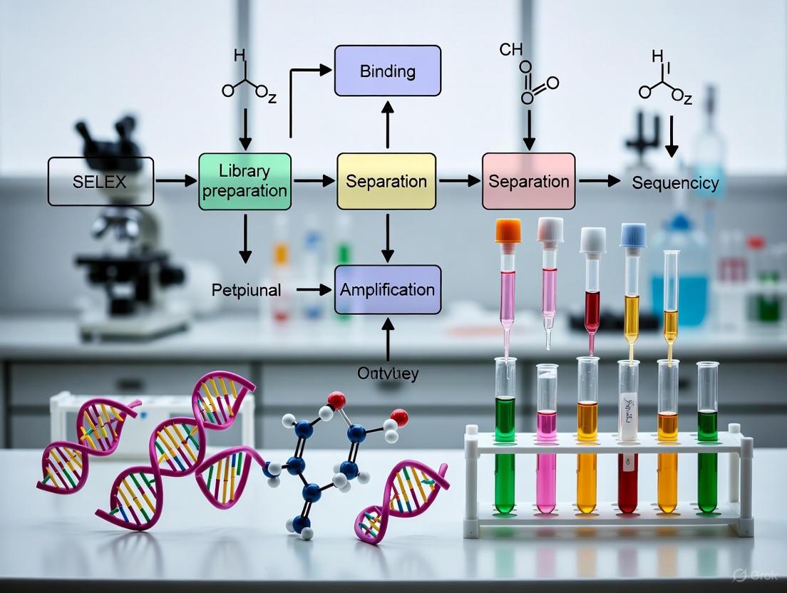
SELEX Workflow: A Step-by-Step Protocol
The standard SELEX protocol comprises multiple iterative cycles of selection and amplification, with careful optimization at each stage to maximize the probability of obtaining high-quality aptamers. The complete process is visualized in Figure 1 below, which outlines the key stages from library synthesis to aptamer characterization.
Figure 1. SELEX Experimental Workflow. The iterative process of aptamer selection through Systematic Evolution of Ligands by Exponential Enrichment.
Generating the Oligonucleotide Library
The first critical step in SELEX involves synthesizing a highly diverse single-stranded oligonucleotide library. This library consists of fully or partially randomized sequences of fixed length (typically 20-80 nucleotides) flanked by constant 5' and 3' ends that serve as primer binding sites for subsequent amplification [1]. The randomized region provides the structural diversity necessary for target recognition, while the constant regions enable efficient amplification throughout the selection process.
Table 1: Oligonucleotide Library Design Considerations
| Parameter | Typical Range | Function | Impact on Selection |
|---|---|---|---|
| Random Region Length | 20-80 nucleotides | Provides structural diversity for target binding | Longer regions increase structural complexity but may reduce binding affinity |
| Constant Primer Regions | 15-25 nucleotides each | Enables PCR amplification and library regeneration | Must be optimized to minimize structural interference with random region |
| Library Diversity | 10^13 - 10^15 unique sequences | Increases probability of containing target-binding sequences | Higher diversity improves chances of finding high-affinity binders |
| Synthesis Method | Chemical synthesis | Generates initial oligonucleotide pool | Synthesis errors can reduce functional diversity |
For a randomly generated region of length n, the theoretical sequence diversity is 4^n, though practical considerations of chemical synthesis typically limit the actual diversity to approximately 10^15 unique sequences [1]. Prior to selection, the oligonucleotide pool is amplified and converted to single-stranded DNA, RNA, or modified nucleotides, depending on the desired aptamer type [1]. For RNA selections, the initial DNA library is transcribed in vitro, while DNA selections use the single-stranded DNA directly.
Target Incubation and Binding Conditions
The single-stranded oligonucleotide library is prepared for target interaction by heating and slow cooling to renature sequences into stable secondary and tertiary structures [1]. This structural folding is essential for generating the complex shapes necessary for specific target recognition. The prepared library is then incubated with the target under carefully controlled conditions that influence the properties of the resulting aptamers.
Target immobilization methods vary depending on the nature of the target and include:
- Affinity chromatography columns for protein targets [1]
- Paramagnetic beads for efficient separation of bound complexes [1]
- Nitrocellulose filter binding for protein-nucleic acid complexes [1]
- Whole cell incubation on culture plates for cell-surface targets [1]
Incubation buffer conditions must be optimized for the specific application of the desired aptamers. For in vivo applications, buffers resembling physiological salt concentrations and homeostatic temperatures are preferred [1]. To minimize non-specific binding, competitors such as tRNA, salmon sperm DNA, or BSA may be added to occupy non-specific binding sites [1]. The relative concentration of target to oligonucleotides represents another critical parameter—excess target increases the probability of binding but provides no selective pressure for affinity, while excess oligonucleotides creates competition that enriches higher-affinity binders [1] [2].
Partitioning, Elution, and Amplification
Following incubation, bound and unbound oligonucleotides are separated through methods appropriate to the immobilization strategy. Unbound sequences are washed away using incubation buffer to maintain binding conditions, while specifically bound sequences are subsequently eluted by creating denaturing conditions that disrupt oligonucleotide-target interactions [1]. Effective elution methods include:
- Flowing deionized water to reduce ionic strength [1]
- Using denaturing solutions containing urea and EDTA [1] [3]
- Applying high heat with physical agitation [1]
- Competitive elution with free target molecules [2]
Eluted sequences are then amplified based on the aptamer type: DNA aptamers proceed directly to PCR amplification, while RNA or modified aptamers require reverse transcription to DNA before amplification [1]. The amplification products must then be converted to single-stranded form for the next selection round, employing methods such as:
- Biotin-streptavidin separation using biotinylated primers followed by alkaline denaturation [1]
- Asymmetric PCR with unequal primer concentrations to favor one strand [1]
- Enzymatic degradation of the unwanted strand using lambda exonuclease [1]
- Spin column purification to separate strands by size or affinity
These methods typically recover 50-70% of the desired single-stranded material, with efficiency varying by technique [1]. The process of incubation, partitioning, elution, and amplification repeats for multiple rounds (typically 5-15 cycles) until the pool shows significant enrichment for target-binding sequences, as measured by the increasing fraction of library binding to the target [1].
Monitoring Progress and Final Characterization
SELEX progression is tracked by measuring the percentage of the oligonucleotide library that binds to the target after each round [1]. As the selection advances, this binding fraction should increase, eventually approaching a plateau indicating successful enrichment of binding species. Quantitative methods for monitoring enrichment include:
- Spectrophotometric quantification of eluted oligonucleotides at 260 nm [1]
- Fluorescent labeling of oligonucleotides with real-time detection [1]
- qPCR monitoring of bound sequences after each round [2]
- Gel electrophoresis to assess pool evolution and diversity
Once sufficient enrichment is achieved (typically when 30-60% of the input library binds target), the final pool is cloned and sequenced to identify individual aptamer candidates [1] [2]. Individual sequences are then synthesized and characterized for binding affinity, specificity, and structural properties. Additional counter-selection steps may be incorporated throughout the process to eliminate sequences with affinity for non-target matrix components or related molecules [1].
Key SELEX Variations and Modifications
The fundamental SELEX methodology has spawned numerous variations designed to address specific challenges or select aptamers with specialized properties. These modifications adapt the core process for particular applications or improve efficiency and success rates.
Table 2: Major SELEX Variants and Applications
| SELEX Variant | Key Modification | Primary Applications | Advantages |
|---|---|---|---|
| Counter-SELEX | Includes negative selection steps against non-targets | Enhancing aptamer specificity | Reduces cross-reactivity with related molecules |
| Toggle-SELEX | Alternates between related targets | Selecting cross-reactive aptamers | Identifies aptamers binding conserved epitopes |
| Capture-SELEX | Immobilizes the library instead of target | Small molecule targets | Avoids target modification that might affect binding |
| Cell-SELEX | Uses whole cells as targets | Cell-surface markers, unknown targets | Identifies aptamers for native cellular structures |
| Automated SELEX | Employs liquid handling systems | High-throughput aptamer discovery | Reduces labor and increases reproducibility |
| FRELEX | Eliminates immobilization of both target and library | Small molecules, fragile targets | Prevents epitope masking from immobilization [1] |
| In vivo SELEX | Conducts selection within living organisms | Therapeutic aptamers with physiological relevance | Identifies aptamers stable and functional in biological systems |
Recent innovations have expanded beyond natural nucleic acids to include chemically modified nucleotides that enhance aptamer stability and binding properties [1] [2]. Modified SELEX approaches incorporate nucleotides with altered sugar moieties (2'-F, 2'-NH₂, 2'-O-methyl), base modifications, or backbone modifications (phosphorothioates) to increase nuclease resistance and structural diversity [1] [2]. Additionally, unnatural base pairs have been incorporated to expand the genetic alphabet, creating aptamers with novel chemical functionalities not possible with standard nucleotides [1] [2].
Critical Experimental Parameters and Optimization
Successful SELEX experiments require careful optimization of multiple parameters throughout the selection process. Key considerations that significantly impact the quality and properties of resulting aptamers include:
Library-to-Target Ratio
The relative concentrations of library and target directly influence selection pressure for binding affinity. Using the target in excess over library sequences increases the probability of recovering binding sequences but provides minimal pressure for high affinity [1]. Conversely, using the library in excess over target binding sites creates competitive conditions that favor the enrichment of higher-affinity binders [1] [2]. Progressive reduction of target concentration in later selection rounds can further drive affinity maturation.
Buffer Conditions
Incubation buffer composition should reflect the intended application environment for the selected aptamers [1]. For diagnostic applications where high specificity is crucial, including specific competitors or adjusted salt concentrations can improve discrimination between related targets. For therapeutic applications, physiological buffer conditions (e.g., PBS at 37°C) help ensure selected aptamers will function under relevant biological conditions [2].
Selection Stringency
Increasing selection stringency throughout the process progressively enriches tighter-binding sequences. Stringency can be modulated through:
- Reduced incubation time to favor faster-binding kinetics
- Increased wash stringency with higher salt or detergent concentrations
- Competitive elution with target analogs to select for specific epitope recognition
- Progressive reduction of target concentration in successive rounds
PCR Amplification Conditions
Amplification must be carefully controlled to prevent the dominance of spurious sequences through PCR bias. Limited cycle PCR helps maintain diversity, while monitoring for amplification artifacts is essential throughout the process [1]. The method for generating single-stranded DNA between rounds significantly impacts library diversity and should be optimized for maximum recovery [1].
The Scientist's Toolkit: Essential Research Reagents
Table 3: Key Research Reagent Solutions for SELEX Experiments
| Reagent Category | Specific Examples | Function in SELEX | Considerations for Use |
|---|---|---|---|
| Oligonucleotide Library | Random ssDNA/RNA library with fixed primer regions | Source of sequence diversity for selection | Diversity, length, and potential modifications should match target properties |
| Amplification Enzymes | Taq polymerase, reverse transcriptase | Amplification of selected sequences between rounds | High-fidelity enzymes reduce mutation rates; optimized buffers maintain diversity |
| Target Molecules | Proteins, small molecules, cells, microorganisms | Selection agent for specific binding | Purity, concentration, and immobilization method critically impact success |
| Separation Matrices | Streptavidin beads, nitrocellulose filters, affinity columns | Partitioning bound from unbound sequences | Matrix choice depends on target properties; minimal non-specific retention is essential |
| Buffer Components | Salts, competitors (tRNA, BSA), detergents | Control binding conditions and stringency | Should mimic intended application environment; competitors reduce non-specific binding |
| Modification Reagents | Biotinylated primers, fluorescent labels | Enable separation and monitoring | Position of modification (5' vs 3') affects efficiency of separation methods |
Applications and Future Perspectives
Aptamers selected through SELEX have found diverse applications across biotechnology, medicine, and basic research. Their unique combination of molecular recognition properties, stability, and design flexibility has enabled several distinct application categories:
Diagnostic Applications
Aptamers serve as recognition elements in biosensors, diagnostic assays, and imaging reagents. Their ability to be chemically synthesized with consistent quality and modified with reporter molecules makes them ideal for diagnostic platforms [2]. Aptamer-based sensors have been developed for targets ranging from small molecules to whole cells, with particular promise in point-of-care testing and multiplexed detection systems [2].
Therapeutic Applications
Therapeutic aptamers represent a promising class of pharmaceutical agents, with one approved drug (pegaptanib for macular degeneration) and several in clinical trials [2]. Their potential advantages include minimal immunogenicity, tissue penetration, and the ability to target proteins considered "undruggable" by conventional approaches. Aptamer therapeutics can function as antagonists, agonists, or targeting moieties in targeted drug delivery systems [2].
Research Tools
In basic research, aptamers serve as specific inhibitors, affinity reagents, and regulatory elements in synthetic biology circuits [2]. Their programmability and compatibility with nucleic acid technologies enable unique applications not possible with conventional antibodies, including direct integration into genetic control systems and combinatorial screening approaches.
The future of SELEX technology continues to evolve with innovations in modified nucleotides that expand chemical diversity, microfluidic selections that enhance efficiency, and computational approaches that complement experimental screening [1] [2]. The integration of artificial genetic systems and automation promises to further accelerate the discovery and optimization of aptamers for increasingly challenging applications. As the field progresses, SELEX remains a versatile and powerful method for generating functional nucleic acids that bridge the molecular recognition properties of biological systems with the engineering flexibility of synthetic chemistry.
{#core-selex-cycle}
The Core SELEX Cycle: Incubation, Partitioning, and Amplification
Systematic Evolution of Ligands by Exponential enrichment (SELEX) is a foundational combinatorial chemistry technique in molecular biology for discovering single-stranded DNA or RNA oligonucleotides, known as aptamers, which exhibit high-affinity binding to specific target molecules [1]. The core SELEX cycle is an iterative process of incubation, partitioning, and amplification that enriches a random oligonucleotide library for target-specific binders over multiple rounds [5]. This application note provides a detailed protocol and key considerations for executing this core cycle, framed within the context of advanced aptamer research and development for therapeutic and diagnostic applications. The procedure follows the rules of directed evolution, leveraging basic molecular biology equipment to isolate rare, high-affinity nucleic acid ligands from a vast pool of random sequences [5].
The SELEX process begins with a synthetic oligonucleotide library containing a central randomized region of fixed length, flanked by constant 5' and 3' ends that serve as primer binding sites for PCR amplification [1] [5]. For a random region of length n, the number of possible sequences is 4n, creating immense diversity from which rare, target-binding sequences are isolated [1]. The elegance of SELEX lies in its simplicity and power; it can generate specific molecular recognition agents using standard biochemistry equipment and techniques accessible to most undergraduate science curricula [5]. The core cycle drives the molecular evolution of the nucleic acid pool, with each round of selection and amplification increasing the proportion of oligonucleotides with strong affinity for the target ligand [5]. The success of a SELEX experiment is measured by the selection of aptamers with high affinity and specificity for their cognate target, often characterized by a low equilibrium dissociation constant (KD) [5].
The Core SELEX Cycle: A Detailed Workflow
The fundamental SELEX cycle consists of three primary stages: incubation of the library with the target, partitioning of bound from unbound sequences, and amplification of the bound sequences to create an enriched library for the subsequent round. The following section and diagram detail this iterative workflow.
Visual Workflow of the Core SELEX Cycle
Diagram 1: The iterative core SELEX cycle. The process begins with a vast library of random sequences and repetitively applies selection pressure to enrich for high-affinity binders. After multiple rounds (typically 5-15), the final enriched pool is sequenced and characterized [1] [5].
Stage 1: Incubation
Objective: To allow oligonucleotides in the library to bind to the immobilized target.
- Library Preparation: The single-stranded oligonucleotide library is first heated and cooled slowly to renature into stable secondary and tertiary structures [1]. For RNA or modified nucleotide selections, the initial DNA library is transcribed into the desired material [5].
- Target Binding: The randomized library is incubated with the immobilized target. Key parameters to consider include:
- Target Immobilization: Common methods include affinity chromatography columns [1], nitrocellulose binding assay filters [1], and paramagnetic beads [1]. Newer methods also use whole cells on culture plates [1].
- Buffer Conditions: Incubation buffer (e.g., salt concentration, pH, presence of non-specific competitors like tRNA or BSA) should be optimized based on the target and the intended application of the aptamer [1]. For in vivo applications, buffer conditions should mimic physiological conditions [1].
- Concentrations: Using the oligonucleotide library in excess over the target introduces competitive pressure, favoring the selection of sequences with higher binding affinity [1].
Stage 2: Partitioning
Objective: To separate target-bound oligonucleotides from the unbound bulk of the library.
- Washing: After incubation, unbound oligonucleotides are washed away using the incubation buffer to preserve specifically bound sequences [1].
- Elution: Specifically bound sequences are recovered by applying denaturing conditions that disrupt the oligonucleotide-target interaction. Common elution methods include using deionized water, denaturing solutions containing urea and EDTA, or applying high heat with physical force [1] [5].
- Counter-Selection: A critical optional step to enhance specificity. The library is incubated with the target immobilization matrix alone or with non-target molecules (e.g., related proteins or non-target cell types) before the main selection. The unbound sequences are retained, thereby depleting the pool of non-specific binders [1].
Stage 3: Amplification
Objective: To amplify the eluted, target-binding sequences to create an enriched library for the next selection round.
- Reverse Transcription (for RNA/modified pools): Eluted RNA or modified base sequences are reverse-transcribed into DNA [1].
- Polymerase Chain Reaction (PCR): The DNA templates (either directly from DNA-SELEX or from RT) are amplified via PCR. The fixed primer regions flanking the random sequence facilitate this amplification [1] [5].
- Generation of Single-Stranded DNA (ssDNA): This is a critical step to regenerate the functional selection pool. Common methods include:
- Biotin-Streptavidin Separation: Using a biotinylated primer during PCR, followed by binding to streptavidin-coated beads and eluting the desired single strand with alkali [1].
- Asymmetric PCR: Performing PCR with an excess of one primer to preferentially produce one strand, though this requires purification from residual double-stranded DNA [1].
- Enzymatic Degradation: Tagging the unwanted strand with a phosphate-probed primer, which is then selectively degraded by enzymes like Lambda exonuclease [1]. These methods typically recover 50-70% of the DNA [1].
Key Experimental Parameters and Reagents
Successful SELEX experimentation requires careful planning and optimization of key parameters. The following tables summarize crucial quantitative data and essential research reagents.
Table 1: Key Parameters to Optimize in a SELEX Experiment
| Parameter | Description | Impact & Consideration |
|---|---|---|
| Number of SELEX Rounds | Total iterations of the core cycle. | Typically requires 5-15 rounds; too few rounds yield insufficient enrichment, while too many can lead to loss of diversity and selection of artifacts [5]. |
| Library Diversity | Number of unique sequences in the initial pool, determined by the length of the random region (e.g., 4n for length n). | A 40 nt region yields 4^40 unique sequences; practical synthesis limits full coverage for n > ~25, but diversity remains vast [5]. |
| Stringency | Selective pressure applied during partitioning. | Can be increased in later rounds by reducing target concentration, increasing wash times, or adding non-specific competitors to select for the tightest-binding aptamers [1] [5]. |
| Dissociation Constant (KD) | Concentration of target at which 50% of the aptamer is bound; measures binding affinity. | Lower KD indicates tighter binding. The desired KD should be guided by the final application (e.g., limit of detection for a sensor) [5]. |
Table 2: Research Reagent Solutions for SELEX
| Reagent / Material | Function in the SELEX Protocol |
|---|---|
| Initial Oligonucleotide Library | A synthetic pool of DNA or RNA with a central random region (e.g., 20-60 nt) flanked by fixed primer binding sites; the source of diversity from which aptamers are selected [1] [5]. |
| Target Molecule | The protein, small molecule, cell, or other ligand against which aptamers are selected. Often immobilized on beads, columns, or plates for partitioning [1]. |
| Selection Buffer | The solution used during incubation. Its composition (ions, pH, additives) is critical for promoting specific binding and can be tailored to the aptamer's intended operational environment [1]. |
| Polymerase Chain Reaction (PCR) Reagents | Enzymes (e.g., Taq polymerase), dNTPs, and primers complementary to the library's fixed regions. Used to amplify the recovered sequences after each partitioning step [1] [5]. |
| Modified Nucleotides | Nucleotides with chemical alterations (e.g., 2'-F, 2'-O-Me pyrimidines). Incorporated into libraries to enhance nuclease resistance, increase binding affinity, and expand structural diversity [5]. |
Monitoring and Analysis
Tracking the progression of SELEX is vital for determining when to stop the selection process.
- Tracking Enrichment: The progress of a SELEX experiment is monitored by comparing the amount of oligonucleotide eluted after target binding to the total input amount for that round. As the pool enriches for binders, this fraction will increase, often converging before reaching 100% [1]. Quantification can be done via UV absorbance at 260 nm or by using fluorescently labeled oligonucleotides [1].
- Sequencing and Bioinformatics: After the final round, the enriched pool is cloned and sequenced, or analyzed by next-generation sequencing (NGS). Bioinformatic analysis is then used to identify sequence families, consensus motifs, and potential secondary structures of the selected aptamers [5].
- Characterization of Individual Aptamers: Individual aptamer candidates must be synthesized and their affinity (KD), specificity, and selectivity characterized using techniques like surface plasmon resonance (SPR), electrophoretic mobility shift assays (EMSA), or isothermal titration calorimetry (ITC) [5].
Concluding Remarks
The core SELEX cycle of incubation, partitioning, and amplification is a robust and adaptable framework for evolving functional nucleic acids. Its success hinges on the careful design and execution of each stage, informed by the nature of the target and the desired properties of the final aptamer. By understanding and strategically optimizing the parameters outlined in this protocol, researchers can effectively harness the power of in vitro evolution to generate high-quality aptamers for a wide range of applications in biomedicine and biotechnology.
This application note provides researchers and drug development professionals with a comprehensive framework for characterizing the critical kinetic and equilibrium parameters of aptamer-target interactions. Aptamers, single-stranded DNA or RNA oligonucleotides selected through Systematic Evolution of Ligands by Exponential Enrichment (SELEX), represent increasingly important tools in therapeutic and diagnostic development [6] [1]. Their binding characteristics are quantitatively defined by the association rate (kON), dissociation rate (kOFF), and equilibrium dissociation constant (KD) [7] [8]. We detail experimental protocols utilizing surface plasmon resonance (SPR) and other biophysical techniques to measure these parameters, supported by quantitative data tables and standardized workflows essential for advancing aptamers from selection to application.
The binding interaction between an aptamer and its target is a dynamic process best described by three fundamental parameters:
- Association Rate Constant (kON): Measures the rate at which the aptamer and target form a complex, expressed in M-1s-1. A higher kON indicates faster complex formation [8].
- Dissociation Rate Constant (kOFF): Measures the rate at which the aptamer-target complex dissociates, expressed in s-1. A lower kOFF indicates a more stable complex with a longer residence time [8] [9].
- Equilibrium Dissociation Constant (KD): Represents the concentration of target at which half of the aptamer binding sites are occupied at equilibrium, expressed in molar units (M). It is calculated as kOFF/kON and provides a direct measure of binding affinity, where a lower KD indicates higher affinity [8] [10] [9].
Understanding these parameters is crucial for developing aptamers for therapeutic use, where both binding strength (KD) and complex stability (kOFF) directly impact efficacy and dosing regimens [6].
Quantitative Characterization of Aptamer Interactions
The following tables summarize kinetic and affinity data for various aptamer-target interactions, providing benchmark values for researchers.
Table 1: Experimentally Determined Kinetic Parameters for Protein-Aptamer Interactions
| Aptamer Target | kON (M-1s-1) | kOFF (s-1) | KD | Measurement Technique | Citation |
|---|---|---|---|---|---|
| Lysozyme | 1.8 × 10⁵ | 2.6 × 10⁻³ | 14.6 nM | SPR (OpenSPR) | [10] |
| Thrombin (TBA) | Varies with conditions | ~Constant across conditions | 0.15 nM - 250 nM | switchSENSE | [8] |
| VV-GMCSF-Lact | Not Specified | Not Specified | ~0.35 µM | Microscale Thermophoresis | [11] |
Table 2: Impact of Cations on Thrombin-Binding Aptamer (TBA) Folding and Affinity
| Cation | Ionic Radius (Å) | Relative Folding Affinity | Resulting KD for Thrombin | Primary Effect |
|---|---|---|---|---|
| K⁺ | 1.33 | Highest | 0.15 nM | Faster kON |
| NH₄⁺ | 1.45 | High | ~2 nM (estimated) | Faster kON |
| Na⁺ | 0.95 | Low | ~20 nM (estimated) | Slower kON |
| Li⁺ | 0.60 | Lowest | 250 nM | Slower kON |
Data adapted from [8]
As illustrated in Table 2, the binding affinity of an aptamer can be profoundly influenced by solution conditions, particularly ions that stabilize its structure. For the thrombin-binding aptamer (TBA), which folds into a G-quadruplex, the correct fold and high affinity for thrombin (low KD) are strongly dependent on the presence of coordinating cations like K⁺ with an appropriate ionic radius [8]. The type of cation primarily influences the association rate (kON), while the stability of the formed complex (kOFF) remains relatively constant across different cations [8].
Experimental Protocols
Protocol 1: Characterizing Aptamer Kinetics via Surface Plasmon Resonance (SPR)
SPR is a powerful, label-free technology for the real-time quantification of biomolecular interactions [7] [9]. The following protocol is adapted for characterizing aptamer-protein kinetics on instruments such as the OpenSPR:
A. Sensor Chip Preparation
- Immobilization: Use a streptavidin-coated sensor chip. Dilute the biotinylated aptamer in the recommended running buffer (e.g., HBS-EP: 10 mM HEPES, 150 mM NaCl, 3 mM EDTA, 0.05% v/v Surfactant P20, pH 7.4).
- Loading: Inject the diluted aptamer over the streptavidin surface for 2-5 minutes at a flow rate of 5 µL/min to achieve a desired immobilization level (typically 100-500 Response Units, RU).
- Stabilization: Wash the sensor with running buffer for 10-15 minutes to establish a stable baseline.
B. Kinetic Measurement via Concentration Series
- Sample Preparation: Prepare a dilution series of the target protein (e.g., 0, 5, 10, 20, 40 nM) in the running buffer. The same buffer must be used for all samples and running buffer to minimize bulk refractive index shifts.
- Binding Cycle: For each concentration, perform the following cycle at a constant flow rate (e.g., 30 µL/min):
- Association Phase: Inject the target solution for 3-5 minutes to monitor binding.
- Dissociation Phase: Switch back to running buffer for 5-10 minutes to monitor complex dissociation.
- Regeneration (if needed): If the aptamer-target complex does not fully dissociate, apply a 30-second pulse of a regeneration solution (e.g., 10 mM Glycine, pH 2.0) to completely remove bound target without damaging the immobilized aptamer.
C. Data Analysis
- Reference Subtraction: Subtract the sensorgram data from a reference flow cell (coated with streptavidin only or a non-specific aptamer) to correct for non-specific binding and bulk effects.
- Kinetic Fitting: Fit the concentration series of sensorgrams globally to a 1:1 binding model using the instrument's software to determine the kinetic rate constants (kON and kOFF).
- Affinity Calculation: The software will calculate the equilibrium dissociation constant (KD = kOFF/kON). An example of the resulting binding curves is shown in the diagram below [10] [9].
Diagram 1: SPR kinetic analysis workflow.
Protocol 2: Assessing the Impact of Ions on Aptamer Folding and Binding
The functional binding of many aptamers is contingent upon their correct folding, which is often stabilized by specific ions in the solution [8]. This protocol uses the switchSENSE platform to characterize this dependency.
A. Pre-conditioning the Aptamer
- Apotamer Preparation: Dilute the aptamer in a chelating buffer (e.g., 10 mM Tris-HCl, 1 mM EDTA, pH 7.5) and heat to 95°C for 5 minutes to denature any pre-existing structures.
- Buffer Exchange: Rapidly exchange the aptamer into the desired cation buffer (e.g., with KCl, NaCl, NH₄Cl, or LiCl) using a size-exclusion spin column. The final ionic strength should be kept consistent across all samples.
B. Real-time Folding and Binding Analysis
- Immobilization: Hybridize the aptamer to a complementary strand on the switchSENSE biosensor chip [8].
- Folding Kinetics: Initiate the experiment by introducing the cation-containing buffer. Monitor the real-time change in the aptamer's hydrodynamic diameter or diffusion speed, which corresponds to its folding into a G-quadruplex or other compact structures.
- Target Binding: Once folding equilibrium is reached, introduce the target protein (e.g., thrombin) in the same cation buffer to measure the binding kinetics (kON, kOFF, KD) under different folding conditions.
C. Data Interpretation
- Compare the folding rates (kF) and unfolding rates (kU) for the aptamer in the presence of different cations.
- Correlate the stability of the folded structure with the resulting binding affinity (KD) and association rate (kON) for the target protein.
The Scientist's Toolkit: Essential Research Reagents and Solutions
Table 3: Key Reagents for SELEX and Aptamer Characterization
| Reagent / Solution | Function / Application | Example Specifications |
|---|---|---|
| SELEX Library | Initial pool of random sequences for in vitro selection. | DNA/RNA library with 30-50 nt random region flanked by constant primer sequences [6] [12]. |
| Modified NTPs | Incorporation into aptamers to enhance nuclease resistance and binding properties. | 2'-Fluoro-pyrimidines (2'-F-dCTP, 2'-F-dUTP); requires mutant T7 RNA polymerase (Y639F) for transcription [6]. |
| Mutant Polymerase | Enzymatic incorporation of modified nucleotides during transcription. | T7 RNA Polymerase (Y639F mutant) for efficient 2'-F-modified RNA transcription [6]. |
| SPR Sensor Chips | Solid support for immobilizing biomolecules to study binding interactions. | Streptavidin-coated chips for capturing biotinylated aptamers [10] [9]. |
| Running Buffers | Maintain consistent pH and ionic strength during binding assays; can be used to study ion effects. | HBS-EP Buffer (10 mM HEPES, 150 mM NaCl, 3 mM EDTA, 0.05% P20, pH 7.4); buffers with specific cations (K⁺, Na⁺) [8]. |
The relationship between the SELEX process, post-selection characterization, and the critical binding parameters is summarized below.
Diagram 2: From SELEX to application via binding parameters.
Rigorous characterization of kON, kOFF, and KD is indispensable for transforming selected aptamer sequences into reliable tools for therapeutics and diagnostics. The experimental protocols and benchmark data provided here serve as a guide for standardizing the evaluation of these critical parameters. As the field advances, integrating kinetic and affinity profiling early in the SELEX process, supported by next-generation sequencing and bioinformatics, will accelerate the development of high-performance aptamer-based reagents and drugs [6].
In the field of nucleic acid research, the in vitro selection of ligands through the Systematic Evolution of Ligands by EXponential enrichment (SELEX) has revolutionized our ability to discover aptamers with high affinity and specificity for therapeutic and diagnostic targets. The success of this process hinges on the capacity of nucleic acids to fold into complex three-dimensional structures, with key structural motifs—G-quadruplexes, hairpins, and loops—serving as critical binding determinants. These motifs provide unique scaffolds that enable molecular recognition by presenting specific chemical groups in defined spatial orientations. This Application Note details the pivotal role these motifs play in facilitating binding interactions, providing researchers with structured data, validated protocols, and analytical frameworks to guide the design and interpretation of SELEX experiments, ultimately enhancing the efficiency of aptamer selection and the development of nucleic acid-based technologies.
Structural Motifs: Definitions and Functional Roles
The following table summarizes the key characteristics, stabilizing forces, and primary functional roles of the three central structural motifs in nucleic acid binding.
Table 1: Key Structural Motifs in Nucleic Acid Binding
| Motif | Structural Description | Key Stabilizing Forces | Primary Functional Role in Binding |
|---|---|---|---|
| G-Quadruplex (G4) | Stacked G-quartets formed by Hoogsteen hydrogen bonding between four guanine bases [13]. | • Hoogsteen H-bonding• Monovalent cations (K⁺ > Na⁺ >> Li⁺) [13] [14]• π-π stacking between quartets | Creates a unique, stable platform for recognizing proteins and small molecules; often found in promoter regions and 5'/3' UTRs for regulatory control [14]. |
| Hairpin | A double-helical stem closed by a single-stranded loop. | • Watson-Crick base pairing in the stem• Base stacking | Presents the loop sequence as a primary recognition element for proteins and ligands; the stem provides stability [15]. |
| Loop | Single-stranded regions connecting secondary structure elements (e.g., helices). | • Backbone flexibility• Specific nucleotide interactions (e.g., GNRA tetraloops) | Provides versatile, accessible binding sites; conformational flexibility allows for induced-fit interactions with diverse targets [16] [17]. |
The Scientist's Toolkit: Essential Research Reagents
Successful investigation of these motifs requires a specific set of reagents to stabilize, probe, and analyze their structures.
Table 2: Key Research Reagent Solutions for Structural Motif Analysis
| Reagent / Material | Function / Application | Example Usage |
|---|---|---|
| KCl / LiCl Buffers | To stabilize (K⁺) or destabilize (Li⁺) G-quadruplex structures during binding assays [13]. | RIP-seq under G4-stabilizing (K⁺) vs. non-stabilizing (Li⁺) conditions to identify G4-dependent protein interactions [13]. |
| G4-Stabilizing Ligands | Small molecules that selectively bind and stabilize G-quadruplex structures. | TMPyP4 (porphyrin derivative) and Bis-4,3 (dimeric carbocyanine dye) used to probe the functional role of G4s in gene regulation (e.g., in the MTOR gene) [14]. |
| CMBL3aL | A water-soluble small molecule used to identify and characterize binding to specific hairpin-loop motifs in pre-miRNA [15] [18]. | HT-SELEX to identify sequence-structure motifs in pre-miRNA hairpin loops essential for binding to CMBL3aL [15]. |
| Thiazole Orange (TO) | A fluorescent dye that exhibits significant fluorescence enhancement upon selective binding to G-quadruplex structures [14]. | Validating the formation and stability of G4 structures in vitro via fluorescence assays [14]. |
Experimental Protocols for Motif Analysis
G4 RIP-seq Protocol for Identifying G-Quadruplex-Dependent Protein Interactions
This protocol is adapted from studies investigating the binding of the FUS protein to RNA targets under different G4-stabilizing conditions [13].
RNA Extraction and Annealing:
- Extract total RNA from your cell line of interest (e.g., SH-SY5Y neuroblastoma cells).
- Divide the RNA into two aliquots and anneal the secondary structures in two different buffers:
- G4-Stabilizing Buffer: 100 mM KCl, 10 mM Tris-HCl (pH 7.4).
- Non-G4-Stabilizing Buffer: 100 mM LiCl, 10 mM Tris-HCl (pH 7.4).
RNA-Protein Binding and Crosslinking:
- Incubate the annealed RNA with the purified recombinant RNA-binding protein (e.g., C-terminal FUS protein, aa 269-526).
- Subject the mixture to UV crosslinking to covalently link the protein to its bound RNA targets.
Immunoprecipitation (IP):
- Perform IP on the complexes using a protein-specific antibody (e.g., anti-His6 tag for His-tagged FUS). Include control IPs with normal IgG and a no-antibody control.
- Wash the beads thoroughly with the respective buffers (K⁺ or Li⁺) to remove non-specifically bound RNA.
RNA Purification and Analysis:
- Purify the immunoprecipitated RNAs by digesting the protein and extracting with phenol-chloroform.
- Convert the purified RNA to cDNA.
- Analyze by RT-qPCR for known targets and prepare next-generation sequencing libraries.
Data Analysis:
- Sequence the Input and IP libraries from both conditions.
- Compare gene enrichment in K⁺ IP vs. Li⁺ IP to identify RNAs whose binding is enhanced by G4 structures (enriched in K⁺) [13].
HT-SELEX for Identifying Small Molecule Binding Motifs in Hairpin Loops
This protocol outlines the process for identifying the sequence-structure motifs in pre-miRNA hairpin loops that bind to a small molecule of interest [15] [18].
Library Design:
- Design a randomized RNA library based on a pre-miRNA scaffold (e.g., pre-miR29a), where the native hairpin loop sequence is replaced with a randomized region (e.g., N11 or a mixed-length N6-11 library) [15].
Immobilization of Target:
- Immobilize the small molecule target (e.g., CMBL3aL) onto a solid support (e.g., resin beads) via a functionalized linker.
Selection Rounds (SELEX):
- Incubate the RNA library with the immobilized target.
- Wash away unbound and weakly bound RNAs.
- Elute the specifically bound RNAs and reverse transcribe them to cDNA.
- Amplify the cDNA by PCR for the next round of selection. Repeat this process for multiple rounds (typically 8-12) to enrich high-affinity binders.
High-Throughput Sequencing and Motif Identification:
- Subject the RNA pools from advanced selection rounds to high-throughput sequencing.
- Use bioinformatic tools (e.g., RaptRanker) to analyze the sequencing data and identify enriched sequence-structure motifs. This analysis often reveals consensus motifs, such as consecutive guanines (GG) flanked by uracils (U) in the context of a hairpin loop [15].
Validation:
- Validate the binding affinity of the identified motifs to the small molecule using Surface Plasmon Resonance (SPR).
- Test the functional consequence of binding, such as the ligand's effect on dicer-mediated cleavage of the aptamers and endogenous pre-miRNAs containing the identified motif [15].
Data Presentation and Analysis
Quantitative data from studies on structural motifs can be systematically organized to facilitate comparison and interpretation. The table below summarizes experimental findings on G-quadruplex-ligand interactions.
Table 3: Quantitative Data on G-Quadruplex-Ligand Interactions in Gene Regulation
| Gene Target | G4-Stabilizing Ligand | Observed Effect on RNA/Protein | Experimental System | Reference |
|---|---|---|---|---|
| MTOR | Bis-4,3 (dimeric carbocyanine) | Downregulation of MTOR RNA and mTOR protein expression | HeLa and SH-SY5Y cells | [14] |
| MTOR | TMPyP4 (porphyrin) | Downregulation of MTOR RNA and mTOR protein expression | HeLa and SH-SY5Y cells | [14] |
| Global FUS targets | K⁺ (ionic condition) | 52 of 56 significantly altered RNAs showed increased FUS binding under G4-stabilizing conditions | In vitro RIP-seq with SH-SY5Y cell RNA | [13] |
Workflow and Pathway Visualizations
G4 RIP-seq Experimental Workflow
The following diagram illustrates the key steps in the G4 RIP-seq protocol, which is used to identify RNA targets whose binding to a protein is dependent on G-quadruplex structures.
HT-SELEX for Motif Discovery
This diagram outlines the iterative HT-SELEX process used to discover specific sequence-structure motifs that bind to a target molecule.
The starting library is a foundational component in the Systematic Evolution of Ligands by EXponential enrichment (SELEX) process, which is used for selecting * oligonucleotide aptamers–highly structured DNA or RNA molecules that bind to specific targets with affinities comparable to antibodies [19]. A typical SELEX library consists of a central *random region flanked by two fixed primer regions. The random region, which can be 20–100 nucleotides long, provides the sequence diversity necessary for binding to a wide array of targets, from small molecules to whole cells [19] [20]. The fixed primer regions are constant sequences essential for the enzymatic amplification (via PCR or RT-PCR) of target-bound sequences throughout iterative selection rounds [19].
Designing this starting library involves critical trade-offs: the random region must be long and diverse enough to fold into complex structures capable of high-affinity binding, while the fixed primer regions must be optimized to minimize interference with the selection process. A significant challenge in traditional SELEX is that these primer sequences can themselves participate in binding, leading to non-specific binding and the enrichment of false-positive sequences [19]. This application note details the principles of library design and provides protocols to overcome these limitations, framed within the broader context of advancing in vitro selection research.
Key Components and Design Principles
The Role of Fixed Primer Regions
The fixed primer regions are not merely amplification handles; their composition and length can significantly influence selection outcomes.
- Function: They provide annealing sites for primers during PCR or RT-PCR amplification between selection rounds. One primer often incorporates a T7 promoter sequence for in vitro transcription when selecting for RNA aptamers [19].
- Design Challenge: The fixed sequences can form secondary structures or directly interact with the target, leading to the selection of primer-derived binders rather than binders from the random region. This "fixed primer interference" can dominate the selected pool and is a major source of failure in SELEX experiments [19].
The Role of the Random Region
The random (N) region is the heart of the library's functional diversity.
- Sequence Diversity: A library with a random region of
nnucleotides has a theoretical diversity of 4ⁿ sequences. A typical library of 40 random nucleotides contains 4⁴⁰ (approximately 1.2 x 10²⁴) unique sequences, though practical library sizes are limited to 10¹⁴–10¹⁵ individual molecules due to synthesis constraints [19]. - Structural Diversity: This sequence diversity translates into a vast array of secondary and tertiary structures (e.g., stem-loops, G-quadruplexes, pseudoknots) that enable specific binding to target molecules [20].
Quantitative Library Specifications
Table 1: Standard Specifications for a SELEX Starting Library
| Component | Typical Length (nt) | Key Function | Design Considerations |
|---|---|---|---|
| 5' Fixed Primer Region | 18-25 | Primer binding for amplification | May include T7 promoter; must avoid self-complementarity. |
| Central Random Region (N) | 20-100 | Generates binding diversity | Longer regions (40-60 nt) allow complex structures but require more sequencing depth. |
| 3' Fixed Primer Region | 18-25 | Primer binding for amplification | Should be designed to minimize dimer formation with the 5' primer. |
| Total Library Length | 60-150 | N/A | Shorter libraries are easier to amplify without errors. |
Overcoming Fixed Primer Interference: Advanced Methodologies
The following protocols address the critical issue of fixed primer interference.
Primer-Switching Genomic SELEX
This method eliminates binding artifacts by completely replacing the fixed sequences and their associated tails partway through the SELEX process [19].
Detailed Protocol:
- Initial SELEX Rounds (Rounds 1-3): Perform standard SELEX cycles against your target (e.g., MS2 coat protein) using the initial genomic library. The library is flanked by fixed sequences (F1 and F2) and 9-nt tails [19].
- Introduction of Restriction Site: After round 3, use RT-PCR to amplify the selected pool. The reverse primer must introduce a FokI restriction endonuclease site. FokI is a type IIS enzyme that cuts at a specific distance (9-13 nt) away from its recognition site [19].
- Digestion and Ligation: Digest the amplified DNA library with FokI. This cleaves off the 3' fixed sequence and tail. Ligate a new, double-stranded DNA adapter with a completely different fixed sequence (F3) to the digested end [19].
- Continued Selection: Use the new library, now flanked by F1 and F3, for subsequent selection rounds (4-6). This switch ensures that sequences whose binding depended on the original fixed regions are no longer amplified [19].
Table 2: Research Reagent Solutions for Primer-Switching SELEX
| Reagent / Material | Function / Description | Example / Specification |
|---|---|---|
| Genomic DNA Library | Source of diverse, physiologically relevant sequences. | E. coli genomic library with ~65 nt inserts [19]. |
| FokI Restriction Enzyme | Type IIS endonuclease for precise cleavage downstream of its binding site. | Cuts 9-13 nt from non-palindromic recognition site [19]. |
| Klenow Fragment | DNA polymerase for filling in overhangs and extending primers. | Used in library construction and adapter ligation [19]. |
| T7 RNA Polymerase | For in vitro transcription to generate RNA libraries. | Required if selecting for RNA aptamers [19]. |
| High-Throughpping Sequencing Platform | Enables deep analysis of library evolution and binding dynamics. | Critical for differential binding analysis; e.g., Illumina [20]. |
Differential Binding Cell-SELEX with HTS Analysis
This workflow uses high-throughput sequencing (HTS) and statistical tools from functional genomics to identify aptamers that bind specifically to target cells over control cells, mitigating issues of non-specific enrichment [20].
Detailed Protocol:
- Cell-SELEX with Negative Selection: Perform standard cell-SELEX for several rounds (e.g., 4 and 11). Use target cells (e.g., RCC-MF for clear cell renal cell carcinoma) for positive selection and control cells (e.g., RC-124 from healthy kidney tissue) for counter-selection [20].
- Differential Binding Round: After a selection cycle, split the enriched aptamer pool and incubate it separately with both the target and control cells. Retrieve the bound sequences from each cell population [20].
- Library Preparation for HTS:
- Bioinformatic Analysis:
- Pre-processing: Use
cutadaptto remove constant primer binding regions from the sequencing reads. Filter reads by length (e.g., 40 nt) [20]. - Sequence Counting: Use the FASTAptamer toolkit to count sequence reads and track enrichment [20].
- Differential Analysis: Use edgeR, a statistical tool designed for RNA-seq data, to identify sequences significantly enriched in the target cell sample compared to the control cell sample. Filter based on log2 fold change and statistical significance (p-value or FDR) [20].
- Pre-processing: Use
The diagram below illustrates the core bioinformatic workflow for the differential binding analysis.
Bioinformatic Workflow for Differential SELEX
Alternative Library Designs and Emerging Approaches
Genomic SELEX
This method replaces the traditional random-sequence library with a library of sheared genomic dsDNA. This approach is particularly useful for identifying physiologically relevant transcription factor binding sites, as the library consists of natural genomic sequences with their native context [21].
DNA Display for Unnatural Nucleic Acids
This technique enables the selection of aptamers composed of unnatural genetic polymers, such as threose nucleic acid (TNA). A key feature is that the TNA molecule is physically linked to its encoding double-stranded DNA template. This phenotype-genotype linkage allows for the selection of functional TNA molecules and the subsequent recovery of their sequence information via PCR amplification [22].
The following diagram outlines the primer-switching method, a direct solution to fixed-region interference.
Primer-Switching SELEX Workflow
Advanced SELEX Methodologies and Expanding Application Landscapes
Systematic Evolution of Ligands by Exponential Enrichment (SELEX) is a combinatorial chemistry technique in molecular biology for producing oligonucleotides of either single-stranded DNA or RNA that specifically bind to a target ligand or ligands; these molecules are commonly referred to as aptamers [1]. Since its introduction in 1990, SELEX has evolved from a basic in vitro selection methodology into a sophisticated family of techniques designed to isolate high-affinity aptamers for a vast range of targets, from small molecules to whole cells [23] [1]. The core principle of SELEX involves the iterative cycles of selection and amplification. Starting with a highly diverse synthetic library of up to 10^15 random nucleic acid sequences, the process involves incubating the library with the target, partitioning bound from unbound sequences, amplifying the bound sequences, and regenerating a single-stranded library for the subsequent round [1] [24]. Through repeated rounds, often with increasing stringency, sequences with the highest affinity and specificity for the target are exponentially enriched.
The estimated success rate of conventional SELEX is generally below 30%, a challenge that has driven the development of specialized SELEX variants [25]. These variants are engineered to overcome specific limitations, such as the accessibility of target epitopes, the replication of complex biological environments, and the overall efficiency of the selection process. This document details the protocols and applications of key SELEX variants—those utilizing immobilized targets, conducted in vivo, performed on whole cells (Cell-SELEX), and automated on advanced platforms—framed within the context of modern aptamer research for therapeutic and diagnostic development.
SELEX with Immobilized Targets
Principle and Application Notes
SELEX variants employing immobilized targets represent the classical approach to aptamer selection. In these methods, the target molecule is fixed to a solid support, which facilitates the separation of target-bound aptamer sequences from the unbound library through simple washing steps [25] [1]. The choice of immobilization matrix is critical, as it can influence the conformation of the target molecule and potentially introduce non-specific binding if the aptamers interact with the matrix itself [25]. Common matrices include nitrocellulose filters, which are simple and affordable but limited to large molecules like proteins; and various beads (e.g., paramagnetic beads), which are more versatile and allow for the immobilization of small molecules via specific coupling chemistries [25] [1]. A significant advantage of this approach is the ease with which negative selection or counter-selection can be incorporated. This involves pre-incubating the oligonucleotide library with the bare immobilization matrix or a related non-target molecule to remove sequences that bind indiscriminately, thereby enhancing the specificity of the enriched aptamer pool [25] [1].
Table 1: Comparison of Common Target Immobilization Methods in SELEX
| Immobilization Method | Target Suitability | Key Advantages | Key Limitations |
|---|---|---|---|
| Nitrocellulose Filter | Proteins, Cells [25] | Simple, affordable [25] | Limited by pore size; not suitable for small molecules [25] |
| Bead-Based (e.g., paramagnetic beads) | Proteins, Small molecules, Cells [25] [1] | Versatile; commercially available with specialized coatings; easy handling via magnet or centrifugation [25] | Coupling process may alter target conformation; matrix can cause non-specific binding [25] |
| Affinity Chromatography Column | Proteins, Small molecules [1] | Established methodology | Potential for non-specific binding to column resin |
Detailed Protocol: Bead-Based SELEX with Counter-Selection
The following protocol outlines a standard bead-based SELEX procedure for a protein target.
I. Research Reagent Solutions
Table 2: Key Reagents for Bead-Based SELEX
| Reagent / Material | Function | Example & Notes |
|---|---|---|
| Oligonucleotide Library | Source of potential aptamer sequences | A ssDNA library with a central 30-40 nt random region flanked by constant primer binding sites [24]. |
| Target Protein | The molecule for which aptamers are being selected | Purified, recombinant protein. Stability under selection conditions is critical. |
| Magnetic Beads with Coupling Chemistry | Solid support for target immobilization | Carboxyl-modified magnetic beads for covalent coupling via EDC/NHS chemistry. |
| Binding/Wash Buffer | Provides physicochemical conditions for binding | Typically PBS with Mg²⁺, and potentially non-specific competitors like tRNA or BSA [1]. |
| Elution Buffer | Dissociates bound aptamers from the target | Denaturing conditions: e.g., 7M urea, 10mM EDTA; or deionized water [1]. |
| PCR Reagents | Amplifies eluted sequences | Primers complementary to library constant regions, dNTPs, thermostable DNA polymerase. |
II. Experimental Workflow
- Target Immobilization: Covalently couple the purified target protein to activated magnetic beads (e.g., using EDC/s-NHS chemistry for carboxylated beads) according to the manufacturer's protocol. Block any remaining active sites on the beads with a blocking agent (e.g., BSA or ethanolamine). Similarly, prepare "negative selection beads" without the target protein but subjected to the same coupling and blocking steps.
- Library Preparation: Denature the initial ssDNA library (e.g., 1 nmol) by heating at 95°C for 5 minutes and snap-cooling on ice. Subsequently, allow it to fold in the binding buffer by warming it to the selection temperature (e.g., 37°C) for 10-20 minutes.
- Counter-Selection (Negative Selection): Incubate the folded ssDNA library with the negative selection beads for 30-60 minutes. Recover the supernatant, which now contains sequences that do not bind to the immobilization matrix or blocking agents. Discard the beads.
- Positive Selection: Incubate the pre-cleared library supernatant with the target-immobilized beads for 30-60 minutes with gentle agitation.
- Washing: Separate the beads using a magnet and carefully remove the supernatant containing unbound sequences. Wash the beads multiple times (e.g., 3-5 times) with the binding/wash buffer to remove weakly or non-specifically bound sequences.
- Elution: Elute the specifically bound sequences by resuspending the beads in a denaturing elution buffer (e.g., 7M urea, 10mM EDTA) or deionized water, and heating at 95°C for 5-10 minutes. Separate the eluate containing the bound ssDNA from the beads.
- Amplification and Regeneration: Amplify the eluted ssDNA by PCR. To regenerate the single-stranded library for the next SELEX round, use a biotinylated reverse primer during PCR. The double-stranded PCR product can then be bound to streptavidin-coated beads and the desired non-biotinylated strand can be eluted with a mild alkaline solution (e.g., 0.1M NaOH) [1]. Purify the resulting ssDNA.
- Repetition: Use the regenerated ssDNA library as the input for the next selection round. Typically, 8-15 rounds are performed, with increasing stringency in later rounds (e.g., increased number of washes, decreased target concentration, or addition of specific competitors) [25].
Cell-SELEX
Principle and Application Notes
Cell-SELEX is a powerful variant where whole, living cells are used as targets for the selection process [23] [26]. This method is particularly valuable for generating aptamers against complex, native cell surface biomarkers, such as receptors or glycoproteins, without prior knowledge of their molecular identity [26]. Since the aptamers are selected against targets in their natural conformation and membrane environment, they often exhibit high functional activity, making them ideal for applications like cancer cell targeting, biomarker discovery, and targeted drug delivery [26]. A defining feature of Cell-SELEX is the use of counter-selection against related non-target cells (e.g., non-malignant cells of the same lineage) to eliminate aptamers that bind to common surface antigens. This step is crucial for achieving high specificity, enabling the generation of aptamers that can distinguish between different cell types, such as cancerous and healthy cells [26].
Detailed Protocol: Cell-SELEX for Cancer Cell Targeting
This protocol is adapted from published methodologies for selecting aptamers against specific cancer cell lines [26].
I. Research Reagent Solutions
Table 3: Key Reagents for Cell-SELEX
| Reagent / Material | Function | Example & Notes |
|---|---|---|
| Target Cells | The cells for which specific aptamers are desired. | A cancer cell line (e.g., EGFR-positive glioblastoma cells). Grown to 80-90% confluence. |
| Counter-Selection Cells | Cells used to remove non-specific binders. | A related non-target cell line (e.g., non-malignant astrocytes). |
| ssDNA Library | Source of potential aptamer sequences. | Can be a standard DNA library or a nuclease-resistant modified library (e.g., 2'-Fluoropyrimidine-modified RNA library) [26]. |
| Cell Culture Media (Serum-Free) | Buffer for selection steps. | DMEM or RPMI, without serum to prevent interference. |
| Wash Buffer | For washing cells to remove unbound sequences. | DPBS (Dulbecco's Phosphate Buffered Saline) or serum-free media. |
| Elution Reagent | To recover cell-bound aptamers. | TRIzol reagent for simultaneous cell lysis and nucleic acid preservation [26]. |
| RT-PCR Reagents | For amplification of eluted sequences. | Required if an RNA library is used. |
II. Experimental Workflow
- Cell Preparation: Culture the target cancer cells and the counter-selection non-target cells to near confluence. On the day of selection, wash the adherent cells gently with serum-free media. Keep the cells viable throughout the process.
- Library Preparation: Denature the modified RNA (or DNA) library (e.g., 300-800 pmol) in serum-free DMEM by heating at 85°C for 5 minutes. Snap-cool on ice for 2 minutes, then allow it to fold at 37°C for 10 minutes.
- Counter-Selection: Incubate the folded RNA library with the non-target (counter-selection) cells in serum-free media for 30 minutes at 37°C. After incubation, carefully recover the supernatant, which contains the unbound sequences. These sequences are now depleted of binders to common surface markers. (The bound fraction from these cells is discarded).
- Positive Selection: Apply the supernatant from the counter-selection step directly to the washed target cancer cells. Incubate for 30 minutes at 37°C.
- Washing: Remove the unbound library supernatant and wash the target cells gently but thoroughly 5-6 times with generous volumes (e.g., 10 mL) of serum-free media to remove weakly associated sequences.
- Elution of Bound Aptamers: Lyse the target cells directly on the plate using TRIzol reagent to recover the cell-bound RNA sequences. Follow the manufacturer's standard protocol for RNA extraction, which includes phase separation with chloroform and precipitation with isopropanol.
- Amplification and Regeneration: Reverse transcribe the recovered RNA into cDNA. Amplify the cDNA by PCR using primers that include the T7 promoter sequence. Use the resulting DNA as a template for in vitro transcription to generate the RNA library for the next round. This transcription step also incorporates the 2'-F-pyrimidine modifications to ensure nuclease resistance in subsequent rounds [26].
- Progression and Monitoring: Repeat the process for multiple rounds (typically 10-20). Increase stringency in later rounds by reducing the number of target cells, incubation time, or library amount. Monitor enrichment by measuring the amount of recovered nucleic acid after each round or by using flow cytometry to detect binding of the pooled library to the cells.
In Vivo SELEX
Principle and Application Notes
In vivo SELEX represents a paradigm shift by conducting the selection process within the complex physiological environment of a living organism [27]. This approach addresses a key limitation of in vitro methods: the failure to account for the complex biological barriers, non-target interactions, and physiological conditions that an aptamer would encounter in therapeutic applications. By injecting the oligonucleotide library into an animal model (e.g., a mouse with a xenografted tumor), the selection pressure inherently favors aptamers that can not only bind to the target tissue but also survive in the bloodstream, evade filtration and immune responses, and efficiently extravasate and penetrate into the target site [27]. The primary outcome is the identification of aptamers with superior in vivo stability, pharmacokinetics, and targeting efficiency, making them highly promising for direct clinical translation.
Automated and Advanced SELEX Platforms
Principle and Application Notes
To address the labor-intensive and time-consuming nature of conventional SELEX, several automated and advanced platforms have been developed. These include Capillary Electrophoresis SELEX (CE-SELEX), Microfluidic SELEX, and the use of High-Throughput Sequencing (HTS) [23] [27].
- CE-SELEX utilizes capillary electrophoresis as both a separation and an analytical tool. It offers high-resolution separation of bound and unbound aptamer complexes based on their charge-to-size ratio in a free solution, without the need for target immobilization [23]. This results in a faster selection process (as few as 4 rounds) and can yield aptamers with very high affinity. Recent innovations like non-equilibrium capillary electrophoresis (NECEEM) and single-step CE-SELEX have further streamlined the process, integrating mixing, reaction, and separation into a single online step [23].
- Microfluidic SELEX (or M-SELEX) employs microfluidic chips to miniaturize and automate the entire SELEX process. The chip's small dimensions reduce reagent consumption and incubation times while allowing for precise control of liquid handling and selection conditions [23] [27]. This technology significantly accelerates the selection timeline and improves efficiency.
- High-Throughput Sequencing (HTS) is not a standalone SELEX variant but a transformative tool used in conjunction with other methods. By applying HTS to the evolving aptamer pool after every round, researchers can monitor the enrichment of specific sequences in real-time, identify promising candidates early, and perform bioinformatic analysis to cluster families of aptamers, moving the process from a "black box" to a data-driven endeavor [23].
Table 4: Comparison of Advanced SELEX Platforms
| SELEX Platform | Core Principle | Key Advantages | Typical Selection Rounds |
|---|---|---|---|
| Capillary Electrophoresis (CE)-SELEX [23] | Separation based on mobility shift of target-aptamer complexes. | High-resolution separation; no immobilization needed; can determine binding parameters; fast and efficient. | 2-4 rounds [23] |
| Microfluidic SELEX [23] [27] | Miniaturization and automation of binding/separation on a chip. | Low reagent consumption; fast cycling; high-throughput; precise fluid control. | Varies, but significantly reduced |
| Capture-SELEX [28] | The library is immobilized, and the target is in solution. | Efficient for small molecule targets; recent quantitative study showed superior enrichment vs. GO-SELEX & Gold-SELEX [28]. | 8-12 rounds |
Workflow Diagram: Integration of Advanced SELEX with HTS
The evolution of SELEX technology from a single, standardized protocol to a diverse toolkit of specialized variants has dramatically expanded the potential of aptamers in biomedical research and drug development. The choice of SELEX method—whether it employs an immobilized target for simplicity, whole cells for target-agnostic discovery, living organisms for physiological relevance, or automated platforms for efficiency—is fundamental to the properties and ultimate utility of the selected aptamers. By understanding the principles, applications, and detailed protocols of these key SELEX variants, researchers can strategically design selection campaigns that are more likely to yield aptamers with the high affinity, specificity, and functional characteristics required for successful diagnostic and therapeutic applications. The integration of advanced tools like HTS and bioinformatics further empowers a data-driven approach, promising to increase the success rate and impact of in vitro selection research.
Nucleic acid aptamers, discovered through the Systematic Evolution of Ligands by EXponential Enrichment (SELEX), are single-stranded DNA or RNA oligonucleotides that bind molecular targets with high affinity and specificity [29] [30]. While offering significant advantages over antibodies—including superior thermal stability, minimal immunogenicity, and reduced production costs—their diagnostic and therapeutic application is often hampered by rapid nuclease-mediated degradation in biological fluids [29] [31]. Incorporating modified nucleotides during or after the SELEX process represents a powerful strategy to overcome this limitation. These modifications, particularly to the sugar moiety of nucleotides, markedly improve the biological stability of aptamers without compromising their binding capabilities [29] [32]. This application note details the use of three prominent modifications—2'-Fluoro (2'-F), 2'-O-Methyl (2'-OMe), and Locked Nucleic Acid (LNA)—within the context of SELEX, providing structured data, protocols, and reagent toolkits for researchers aiming to generate stable, drug-ready aptamers.
Properties and Applications of Key Modifications
The primary strategy for enhancing nuclease resistance involves chemical alteration of the ribose sugar's 2'-position. The table below summarizes the key characteristics of the three modifications.
Table 1: Comparison of Key Modified Nucleotides for SELEX
| Modification | Chemical Description | Primary Application in SELEX | Key Advantages | Notable Examples |
|---|---|---|---|---|
| 2'-Fluoro (2'-F) | Substitution of the 2'-OH group with fluorine [29]. | De novo SELEX for RNA aptamers; often used for pyrimidines [29] [31]. | High nuclease resistance; good acceptance by engineered polymerases [32]. | FDA-approved drug Pegaptanib (Macugen) was selected from a 2'-F-pyrimidine library [31]. |
| 2'-O-Methyl (2'-OMe) | Modification of the 2'-OH with a methyl group [29]. | Both de novo and post-SELEX modification [29] [33]. | Excellent nuclease resistance; naturally occurring; less immunogenic [31]. | Used to generate stable aptamers to VEGF and other targets [29] [31]. |
| Locked Nucleic Acid (LNA) | 2'-O and 4'-C linked via a methylene bridge, locking the sugar [29] [34]. | Primarily post-SELEX optimization; some de novo applications [29] [33]. | Dramatically increased thermal stability (Tm increase of +2 to +8°C per monomer); superior mismatch discrimination [34]. | Used in aptamers and qPCR probes for SNP detection and pathogen identification [34]. |
These modifications can be introduced via two principal approaches: de novo SELEX, where a modified nucleotide library is used from the first selection cycle, and post-SELEX modification, where a native aptamer sequence is synthesized with strategic substitutions after selection [29] [33]. The choice of strategy involves a trade-off between achieving maximum stability and maintaining the binding affinity evolved during the selection process.
Experimental Protocols
De Novo SELEX with Modified Nucleotides
This protocol is designed for selecting aptamers using an initial library composed of 2'-F and 2'-OMe nucleotides, as demonstrated for the generation of aptasensors against Bacillus cereus metallo-β-lactamase [32].
Workflow Overview:
Detailed Procedure:
Library Synthesis and Transcription:
- Synthesize a single-stranded DNA (ssDNA) library containing a central random region (e.g., 30-40 nucleotides) flanked by constant primer sequences.
- Critical Step: Convert the dsDNA template into a modified RNA library by in vitro transcription. Use a laboratory-evolved polymerase (e.g., a mutant T7 RNA polymerase like Y639F/H784A) capable of incorporating 2'-modified nucleotides [29] [31].
- Reaction Mix:
- dsDNA template (1 µg)
- Mutant T7 RNA Polymerase (20 U)
- Transcription Buffer (as supplied)
- NTPs: 2'-F-UTP, 2'-F-CTP, 2'-OMe-ATP, 2'-OMe-GTP (3.5 mM each) [32]
- Incubate at 37°C for 4-16 hours.
Selection Rounds:
- Incubation: Denature the modified RNA library (95°C for 2 min, snap-cool on ice) and incubate with the immobilized target protein in selection buffer (e.g., 20 mM Tris-HCl, pH 7.5, 150 mM NaCl, 5 mM KCl, 1 mM MgCl₂) for 30-60 minutes at room temperature [32] [35].
- Partitioning: Remove unbound sequences by extensive washing with the selection buffer.
- Elution: Elute specifically bound sequences using a denaturing buffer (e.g., 95% formamide, 10 mM EDTA) or by heating.
Amplification:
- Reverse transcribe the eluted RNA into cDNA using a reverse transcriptase.
- Amplify the cDNA using PCR with primers complementary to the constant regions.
- Purify the PCR product to serve as the template for the next round of transcription and selection.
Monitoring and Completion:
- Typically, 8-15 selection rounds are required. Monitor enrichment by quantifying the amount of eluted nucleic acid after each round [1]. When significant enrichment is observed, clone and sequence the final pool from the last round to identify individual aptamer candidates.
Post-SELEX Modification with LNA
This protocol outlines the stabilization of a pre-selected, native DNA or RNA aptamer by substituting specific nucleotides with LNA monomers [34] [33].
Workflow Overview:
Detailed Procedure:
Aptamer Analysis:
- Use structure prediction software (e.g., Mfold, RNAfold) to model the secondary structure of the native aptamer. Identify nucleotides involved in key tertiary contacts or those known to be critical for target binding from mutation studies.
Strategic LNA Incorporation:
- Design Rule: Prioritize the substitution of residues in loop regions or flexible linkers that are not directly involved in base-specific hydrogen bonding with the target. LNA incorporation in these areas stabilizes the overall aptamer fold without disrupting critical interactions [34] [33].
- Synthesis: Chemically synthesize a series of aptamer variants where different subsets of nucleotides (e.g., all pyrimidines in loops, or every third nucleotide) are replaced with their LNA counterparts. A typical starting point is to incorporate 3-5 LNA monomers in a 20-30 nt aptamer.
Characterization of Modified Aptamers:
- Binding Affinity: Determine the equilibrium dissociation constant (Kd) of each LNA variant using a technique like surface plasmon resonance (SPR) or fluorescence anisotropy. Compare it to the Kd of the unmodified aptamer. A successful modification will retain a Kd in the same order of magnitude (e.g., nM or pM range) [33].
- Stability Assay: Incubate the modified and unmodified aptamers (e.g., 1 µM) in 50-100% fetal bovine serum (FBS) or human plasma at 37°C. Withdraw aliquots at various time points (0, 1, 2, 4, 8, 24 hours), denature proteins, and run the samples on a denaturing polyacrylamide gel. Quantify the remaining intact aptamer band to determine the half-life. LNA-modified aptamers typically show significantly extended half-lives compared to their native counterparts [32] [34].
The Scientist's Toolkit: Research Reagent Solutions
Table 2: Essential Reagents for SELEX with Modified Nucleotides
| Reagent / Material | Function / Application | Example Specifications & Notes |
|---|---|---|
| 2'-F-dNTPs / 2'-OMe-NTPs | Building blocks for de novo SELEX or in vitro transcription of modified libraries. | ≥ 95% purity (HPLC); 100 mM solution in water; store at -20°C [29] [36]. |
| LNA Phosphoramidites | Chemical synthesis of LNA-modified oligonucleotides for post-SELEX optimization. | Used in solid-phase oligonucleotide synthesizers. |
| Engineered Polymerases | Enzymes capable of recognizing and incorporating modified nucleotides during transcription or PCR. | Mutant T7 RNA Polymerase (e.g., Y639F/H784A) for 2'-F/OMe-NTPs [29] [31]. |
| Selection Target | The molecule against which the aptamer is selected. | High-purity, recombinant protein; can be immobilized on beads or plates. |
| ssDNA Library | The starting pool of diverse sequences for SELEX. | A typical design: 5'-fixed primer - 40N random region - 3'-fixed primer. |
| PCR Purification Kit | Purification of amplification products between SELEX rounds. | Silica-membrane based kits for efficient recovery of dsDNA. |
| Affinity Resin | For target immobilization and partitioning of bound/unbound sequences. | Ni-NTA resin for His-tagged proteins; streptavidin-coated magnetic beads for biotinylated targets. |
The strategic incorporation of 2'-F, 2'-OMe, and LNA modified nucleotides is a cornerstone of modern aptamer development, directly addressing the critical challenge of nuclease stability. As demonstrated by successful applications like 2'-F/OMe-RNA aptasensors for pathogen detection [32], these modifications enable the transition of aptamers from research tools to robust diagnostic and therapeutic agents. The choice between de novo SELEX and post-SELEX modification depends on project goals, resource availability, and the nature of the target. By leveraging the protocols and reagents outlined in this application note, researchers can systematically engineer next-generation aptamers with the enhanced stability required for real-world applications.
The in vitro selection of nucleic acid aptamers through the Systematic Evolution of Ligands by EXponential Enrichment (SELEX) process has revolutionized molecular recognition, providing powerful alternatives to antibodies for therapeutic and diagnostic applications [1]. Aptamers are short, single-stranded DNA or RNA oligonucleotides that bind specific targets with high affinity and specificity by folding into defined three-dimensional structures [37]. The functional diversity of naturally occurring nucleic acids, limited to only four nucleotides, often results in aptamers with suboptimal binding characteristics and poor stability in biological environments [37] [38]. Chemical modification of aptamers addresses these limitations by enhancing their binding properties, nuclease resistance, and pharmacokinetic profiles, with these modifications introduced either before (pre-SELEX) or after (post-SELEX) the selection process [37] [39]. This application note details the strategic implementation of pre- and post-SELEX modification strategies within the context of SELEX research, providing structured protocols, quantitative comparisons, and essential workflows to guide researchers in developing high-performance modified aptamers.
Pre-SELEX Modification Strategies
Pre-SELEX modification involves incorporating chemically altered nucleotides during the initial library synthesis, prior to the selection process. This approach expands the chemical diversity of the library, potentially yielding aptamers with novel functional groups that engage in enhanced interactions with the target [37] [39].
Common Pre-SELEX Modifications and Their Characteristics
Table 1: Overview of Common Pre-SELEX Modification Types and Their Properties.
| Modification Type | Description | Key Advantages | Compatibility Considerations |
|---|---|---|---|
| Ribose Modifications (e.g., 2'-F, 2'-OMe, 2'-NH₂) [39] | Replacement of the 2'-OH group on the ribose sugar. | Greatly improved nuclease resistance; enhanced binding affinity for some targets. | Requires engineered polymerase (e.g., T7 RNA polymerase Y639F mutant for 2'-F-pyrimidine incorporation) [39]. |
| Nucleobase Modifications [37] [38] | Introduction of novel functional groups to the natural bases (A, C, G, T/U). | Expands chemical diversity for interaction; can introduce hydrophobic or charged moieties. | Polymerase must accept the modified nucleobase triphosphates as substrates during PCR and/or transcription. |
| Phosphate Backbone Modifications (e.g., phosphorothioate) [39] | Replacement of a non-bridging oxygen in the phosphate backbone with sulfur. | Increases resistance to nuclease degradation; can improve pharmacokinetics. | Can sometimes reduce binding affinity if not strategically placed [39]. |
| Unnatural Base Pairs (AEGIS) [38] | Incorporation of additional, orthogonal nucleotide pairs beyond A-T and G-C. | Dramatically increases the sequence and functional diversity of the library. | Requires specialized polymerases capable of replicating the unnatural base pairs with high fidelity. |
Protocol: Initiating a Pre-SELEX Modified RNA Aptamer Selection
This protocol outlines the key steps for selecting RNA aptamers using a library where pyrimidines (C and U) are replaced with their 2'-fluoro (2'-F) counterparts, a common and robust pre-SELEX strategy.
1. Library Design and Synthesis:
- Design a single-stranded DNA (ssDNA) template library featuring a central random region (e.g., 30-50 nucleotides) flanked by constant primer binding sites for amplification.
- In vitro transcription is performed using the ssDNA template and a mutant T7 RNA polymerase (e.g., Y639F) in the presence of nucleotide triphosphates (NTPs) where Cytidine Triphosphate (CTP) and Uridine Triphosphate (UTP) are replaced by 2'-Fluoro-CTP and 2'-Fluoro-UTP [39].
- Purify the resulting 2'-F-modified RNA library using denaturing polyacrylamide gel electrophoresis (PAGE) or appropriate purification kits.
2. SELEX Process with Modified Library:
- Incubation: Incubate the 2'-F-modified RNA library with the immobilized target molecule. Utilize negative selection steps against the immobilization matrix (e.g., uncoupled beads) to remove non-specific binders [40] [1].
- Partitioning and Elution: Wash away unbound sequences. Elute specifically bound RNAs using a method appropriate for the target, such as competitive elution with free target or denaturing conditions [1].
- Amplification: Reverse transcribe the eluted RNA into cDNA. Amplify the cDNA using PCR. The resulting DNA template is then used for the next round of transcription with 2'-F NTPs, repeating the cycle.
- Monitoring and Sequencing: Monitor enrichment of binding sequences over successive rounds (e.g., 8-15 rounds). Once significant enrichment is observed, subject the final pool to High-Throughput Sequencing (HTS) to identify candidate aptamers [38].
Figure 1: Workflow for a typical pre-SELEX modification strategy, exemplified by 2'-Fluoro-modified RNA selection.
Post-SELEX Modification Strategies
Post-SELEX optimization involves introducing chemical changes to a pre-selected, unmodified aptamer sequence. This approach is ideal for improving aptamer stability and pharmacokinetics without risking the failure of the primary SELEX process due to polymerase incompatibility [37] [39].
Common Post-SELEX Modification Approaches
Table 2: Common Post-SELEX Modification Strategies and Their Primary Applications.
| Modification Strategy | Methodology | Primary Application & Rationale |
|---|---|---|
| Ribose 2'-O-Methyl (2'-OMe) Substitution [39] | Solid-phase synthesis of the aptamer sequence with 2'-OMe nucleotides replacing native riboses. | Enhanced Nuclease Resistance: A widely used strategy to protect against nucleases, often applied systematically or to specific vulnerable nucleotides. |
| Phosphorothioate Backbone Linkage [39] | Replacing a non-bridging oxygen with sulfur at specific phosphate groups in the backbone. | Improved Stability & Pharmacokinetics: Increases resistance to nucleases; can be used to slow renal clearance by increasing binding to serum proteins. |
| Terminal Modifications [39] | Adding large molecules (e.g., PEG, cholesterol) or inverted nucleotides (3'-3' linkage) to the 5' or 3' end. | Prolonged Circulating Half-life: Shields ends from exonuclease activity; PEGylation increases hydrodynamic radius to reduce renal filtration. |
| Spiegelmer Creation [39] | Chemical synthesis of the aptamer sequence entirely using L-nucleotides (mirror-image). | Exceptional Biostability: The L-oligonucleotide is unrecognizable by natural D-specific nucleases, conferring extreme stability in biological fluids. |
| Truncation & Minimization | Identifying the minimal functional sequence within the selected aptamer through systematic deletion. | Cost Reduction & Optimization: Shorter sequences are cheaper to synthesize and may have improved folding or binding characteristics. |
Protocol: Optimizing an Aptamer via Post-SELEX 2'-OMe Stabilization
This protocol describes a standard method for enhancing the biostability of a DNA or RNA aptamer by incorporating nuclease-resistant 2'-O-methyl (2'-OMe) modifications.
1. Aptamer Sequence Analysis:
- Use secondary structure prediction software (e.g., Mfold, RNAfold) to identify structural elements (stems, loops, bulges) of the selected aptamer [40].
- If available, review mutation or truncation data to pinpoint nucleotides critical for binding.
2. Strategic 2'-OMe Incorporation:
- Stem Regions: Prioritize the substitution of nucleotides in predicted double-stranded stem regions with 2'-OMe analogues. This generally stabilizes the structure without significantly disrupting binding motifs.
- Loop Regions: Be cautious when modifying loop regions, especially those involved in direct target contact. It is often advisable to leave key loops unmodified initially or to test several variants.
- Systematic Screening: Chemically synthesize a panel of aptamer variants with different 2'-OMe substitution patterns (e.g., full 2'-OMe, only purines 2'-OMe, only stem regions 2'-OMe).
3. Binding and Stability Assay:
- Binding Affinity Measurement: Determine the dissociation constant (Kd) of each modified variant versus the original aptamer using a suitable method (e.g., surface plasmon resonance, EMSA, or the gel-based diffusion method [41]). The goal is to identify variants that retain a Kd value within the same order of magnitude as the original.
- Nuclease Stability Test: Incubate the original aptamer and its modified variants in fetal bovine serum (FBS) or a defined nuclease solution at 37°C. Withdraw aliquots at various time points and analyze the intact oligonucleotide by denaturing PAGE or UPLC-MS. The half-life of the modified aptamers should be significantly prolonged compared to the unmodified parent.
Figure 2: A strategic workflow for post-SELEX optimization of aptamers, focusing on 2'-OMe modification.
Performance Comparison and Selection Guidelines
The choice between pre- and post-SELEX strategies depends on the project's goals, the available polymerases, and the required aptamer properties. A quantitative comparison of SELEX methodologies highlights the varying efficiency of different selection platforms.
Table 3: Quantitative Comparison of SELEX Method Enrichment Efficiency. Data from a study using a spiked library with known aptamers against adenosine, analyzed by deep sequencing [28].
| SELEX Method | Description | Reported Enrichment Factor | Key Finding |
|---|---|---|---|
| Capture-SELEX | Library is immobilized; elution is induced by target binding. | 30 to 50-fold | Most efficient method for enriching specific aptamers in this study. |
| GO-SELEX | Graphene oxide is used to adsorb unbound ssDNA; target binding releases aptamers. | < 1-fold (up to 14% enrichment after blocking primers) | Low efficiency due to nonspecific retention; performance can be improved. |
| Gold-SELEX | Uses gold nanoparticles for library immobilization. | < 1-fold | Minimal target-induced release was observed. |
Strategic Guidelines for Method Selection
- Use Pre-SELEX Modification When: The target is challenging (e.g., small molecules with few functional groups), and you need to access a broader chemical space for binding. This is also suitable when the goal is a direct therapeutic candidate and compatible polymerases for the desired modifications are available [37] [38].
- Use Post-SELEX Modification When: You have already identified a functional aptamer with good binding affinity but need to enhance its stability, half-life, or other drug-like properties for in vivo applications. This is a lower-risk approach for stabilizing a known performer [39].
- Combined Approach: For the best results, a combined strategy can be employed. An aptamer selected with a basic pre-SELEX modification (e.g., 2'-F) can be further optimized post-SELEX with additional alterations (e.g., 3'-inverted thymidine, PEGylation) to create a final candidate with superior overall properties [37] [39].
The Scientist's Toolkit: Essential Reagents and Materials
Table 4: Key Research Reagent Solutions for Generating Modified Aptamers.
| Reagent / Material | Function / Application | Example & Notes |
|---|---|---|
| Modified Nucleotide Triphosphates | Building blocks for creating pre-SELEX modified libraries during transcription or PCR. | 2'-F-dNTPs, 2'-OMe-NTPs, dUTP-Biotin [39]. |
| Engineered Polymerases | Enzymes capable of incorporating modified nucleotides during amplification or transcription. | T7 RNA Polymerase Y639F mutant for 2'-F-pyrimidine incorporation [39]. |
| Solid Support for Immobilization | Matrices for partitioning target-bound aptamers from the library. | Streptavidin-coated magnetic beads, Epoxy-activated Sepharose, Nitrocellulose filters [40] [1]. |
| Next-Generation Sequencing (NGS) Platform | For high-throughput analysis of enriched pools to identify aptamer candidates. | Illumina, PacBio. Essential for modern SELEX to monitor enrichment and discover leads [38]. |
| Surface Plasmon Resonance (SPR) | A label-free technique for quantifying binding kinetics (association/dissociation rates) and affinity (Kd). | Biacore systems. Used for characterizing selected aptamers and their modified variants. |
| Gel-Based Diffusion Method (GBDM) | A simple, low-cost method to monitor aptamer-target binding during SELEX or for initial validation [41]. | Custom mini-gel cassettes. Useful for quickly assessing binding without specialized equipment. |
The strategic application of chemical modifications is paramount for unlocking the full potential of aptamers in research, diagnostics, and therapeutics. Pre-SELEX and post-SELEX strategies offer complementary pathways to this goal: pre-SELEX expands the chemical diversity of the library to find more potent binders from the outset, while post-SELEX refines and stabilizes a known aptamer for practical application. The choice of SELEX methodology itself, such as the efficient Capture-SELEX, significantly impacts the success rate of aptamer isolation [28]. By integrating the structured protocols, performance data, and strategic guidelines outlined in this application note, researchers can make informed decisions to systematically generate high-affinity, stable, and specific modified aptamers tailored to their specific experimental and developmental needs.
Nucleic acid aptamers are short, single-stranded DNA or RNA oligonucleotides (typically 20–100 nucleotides in length) that fold into defined three-dimensional structures to specifically bind molecular targets such as proteins, small molecules, and whole cells [42]. These synthetic molecules, often termed "chemical antibodies," are generated through an iterative combinatorial chemistry process known as Systematic Evolution of Ligands by EXponential enrichment (SELEX) [1] [5]. The therapeutic application of aptamers represents a rapidly advancing field with significant clinical potential, leveraging unique advantages including relatively small physical size, flexible structure, cost-effective chemical production, versatile chemical modification, high stability, and low immunogenicity compared to traditional protein-based biologics [42]. This application note details the progression of aptamer therapeutics from the pioneering example of Macugen to contemporary clinical candidates, providing researchers with essential protocols and analytical frameworks for developing aptamer-based therapeutics within the context of in vitro selection (SELEX) research.
The SELEX Process: Generating Therapeutic Aptamers
Fundamental Principles and Workflow
The SELEX methodology employs combinatorial chemistry and directed evolution principles to isolate high-affinity binding oligonucleotides from immensely diverse libraries [5]. A typical SELEX library features a central randomized region (30-60 nucleotides) flanked by constant primer sequences for amplification [1] [5]. The theoretical diversity of a library with a randomized region of length n is 4^n sequences, though practical limitations of DNA synthesis mean libraries with >25 random nucleotides cannot contain every possible sequence [5]. The process iterates through three core stages: (1) incubation of the library with the target, (2) partitioning of bound from unbound sequences, and (3) amplification of bound sequences for subsequent selection rounds [5]. Successive rounds increase stringency to enrich sequences with the highest affinity and specificity.
Table 1: Key Steps in the SELEX Process
| Step | Description | Key Considerations |
|---|---|---|
| Library Design | Synthetic oligonucleotides with random central region flanked by fixed primer sequences | Random region length (typically 35-60 nt); chemical modifications for stability/diversity [5] |
| Target Incubation | Library exposed to target under controlled conditions | Buffer composition, temperature, time, target vs. oligonucleotide concentration [1] |
| Partitioning | Separation of target-bound sequences from unbound | Methods: affinity chromatography, nitrocellulose filters, paramagnetic beads [1] [43] |
| Amplification | PCR amplification of bound sequences | Efficient conversion to single-stranded DNA for next round [1] |
| Counter-Selection | Removal of non-specific binders | Incubation with non-target molecules/cells to increase specificity [1] |
SELEX Experimental Protocol
Design and Synthesis of DNA Library and Primers
- Design the initial ssDNA library with a central random region of 35-60 nucleotides flanked by two constant sequences (18-20 nt each) that serve as primer binding sites [44].
- Incorporate a biotin modification on the reverse primer to enable separation of ssDNA from amplified double-stranded PCR products using streptavidin-coated beads [44].
- Purify the ssDNA library and primers by 10% denaturing polyacrylamide gel electrophoresis (dPAGE) followed by ethanol precipitation [44]. Determine concentration by UV absorption at 260 nm.
Target Incubation and Binding Conditions
- Prepare SELEX buffer appropriate for the intended application. For therapeutic aptamers targeting physiological conditions, use buffers mimicking in vivo parameters (e.g., 1x PBS, 2.5 mM MgCl₂, 0.5 mM CaCl₂, pH 7.4) [44].
- Prior to target introduction, heat the single-stranded oligonucleotide library and cool slowly to renature oligonucleotides into stable secondary and tertiary structures [1].
- Incubate the randomized library with immobilized target. For protein targets like VEGF, immobilization methods may include affinity chromatography columns or paramagnetic beads [1]. Use appropriate target-to-library ratios—excess oligonucleotide library creates competitive pressure for higher affinity binders [1].
Partitioning, Elution, and Amplification
- Wash away unbound oligonucleotides using incubation buffer while retaining specifically bound sequences on the immobilized target [1].
- Elute specifically bound sequences under denaturing conditions (e.g., deionized water, urea/EDTA solutions, or high heat) [1].
- Amplify eluted sequences via PCR. For RNA selections, include reverse transcription to DNA before PCR amplification [1].
- Generate single-stranded DNA for subsequent selection rounds using methods such as biotin-streptavidin separation, asymmetric PCR, or enzymatic degradation of the unwanted strand [1].
- Repeat selection cycles typically 8-15 rounds until target-binding sequences dominate the pool, monitoring enrichment through increased binding in each round [1] [5].
Critical Considerations for Therapeutic Aptamer Selection
- Specificity Enhancement: Implement counter-selection steps by incubating the oligonucleotide library with non-target molecules, non-target cell types, or target immobilization matrix components to eliminate non-specific binders [1] [44].
- Stability Modifications: For therapeutic applications, incorporate modified nucleotides during SELEX to enhance nuclease resistance and serum stability, such as 2'-F, 2'O-Me pyrimidines, or Xeno Nucleic Acid (XNA) bases [5] [42].
- Functional Target Selection: Use whole cells or native protein conformations as targets to ensure selected aptamers recognize biologically relevant structures [44].
- Binding Characterization: Determine apparent dissociation constant (KD) values for selected aptamers using quantitative methods, ensuring KD values align with therapeutic requirements based on target concentration in vivo [5].
Macugen: The First Approved Aptamer Therapeutic
Mechanism of Action and Clinical Significance
Macugen (pegaptanib sodium) holds distinction as the first therapeutic aptamer approved by the US FDA in 2004 for treating neovascular age-related macular degeneration (AMD) [45] [42]. This RNA aptamer specifically targets vascular endothelial growth factor (VEGF)-165, the VEGF isoform primarily responsible for pathological angiogenesis in ocular vascular diseases [46] [45]. Macugen inhibits angiogenesis by binding to the heparin binding domain (HBD) of VEGF165, preventing its interaction with cellular receptors [46]. Structural studies utilizing molecular dynamics simulations reveal that Macugen recognizes HBD through an induced-fit mechanism with major conformational changes in the aptamer, while HBD recognizes Macugen by conformational selection [46]. Although Macugen demonstrated efficacy in treating neovascular AMD, it was eventually overshadowed by more potent anti-VEGF therapies with broader specificity (e.g., ranibizumab, aflibercept) that emerged subsequently [45].
Molecular Recognition Mechanism
The molecular mechanism underlying Macugen's binding to VEGF represents a sophisticated example of aptamer-target recognition. Research reveals an asymmetric binding process where Macugen undergoes significant conformational changes to adapt to the VEGF protein (induced-fit mechanism), while the VEGF HBD maintains its structure and selectively binds pre-existing Macugen conformations (conformational selection) [46]. This dual mechanism results in high specificity for the VEGF165 isoform, highlighting how aptamers can achieve targeted therapeutic effects through complex binding dynamics distinct from traditional antibody interactions.
Diagram 1: Macugen Molecular Mechanism of Action
Current Clinical Candidates and Second-Generation Agents
Evolving Therapeutic Landscape
The clinical aptamer landscape has expanded significantly beyond Macugen, with multiple candidates advancing through clinical development. Current therapeutic aptamers typically exploit one of three strategic approaches: (1) antagonist function for blocking disease-associated target interactions, (2) agonist function for activating target receptors, or (3) cell-type-specific targeting for delivering therapeutic agents to target cells [42]. To date, most clinical-stage aptamers function as antagonists, reflecting their natural suitability for inhibition protein-protein interactions [42]. Advancements in SELEX technology, including incorporation of modified nucleotides and sophisticated counter-selection strategies, have enabled development of these next-generation therapeutics with improved stability, specificity, and pharmacokinetic properties.
Table 2: Clinical-Stage Aptamer Therapeutics
| Aptamer (Drug Name) | Target | Indication | Stage | Key Features |
|---|---|---|---|---|
| Pegaptanib (Macugen) | VEGF165 | Neovascular AMD | Approved (2004) | First FDA-approved aptamer; specific VEGF isoform targeting [45] [42] |
| Zimura (ACR-1905) | C5 | AMD | Phase III | Complement pathway inhibition; used in combination therapy [42] |
| Fovista (E-10030) | PDGF-B | AMD | Phase III | Anti-platelet-derived growth factor; combination therapy [42] |
| REG1 Anticoagulation System | Factor IXa | Coronary Artery Disease | Phase III | Antidote-controllable anticoagulation system [42] |
| ARC1779 | vWF | Thrombotic Microangiopathy | Phase II | Von Willebrand Factor antagonist [42] |
| AS1411 | Nucleolin | Acute Myeloid Leukemia | Phase II | G-quadruplex DNA aptamer; nucleolin targeting [42] |
| NOX-A12 | CXCL12 | Multiple Myeloma | Phase II | Spiegelmer (L-oligonucleotide) [42] |
| NOX-H94 | Hepcidin | Anemia of Chronic Disease | Phase II | Spiegelmer targeting iron metabolism [42] |
| ARC19499 | Tissue Factor Pathway Inhibitor | Hemophilia | Phase II | Modulation of coagulation pathway [42] |
| EYE001 | VEGF | Diabetic Retinopathy | Phase II | Anti-VEGF RNA aptamer [42] |
Second-Generation Anti-VEGF Agents
In ophthalmology, second-generation anti-VEGF agents have significantly altered the treatment landscape for neovascular AMD, building upon the foundation established by Macugen [45]. These newer agents extend durability, improve fluid reduction, and decrease injection frequency while maintaining visual outcomes and safety. Notable second-generation agents include brolucizumab (Beovu), faricimab-svoa (Vabysmo), and high-dose aflibercept (Eylea HD), each offering distinct pharmacological advantages [45].
- Brolucizumab: A humanized single-chain variable fragment (scFv) monoclonal antibody with molecular weight of 26 kDa, enabling 12.7 times the molar concentration of aflibercept and potentially deeper retinal penetration [45].
- Faricimab: A bispecific antibody representing the first of its kind for intraocular use, targeting both VEGF-A and angiopoietin-2 (Ang-2) to enhance fluid control and improve clinical outcomes through dual pathway inhibition [45].
- Aflibercept (8 mg): A high-dose formulation offering 4 times the molar dose of conventional aflibercept (2 mg), acting as a decoy receptor for VEGF-A, VEGF-B, and placental growth factor (PlGF) with much higher binding affinity for VEGF-A than other anti-VEGF agents [45].
Clinical trials of these second-generation agents demonstrate non-inferiority to earlier treatments with extended dosing intervals. The HAWK and HARRIER trials showed brolucizumab maintained visual acuity gains with potential for 12-week dosing, while the TENAYA and LUCERNE trials established faricimab's efficacy with 8-16 week intervals [45]. The PULSAR trial demonstrated aflibercept 8 mg's non-inferiority with 12-16 week dosing [45].
Research Reagent Solutions for Aptamer Development
Table 3: Essential Research Reagents for SELEX and Aptamer Characterization
| Reagent/Category | Function | Examples/Specifications |
|---|---|---|
| Oligonucleotide Library | Source of sequence diversity for selection | Central random region (35-60 nt) flanked by fixed primer sequences; chemical modifications (2'-F, 2'-O-Me) for stability [5] [44] |
| Modified Nucleotides | Enhance nuclease resistance and binding diversity | 2'-F, 2'O-Me, 2'-NH₂, LNA, TNA, FANA, HNA monomers; hydrophobic residues for protein-like binding [5] |
| Partitioning Matrices | Separation of bound and unbound sequences | Glutathione Sepharose (GST-tagged targets); streptavidin-coated magnetic beads; nitrocellulose filters; affinity columns [1] [43] |
| Amplification Reagents | PCR amplification of selected sequences | High-fidelity DNA polymerase; biotinylated primers for ssDNA separation; appropriate buffer systems [1] |
| Binding Assay Systems | Characterization of aptamer-target interactions | Fluorescence polarization; surface plasmon resonance (SPR); electrophoretic mobility shift assays (EMSA) [43] [5] |
| Cell-Based Assay Systems | Validation of therapeutic activity in biological context | Cell culture models; receptor binding assays; functional response measurements [42] |
Therapeutic aptamers have evolved substantially from the initial approval of Macugen to a diverse landscape of clinical candidates targeting various diseases. The SELEX methodology continues to advance with incorporation of novel nucleotide chemistries, sophisticated partitioning strategies, and increasingly complex target systems including whole cells and pathogens [44]. For researchers developing aptamer therapeutics, careful consideration of SELEX conditions—including library design, binding stringency, counter-selection strategies, and appropriate modification for enhanced stability—remains crucial for success [5]. The unique advantages of aptamers, including their chemical synthesis, tunable affinity, and versatility as targeted delivery agents, position them as compelling therapeutic modalities alongside traditional biologics. As the field progresses, integration of aptamers with other therapeutic modalities (e.g., siRNA conjugates, drug delivery systems) promises to further expand their clinical utility in precision medicine applications.
Diagram 2: SELEX Workflow for Therapeutic Aptamer Development
Aptamers, often termed "chemical antibodies," are single-stranded DNA or RNA oligonucleotides that bind to specific targets with high affinity and selectivity by folding into defined three-dimensional structures [47] [48]. These molecules are developed through an in vitro selection process known as Systematic Evolution of Ligands by EXponential enrichment (SELEX) [49] [30]. Their synthetic nature, low immunogenicity, thermal stability, and ease of chemical modification make them powerful alternatives to antibodies in diagnostics and biomarker discovery [47] [30] [48]. This document details the practical applications of aptamers within the context of SELEX research, providing structured protocols and resources for developing aptasensors, delivery systems, and biomarker discovery platforms.
Application Notes: Core Functions and Implementations
Aptamer technology has diversified into several key application areas, each with distinct experimental considerations and performance outcomes. The table below summarizes the primary functions, targets, and performance metrics of aptamer-based tools.
Table 1: Overview of Aptamer Applications and Performance
| Application Area | Specific Target | Aptamer Function | Key Performance Metrics | Reference |
|---|---|---|---|---|
| Diagnostic Aptasensors | Low-Density Lipoprotein (LDL) | Recognition element in electrochemical sensor | LOD: 0.095 μmol/LLinear Range: 0.1-4.0 μmol/LSample: Human serum | [50] |
| Diagnostic Aptasensors | C-reactive Protein (CRP) | Recognition element in FRET-based sensor | LOD: 2.27 fg/mLLinear Range: 33-82 fg/mL and 114-207 fg/mLSample: Human serum | [51] |
| Diagnostic Aptasensors | Legionella pneumophila SG1 | Recognition element in electrochemical sensor | LOD: 5 CFU/mLSample: Spiked water | [52] |
| Biomarker Discovery | Proteome-wide analysis | Capture reagent in SomaScan platform | Multiplexing: Up to 7,000 proteins simultaneously | [47] [49] |
| Biomarker Discovery | Cell surface targets | Probe in Cell-SELEX | Target: Whole live cellsOutput: Identification of unknown surface biomarkers | [49] |
| Biomarker Discovery | Secreted proteins | Probe in Secretome-SELEX | Target: Proteins secreted from specific cells | [49] |
Aptasensors for Clinical Diagnostics
Aptasensors integrate aptamers with transducers to detect targets in complex samples. Recent advances focus on enhancing sensitivity and point-of-care applicability.
- Electrochemical Aptasensors leverage electrical signals for detection. A representative example is the LDL sensor, which uses a gold nanoparticle-modified electrode for rapid cardiovascular risk assessment [50].
- Optical Aptasensors utilize light-based signals. The FRET-based CRP sensor demonstrates ultra-sensitive detection of inflammatory markers by leveraging graphene oxide as a quencher, enabling detection in biologically relevant matrices like serum [51].
Aptamers in Biomarker Discovery
Aptamers facilitate the discovery of novel biomarkers through their ability to probe complex proteomic landscapes without prior knowledge of target identity.
- Multiplexed Proteomic Analysis: The SomaScan platform uses slow off-rate modified aptamers (SOMAmers) to measure thousands of proteins in a single assay, enabling comprehensive biomarker signature discovery [47] [49].
- Cell-SELEX: This method uses whole living cells as targets to generate aptamers that recognize disease-specific cell surface markers, often revealing previously unknown biomarkers [49] [52].
- Secretome SELEX: Targets proteins secreted or shed from cells, identifying potential biomarkers in circulation [49].
Experimental Protocols
This section provides detailed methodologies for key experimental procedures in aptamer application.
Protocol: Electrochemical Aptasensor for LDL Detection
This protocol details the construction of a label-free electrochemical aptasensor for detecting Low-Density Lipoprotein (LDL) in plasma, based on [50].
1. Reagents and Materials
- Screen-printed carbon electrode (SPCE)
- Gold salt solution (e.g., HAuCl₄) for nanoparticle electrodeposition
- Titanium carbide-carboxymethyl chitosan-hemin (MXene-CMCS-Hemin) nanocomposites
- LDL-specific DNA aptamer (LDLApt)
- Phosphate Buffered Saline (PBS), pH 7.4
- Human plasma or serum samples
2. Sensor Fabrication and Modification
- Step 1: Electrode Pretreatment. Clean the SPCE according to manufacturer's instructions.
- Step 2: Gold Nanoparticle Electrodeposition. Electrochemically deposit gold nanoparticles (Au NPs) onto the SPCE surface using a constant potential or cyclic voltammetry in a gold salt solution. This creates a conductive, high-surface-area substrate.
- Step 3: Nanocomposite Immobilization. Anchor the synthesized MXene-CMCS-Hemin nanocomposites onto the Au NPs/SPCE surface. This layer acts as the electrochemical signal probe.
- Step 4: Aptamer Immobilization. Incubate the electrode with the LDLApt solution to immobilize the aptamer on the nanocomposite surface, forming the final biosensor (LDLApt/MXene-CMCS-Hemin/Au NPs/SPCE).
3. Measurement Procedure
- Step 1: Baseline Measurement. Place the fabricated aptasensor in a blank binding buffer and record the differential pulse voltammetry (DPV) signal of Hemin.
- Step 2: Sample Incubation. Incubate the sensor with the sample (calibrator or plasma) for a fixed time (e.g., 10-15 minutes).
- Step 3: Signal Measurement. Wash the sensor gently and record the DPV signal again in a clean buffer. The specific binding of LDL to the aptamer forms a complex that hinders electron transfer, resulting in a decrease in the Hemin oxidation current.
- Step 4: Quantification. The change in current (ΔI) is proportional to the LDL concentration. Plot ΔI against LDL standard concentrations to generate a calibration curve for determining unknown sample concentrations.
Protocol: FRET-based Aptasensor for CRP Detection
This protocol describes a homogeneous, turn-on fluorescent aptasensor for ultra-sensitive detection of C-reactive protein (CRP), based on [51].
1. Reagents and Materials
- FAM-labeled CRP-specific DNA aptamer
- Synthesized Graphene Oxide (GO) sheets
- CRP protein standard
- Bovine Serum Albumin (BSA)
- Phosphate Buffered Saline (PBS), pH 7.4
- Human serum samples
- Fluorescence spectrophotometer
2. Sensor Preparation and Optimization
- Step 1: Solution Preparation. Prepare a stock solution of FAM-aptamer (e.g., 100 µM) in ultrapure water and a homogeneous dispersion of GO (4 mg/mL) in Milli-Q water via sonication.
- Step 2: Optimization. Optimize the GO concentration and incubation times for quenching and recovery to achieve maximum signal-to-noise ratio. The cited study found 0.03 mg/mL GO and a 5-minute incubation to be optimal.
3. Fluorescent Assay Procedure
- Step 1: Quenching ("Turn-off" State). Mix 1 µL of FAM-aptamer (330 nM) with 0.5 µL of GO solution (0.03 mg/mL). Dilute the mixture to 300 µL with binding buffer. Incubate for 5 minutes at room temperature. During this step, the aptamer adsorbs onto the GO surface via π-π stacking, and the FAM fluorescence is quenched via FRET.
- Step 2: Recovery ("Turn-on" State). Add the sample containing CRP to the FAM-aptamer-GO mixture. Incubate for 5 minutes at room temperature with gentle shaking. The binding of CRP to the aptamer causes a conformational change, releasing the FAM-aptamer from the GO surface and recovering the fluorescence.
- Step 3: Signal Detection. Transfer the solution to a quartz cuvette. Using a fluorescence spectrophotometer, measure the fluorescence intensity at an emission wavelength of 520 nm, with an excitation wavelength of 450 nm.
- Step 4: Quantification. The recovered fluorescence intensity is proportional to the CRP concentration. Construct a calibration curve using CRP standards to interpolate sample concentrations.
Protocol: Cell-SELEX for Aptamer Discovery
This protocol outlines the process for selecting aptamers against specific whole cells, enabling the discovery of unknown cell surface biomarkers, based on [49] [52].
1. Reagents and Materials
- Initial ssDNA library (e.g., 40-nt random region flanked by fixed primer sequences)
- Target cells (e.g., specific bacterial strain or cancer cell line)
- Counter-selection cells (e.g., related but non-target strain or healthy cells)
- Binding buffer (e.g., 50 mM Tris, 2 mM MgCl₂, 150 mM NaCl, pH 7.5)
- Elution buffer (e.g., 7 M urea in binding buffer)
- PCR reagents (Taq polymerase, dNTPs, primers)
- Cell culture media and reagents
2. SELEX Process
- Step 1: Incubation. Incubate the ssDNA library with the counter-selection cells. Collect the unbound sequences. This negative selection removes sequences that bind to common or non-specific cell surface components.
- Step 2: Positive Selection. Incubate the pre-cleared library with the target cells. Gently wash away unbound and weakly bound sequences.
- Step 3: Elution. Elute the cell-bound sequences using a denaturing elution buffer (e.g., 7 M urea) or by heating.
- Step 4: Amplification. Amplify the eluted sequences using PCR. For DNA aptamers, use symmetric PCR. For RNA aptamers, include a reverse transcription step. The PCR product is then converted to single-stranded DNA for the next selection round.
- Step 5: Iteration. Repeat Steps 1-4 for 5-15 rounds, increasing the selection stringency in later rounds (e.g., by reducing incubation time, increasing wash stringency, or decreasing target cell number).
- Step 6: Cloning and Sequencing. After the final round, clone and sequence the enriched pool. Analyze the sequences for consensus motifs and test individual aptamer candidates for binding affinity (e.g., by measuring dissociation constant, Kd) and specificity.
Visualization of Workflows and Pathways
To aid in experimental planning and understanding, the following diagrams illustrate key procedural and conceptual pathways.
Cell-SELEX Workflow for Aptamer Discovery
This diagram outlines the iterative cycle of Cell-SELEX, used to generate aptamers against specific cell types.
FRET-based "Turn-on" Aptasensor Mechanism
This diagram illustrates the signal transduction mechanism of the graphene oxide-based FRET aptasensor for CRP.
The Scientist's Toolkit: Research Reagent Solutions
Successful implementation of the protocols requires key reagents and materials. The following table catalogs essential components and their functions.
Table 2: Essential Research Reagents for Aptamer-Based Research
| Item Name | Function/Application | Key Characteristics | Example Use Case |
|---|---|---|---|
| ssDNA Oligonucleotide Library | Starting point for SELEX; diverse pool of random sequences. | 40-60 nt random region, fixed primer binding sites. | Cell-SELEX for novel aptamer selection [49] [52]. |
| Slow Off-rate Modified Aptamer (SOMAmer) | Engineered aptamer with enhanced affinity for proteomic profiling. | Contains modified nucleotide bases (e.g., Bn-dU, Nap-dU). | Multiplexed protein measurement in SomaScan platform [47] [49]. |
| MXene-CMCS-Hemin Nanocomposite | Electrochemical signal probe and immobilization matrix. | High conductivity, large surface area, intrinsic redox activity. | Signal amplification in electrochemical LDL aptasensor [50]. |
| Graphene Oxide (GO) | Fluorescence quencher in FRET-based assays. | High surface area, efficient energy transfer, water dispersibility. | FRET-based "turn-on" detection of CRP [51]. |
| Screen-Printed Electrode (SPE) | Disposable electrochemical sensing platform. | Integrated working, counter, and reference electrodes; cost-effective. | Point-of-care electrochemical aptasensors [50] [52]. |
| Hexamethylene Glycol (HEG) Spacer | Blocks polymerase extension during PCR. | Used in primer design for SELEX. | Generation of single-stranded DNA library during SELEX [52]. |
Troubleshooting SELEX: Critical Factors and Optimization Strategies for Success
Within the broader context of in vitro selection of nucleic acids (SELEX) research, the design of the initial oligonucleotide library is a critical determinant of success. The library serves as the foundational genetic reservoir from which specific aptamers are evolved, and its optimization directly influences the affinity, specificity, and functional utility of the selected molecules [25] [53]. A well-designed library maximizes the probability of housing rare, high-affinity binding sequences, while a poorly constructed one can lead to selection failure or the identification of suboptimal aptamers [53]. This application note details the core principles and methodologies for optimizing library design, focusing on the two pillars of success: maximizing sequence diversity and the strategic incorporation of chemical modifications. These elements work in concert to expand the structural and functional space available for in vitro evolution, thereby enhancing the potential for discovering aptamers with novel properties and robust performance in diagnostic and therapeutic applications [54].
Optimizing Sequence Diversity
Sequence diversity is the cornerstone of a potent SELEX library. A highly diverse library increases the likelihood of containing nucleic acid species that can adopt three-dimensional structures capable of tightly and specifically binding to a target of interest.
Key Parameters for Library Design
- Random Region Length: The length of the random core region is a primary factor defining theoretical diversity. While a library with a random region of 25 nucleotides (nt) can theoretically encompass ~1015 unique sequences, practical considerations support the use of longer regions. Evidence from the selection of isoleucine-binding RNA aptamers demonstrated that libraries with 50-70 nt random regions yielded the target motif with maximum speed, whereas a shorter 16 nt library failed to isolate the motif after 11 selection cycles [53]. Longer libraries provide a greater probability of containing complex structural motifs, though they also present challenges in synthesis and amplification [53].
- Nucleotide Distribution: Achieving equal representation of all four nucleotides (A, C, G, T/U) in the random region is crucial for structural diversity. Chemical synthesis inherently incorporates nucleotides at unequal rates, often with a bias toward G and T. To counteract this, the molar ratio of phosphoramidites during synthesis should be optimized, for example, to a 1.5:1.5:1.0:1.2 (A:C:G:T) ratio [53]. High-throughput sequencing analysis has revealed that initial libraries from different manufacturers can exhibit distinct sequence biases and nucleotide distributions, such as guanine-rich or thymidine-rich shifts, which can subsequently influence the selection outcome [55].
- Primer-Binding Sites: The constant flanking regions, necessary for amplification, can comprise 40-70% of the total library sequence and significantly influence the overall structure. These regions can form self-dimers or primer-dimers, which reduce the effective diversity of the library by predefining structures or interfering with the folding of the random core [53]. Mfold or similar computer modeling algorithms can be used to predict and minimize such adverse secondary structures during the design phase [53].
Table 1: Impact of Library Design Parameters on SELEX Outcomes
| Parameter | Optimal Design Consideration | Influence on Selection Process |
|---|---|---|
| Random Region Length | 40-70 nucleotides | Balances structural complexity with practical synthesis and amplification efficiency. Longer regions increase the probability of finding complex motifs [53]. |
| Nucleotide Distribution | Optimized phosphoramidite ratios (e.g., 1.5:1.5:1.0:1.2 A:C:G:T) | Promotes equal representation of all nucleotides, maximizing structural diversity and minimizing inherent synthesis bias [53]. |
| Primer-Binding Sites | 18-21 nt; designed to minimize self-complementarity | Reduces formation of primer-dimers and unwanted structures that can deplete library diversity and complicate PCR amplification [53]. |
| Theoretical Diversity | >1015 unique molecules | Increases the probability that high-affinity binders for the target are present in the initial pool [53]. |
Protocol: Assessing Initial Library Diversity
Purpose: To evaluate the sequence heterogeneity and nucleotide composition of a synthesized oligonucleotide library prior to initiating SELEX, ensuring it possesses sufficient diversity for a successful selection campaign.
Materials:
- Single-stranded DNA (ssDNA) template library
- High-fidelity DNA polymerase (e.g., Phusion)
- dNTPs
- Forward and Reverse primers
- PCR purification kit
- High-throughput sequencing platform (e.g., Illumina)
- Bioinformatics software for sequence analysis
Method:
- Amplification: Convert the ssDNA template into double-stranded DNA (dsDNA) using a high-fidelity PCR protocol. Limit the number of amplification cycles (e.g., 5-8 cycles) to prevent the introduction of PCR bias [55].
- Purification: Purify the amplified dsDNA library using a commercial PCR purification kit. Verify the correct length and purity of the library by capillary electrophoresis or a 10% polyacrylamide gel [35].
- Sequencing: Prepare the sequencing library according to the manufacturer's instructions for your high-throughput sequencing platform. Aim for a sequencing depth of 30-50 million reads to ensure comprehensive coverage [55].
- Bioinformatic Analysis:
- Nucleotide Distribution: Calculate the percentage of each nucleotide (A, C, G, T) at every position in the random region. The distribution should be close to 25% for each base. Significant deviations indicate a synthesis bias [55].
- Sequence Complexity: Extract the top 1000 most frequent unique sequences and calculate their individual frequencies. A high-quality, complex library will show low frequencies for these top sequences (e.g., <0.01%), indicating no single sequence dominates the pool [55].
- Clustering Analysis: Perform clustering on the top unique sequences to identify predominant sequence families or "biased sequences." The presence of few, highly abundant clusters suggests lower complexity [55].
Strategic Incorporation of Chemical Modifications
The natural chemical diversity of nucleic acids is limited to four nucleotides, which can restrict the affinity and stability of selected aptamers. Incorporating chemically modified nucleotides addresses these limitations by introducing novel functional groups that can enhance interactions with targets and confer nuclease resistance.
Types and Benefits of Chemical Modifications
Chemical modifications can be introduced into three parts of a nucleotide: the base, the sugar, or the phosphate moiety. The choice of modification depends on the desired property for the final aptamer.
- Base Modifications: Pyrimidine analogs substituted at the 5-position (e.g., 5-benzyl-aminocarbonyl-dUTP) and purine analogs substituted at the 7- or 8-positions are commonly used. These modifications can enhance hydrophobic interactions, stackling, and hydrogen bonding with target molecules, potentially leading to higher affinity binders [54].
- Sugar Modifications: Modifications at the 2'-position of the ribose sugar, such as 2'-fluoro (2'-F) or 2'-amino (2'-NH2) groups, are highly effective. They dramatically increase the nuclease resistance of RNA aptamers, which is critical for therapeutic applications in serum-rich environments [54] [56]. The 2'-O-methyl modification is also used to improve stability and can help reduce 3'-end heterogeneity during in vitro transcription [56].
- Phosphate Backbone Modifications: Replacing a non-bridging oxygen in the phosphate backbone with sulfur (resulting in phosphorothioates) can enhance nuclease resistance and alter binding properties [54].
Table 2: Common Chemical Modifications for Functional Nucleic Acids
| Modification Type | Example | Key Function and Benefit |
|---|---|---|
| Base Modification | 5-position modified pyrimidines | Expands chemical diversity for enhanced hydrophobic and stacking interactions with target proteins [54]. |
| Sugar Modification | 2'-Fluoro (2'-F) | Confers high nuclease resistance, crucial for in vivo stability of RNA aptamers [54] [56]. |
| Sugar Modification | 2'-O-Methyl (2'-OMe) | Improves biostability and can enhance transcript homogeneity by reducing non-templated nucleotide addition [56]. |
| Backbone Modification | Phosphorothioate | Increases resistance to nucleases and can improve pharmacokinetic properties [54]. |
Protocol: Enzymatic Synthesis of a Modified RNA Library
Purpose: To generate an RNA library where specific natural nucleotides are replaced with their chemically modified counterparts, creating a pool of nuclease-resistant and functionally enhanced oligonucleotides for SELEX.
Materials:
- ssDNA template library
- T7 RNA polymerase mutant (Y639F) for incorporating modified nucleotides [56]
- Natural NTPs (rATP, rGTP)
- Modified NTPs (e.g., 2'-F-dCTP, 2'-F-dUTP)
- DNase I (RNase-free)
- PCR purification kit
- Phenol:chloroform:isoamyl alcohol
- 3M sodium acetate
- 100% ethanol
Method:
- dsDNA Template Preparation: Amplify the ssDNA template library via PCR and purify the dsDNA product as described in Section 2.2.
- In Vitro Transcription:
- Set up a transcription reaction containing:
- 0.5 µM dsDNA template
- 1x transcription buffer
- T7 RNA polymerase (Y639F mutant) [56]
- 1.5 mM of each NTP: rATP, rGTP, 2'-F-dCTP, and 2'-F-dUTP.
- Optional: Supplement the reaction with anhydrous DMSO to a final concentration of 20% (v/v) to improve yield [56].
- Incubate at 37°C for 4-16 hours.
- Set up a transcription reaction containing:
- DNase Treatment and Purification:
- Add DNase I and incubate for 15-30 minutes at 37°C to digest the DNA template.
- Add a volume of phenol:chloroform:isoamyl alcohol, vortex, and centrifuge. Transfer the upper aqueous phase to a new tube.
- Precipitate the transcribed RNA by adding 0.1 volumes of 3M sodium acetate and 2.5 volumes of 100% ethanol. Incubate at -20°C for 1 hour, then centrifuge to pellet the RNA.
- Wash the pellet with 70% ethanol, air-dry, and resuspend in nuclease-free water.
- Quality Control: Quantify the RNA by measuring absorbance at 260 nm. Analyze the integrity and length of the transcripts by denaturing polyacrylamide gel electrophoresis (PAGE).
The Scientist's Toolkit: Key Research Reagent Solutions
The following table lists essential reagents and their critical functions in the process of library design and optimization for SELEX.
Table 3: Essential Reagents for Library Optimization
| Research Reagent | Function in Library Optimization |
|---|---|
| Chemically Modified Nucleotides (e.g., 2'-F NTPs) | Incorporated during in vitro transcription to create nuclease-resistant RNA libraries, enhancing aptamer stability for biological applications [54] [56]. |
| Mutant T7 RNA Polymerase (Y639F) | An engineered polymerase capable of efficiently utilizing modified nucleoside triphosphates (e.g., 2'-F NTPs) as substrates for transcription, enabling the synthesis of modified RNA libraries [56]. |
| High-Fidelity DNA Polymerase (e.g., Phusion) | Used for the amplification of library DNA with low error rates, minimizing the introduction of random mutations that could corrupt the library pool during preparation and SELEX cycles [35]. |
| Next-Generation Sequencing (NGS) Kit | Enables high-throughput analysis of library composition and diversity before, during, and after SELEX, providing deep insight into sequence enrichment and bias [25] [55]. |
| Droplet Digital PCR (ddPCR) System | A partitioning technology that reduces PCR bias by creating thousands of individual reaction droplets, helping to maintain library diversity during amplification steps [55]. |
Workflow and Strategy Visualization
The following diagram illustrates the integrated workflow for optimizing library design, from initial parameter selection to the generation of a modified nucleic acid pool ready for SELEX.
Diagram 1: Library Design and Optimization Workflow. This flowchart outlines the key decision points and processes for creating an optimized SELEX library, integrating both sequence diversity checks and chemical modification strategies.
The strategic relationship between the different modification types and their collective contribution to creating advanced functional nucleic acids is summarized in the following concept map.
Diagram 2: Chemical Modification Strategy Map. This diagram categorizes the primary goals of incorporating chemical modifications and links them to specific modification types and their resulting functional outcomes.
In the Systematic Evolution of Ligands by Exponential Enrichment (SELEX), the polymerase chain reaction (PCR) serves as the essential engine that amplifies target-binding nucleic acid aptamers over successive selection rounds [57]. However, PCR amplification is imperfect and introduces multiple forms of bias that can significantly skew sequence representation in the final aptamer pool [58]. When PCR preferentially amplifies certain sequences regardless of their binding affinity for the target, it compromises the entire selection process, potentially leading to the dominance of non-binding sequences or the loss of rare, high-affinity aptamers [25]. The stochastic nature of early PCR cycles has been identified as the most significant source of skewed sequence representation, with effects surpassing those of GC-content bias and polymerase errors [58]. For researchers and drug development professionals working with in vitro nucleic acid selection, understanding, measuring, and mitigating PCR bias is therefore not merely an optimization step but a fundamental requirement for generating faithful, reproducible results.
PCR bias in SELEX originates from multiple distinct processes, each contributing differently to the final distortion of sequence abundances. A comprehensive understanding of these sources enables more targeted mitigation strategies.
Table 1: Primary Sources of PCR Bias in Amplification-Based Methods
| Bias Source | Impact on SELEX | Key Characteristics |
|---|---|---|
| Amplification Stochasticity [58] | Major skewing of sequence representation in low-input libraries. | Most significant in early PCR cycles; binomial distribution of offspring molecules. |
| Polymerase Errors [58] | Introduction of erroneous sequences; minimal impact on overall distribution. | Becomes common in later PCR cycles; confined to small copy numbers. |
| GC-Content Bias [59] [58] | Poor amplification of GC-rich (>80%) targets; incomplete denaturation. | Considered a major source of error, but experiments show minor effect compared to stochasticity. |
| Template Switching [58] | Generation of novel chimeric sequences. | Rare event; confined to low copy numbers. |
Quantitative studies utilizing Illumina sequencing of defined amplicon pools reveal that PCR stochasticity is the dominant force distorting sequence representation after amplification [58]. This stochasticity refers to the random, binomial distribution of offspring molecules in each amplification cycle, which is particularly impactful when amplifying the minimal template quantities typical of early SELEX rounds. Meanwhile, polymerase errors, while frequent in later cycles, typically remain at low copy numbers and thus have less impact on the overall sequence distribution [58]. GC-content bias, often considered a major factor, appears to have a more minor effect on final sequence representation compared to stochastic fluctuations [58].
Experimental Protocols for Bias Mitigation
Faithful amplification in SELEX requires integrated strategies targeting both the biochemical reaction conditions and the amplification hardware. The following protocols provide detailed methodologies for key mitigation approaches.
Protocol 1: Optimization of PCR Amplification Conditions
This protocol outlines steps to minimize bias through careful adjustment of reaction components and thermal cycling parameters [25].
Reagent Preparation:
- High-Fidelity Polymerase: Select a polymerase blend with proofreading capability (e.g., Accuprime Pfx SuperMix [58]).
- Primers: Design and synthesize primers with balanced GC content. Resuspend to 10 µM working concentration in nuclease-free water.
- Modified Nucleotides: For artificial nucleic acid aptamers, prepare base-modified dNTPs (e.g., dUtrpTP, dCaaTP [60]).
- Template: Purify ssDNA library from previous SELEX round using denaturing PAGE.
PCR Setup:
- Prepare a master mix for multiple reactions to minimize pipetting error.
- For a 50 µL reaction: 1x High-Fidelity PCR Buffer, 0.2 mM each dNTP (or modified dNTP), 0.4 µM forward primer, 0.4 µM reverse primer, 1-2 µL template ssDNA, 1.25 U High-Fidelity DNA Polymerase.
- Include a no-template control (NTC) to detect contamination.
Thermal Cycling with Modified Parameters:
- Initial Denaturation: 95°C for 2-5 minutes.
- Amplification Cycles (limit to 8-12 cycles):
- Denaturation: 95°C for 20-30 seconds. For GC-rich targets (>80% GC), extend to 6-8 times the standard duration or reduce ramping speed to ensure complete denaturation [59].
- Annealing: Temperature gradient (e.g., 50-65°C) for 30 seconds to determine optimal specificity.
- Elongation: 68-72°C; 15-30 seconds per kilobase.
- Final Elongation: 72°C for 5-10 minutes.
Post-Amplification Analysis:
- Analyze 5 µL of PCR product by agarose gel electrophoresis.
- Purify the remaining product using silica-membrane columns or Agencourt RNAClean XP beads [58].
Protocol 2: Emulsion PCR (ePCR) for Clonal Amplification
ePCR physically separates individual template molecules in water-in-oil microdroplets, effectively eliminating inter-template competition and dramatically reducing bias and chimeras [25].
Emulsion Formulation:
- Prepare the aqueous phase: 1x PCR buffer, 0.2 mM dNTPs, 0.4 µM primers, 0.5 U/µL polymerase, and template DNA at a concentration optimized for single-molecule encapsulation (typically <10 copies per droplet).
- Prepare the oil phase: 4.5% (w/w) Span 80, 0.4% (w/w) Tween 80, 0.05% (w/w) Triton X-100 in light mineral oil.
- Generate Emulsion: Add 100 µL of aqueous phase to 400 µL of oil phase in a 2 mL microtube. Vortex vigorously for 1 minute to form a stable water-in-oil emulsion.
Amplification:
- Aliquot 50 µL of emulsion into PCR strips or plates.
- Perform thermal cycling using standard PCR conditions (as in Protocol 1).
Emulsion Breaking and Recovery:
- Post-amplification, pool emulsion aliquots into a 1.5 mL tube.
- Add 500 µL of n-hexane, vortex for 10 seconds, and centrifuge at 12,000 x g for 2 minutes.
- Carefully remove the upper oil phase and lower aqueous phase for product purification.
- Purify the DNA product from the aqueous phase using a DNA cleanup kit.
Protocol 3: Computational Correction of PCR Bias
This bioinformatics protocol uses log-ratio linear models to measure and correct for bias directly from sequencing data, without requiring mock communities [61].
Data Generation:
- Perform the same SELEX PCR amplification at different cycle numbers (e.g., 10, 15, 20 cycles) using the same template.
- Sequence all samples on a high-throughput platform (e.g., Illumina HiSeq).
- Preprocess reads: merge paired-end reads, quality filter, and cluster into operational taxonomic units (OTUs) or amplicon sequence variants (ASVs).
Model Fitting:
- Let
wijrepresent the observed read count of transcriptjafterxicycles of PCR. - The model assumes:
log(wi1/wi2) = log(a1/a2) + xi * log(b1/b2), wherea1/a2is the true initial ratio andb1/b2is the ratio of amplification efficiencies [61]. - Using a statistical software environment (e.g., R, SAS), fit this log-ratio linear model to the data from different cycle numbers to infer the true relative abundance (
a1/a2) and the relative amplification efficiency (b1/b2).
- Let
Bias Correction:
- Use the fitted model parameters to calculate corrected counts that estimate the original template proportions prior to amplification.
Table 2: Strategic Comparison of PCR Bias Mitigation Approaches
| Strategy | Core Principle | Best Suited For | Technical Complexity |
|---|---|---|---|
| PCR Optimization (Protocol 1) | Biochemical and physical parameter adjustment. | Standard SELEX procedures; GC-rich target amplification. | Low (standard lab skills) |
| Emulsion PCR (Protocol 2) | Physical separation of templates to prevent competition. | Early SELEX rounds with highly diverse, low-abundance libraries. | High (specialized skills needed) |
| Computational Correction (Protocol 3) | Mathematical modeling of bias from multi-cycle data. | Any SELEX protocol with NGS monitoring; post-selection data refinement. | Medium (requires bioinformatics) |
Integrated Workflow for Bias Management in SELEX
The following diagram illustrates how the described strategies are integrated into a complete SELEX workflow to manage PCR bias at critical points.
Diagram 1: An integrated workflow for managing PCR bias throughout the SELEX process. The feedback loop from sequence analysis to PCR optimization is critical for iterative improvement.
The Scientist's Toolkit: Essential Reagents and Solutions
Table 3: Key Research Reagent Solutions for Faithful PCR in SELEX
| Reagent / Material | Function & Importance in Bias Reduction | Example Specifications |
|---|---|---|
| High-Fidelity DNA Polymerase | Catalyzes DNA synthesis with proofreading activity to minimize polymerase errors, a key source of bias [58]. | Proofreading enzyme (e.g., KOD Dash, AccuPrime Pfx); high processivity. |
| Base-Modified dNTPs | Increases chemical diversity of nucleic acid libraries; can enhance binding affinity and stability, potentially reducing selection of non-specific binders [60]. | dUtrpTP, dCaaTP; C5-position modifications mimicking amino acid side chains. |
| Magnetic Beads (Streptavidin) | Facilitates efficient partitioning of target-bound sequences and regeneration of single-stranded DNA (ssDNA) for subsequent rounds, reducing by-products [25]. | 1-5 µm diameter; used for target immobilization or ssDNA separation. |
| Next-Generation Sequencing (NGS) Platform | Enables high-resolution analysis of aptamer pool diversity after PCR; essential for monitoring bias and identifying enrichment patterns [25]. | Illumina HiSeq/MiSeq; enables monitoring of pool diversity and bias. |
| Artificial Nucleic Acid Library | Starting pool of modified oligonucleotides; a diverse library is fundamental for selecting high-affinity binders and can influence PCR efficiency [60]. | 10^14 - 10^15 unique sequences; 40-80 nt random region; base-modified (e.g., Library B, C [60]). |
Effective management of PCR bias is not a single intervention but a continuous process integrated throughout the SELEX pipeline. By combining limited-cycle amplification, emulsion-based physical separation, and advanced computational correction, researchers can significantly improve the fidelity of aptamer enrichment. The strategic application of these protocols, supported by the essential reagents outlined, empowers the development of nucleic acid aptamers with high affinity and specificity, thereby accelerating their translation into robust diagnostic and therapeutic agents.
The Systematic Evolution of Ligands by EXponential enrichment (SELEX) is a powerful in vitro selection technique for identifying nucleic acid aptamers with high affinity and specificity for a given target. The success of SELEX hinges on the precise control of selection pressure across iterative rounds to enrich populations of oligonucleotides that are high-affinity binders. This application note details the core principles and practical methodologies for modulating stringency during SELEX, providing a structured protocol and analytical tools to guide researchers in the efficient development of aptamers for therapeutic, diagnostic, and drug development applications.
Aptamers are single-stranded DNA or RNA oligonucleotides that fold into defined three-dimensional structures, enabling them to bind to diverse targets—from small molecules to entire cells—with affinity and specificity rivaling antibodies [62]. The SELEX process involves repeated cycles of selection, partitioning, and amplification from a vast random nucleic acid library (typically containing 10^14-10^15 sequences) to isolate these high-performance aptamers [62].
A critical, yet often challenging, aspect of SELEX is the deliberate application and gradual increase of selection pressure. By controlling stringency, researchers can steer the evolution of the oligonucleotide pool toward aptamers with the desired binding characteristics, thereby overcoming issues like low success rates and the enrichment of non-specific binders [41]. This document frames the control of selection pressure within the broader context of advanced SELEX research, offering quantitative guidelines and detailed protocols to systematically enhance the yield of high-affinity binders.
Core Principles of Selection Pressure
Selection pressure in SELEX refers to the experimental conditions that favor the binding and subsequent recovery of oligonucleotides with the highest affinity for a target, while discarding low-affinity or non-specific sequences.
- Defining Stringency: Stringency is the practical implementation of selection pressure. It can be defined and controlled through several quantifiable parameters, summarized in Table 1.
- The Role of Washing: Stringent washing is a primary tool for controlling pressure. Insufficient washing may fail to remove weakly bound sequences, while excessive washing can desorb even high-affinity aptamers. A balanced, progressively rigorous approach is key.
- Objective of Progressive Stringency: The goal is to mimic natural evolutionary pressure, beginning with lower stringency in early rounds to capture a diverse set of potential binders and systematically increasing it in subsequent rounds to refine the pool and discriminate the best performers.
Table 1: Key Parameters for Controlling Selection Pressure in SELEX
| Parameter | Objective | Method of Control & Quantitative Range |
|---|---|---|
| Target Concentration | To favor binders with lower Kd by reducing target availability. | Gradually decrease from 1 µM - 100 nM over 5-15 rounds [62]. |
| Incubation Time | To favor binders with faster association kinetics. | Reduce from 60 minutes to 15 minutes or less in later rounds. |
| Wash Stringency | To remove weakly associated sequences through disruptive conditions. | Increase wash volume (100 µL - 1 mL) and number (2 to 5 washes); add mild denaturants (e.g., 0.01-0.1% Tween-20). |
| Counter-Ligand Selection (Counter-SELEX) | To eliminate cross-reactive binders and improve specificity. | Pre-clear library against non-target molecules (e.g., counter-proteins, cell types) for 15-30 minutes prior to positive selection [62]. |
| Salt & pH Conditions | To disrupt electrostatic interactions and favor binders with specific structural interactions. | Vary [Mg²⁺] from 0.1 - 5 mM; adjust pH within ±1 unit of physiological pH. |
Detailed Experimental Protocol
This protocol outlines a standard protein-target SELEX procedure with integrated steps for modulating stringency.
Materials and Reagents
The Scientist's Toolkit: Essential Research Reagents
| Reagent / Material | Function in SELEX |
|---|---|
| Synthetic ssDNA or RNA Library | The starting pool of random sequences (e.g., 40-nt random core flanked by constant primer regions) from which aptamers are selected [62]. |
| Purified Target Protein | The molecule against which high-affinity binders are to be selected. Purity is critical for specificity. |
| Immobilization Matrix (e.g., Ni-NTA beads for His-tagged proteins, Streptavidin beads for biotinylated targets) | Provides a solid support to partition target-bound oligonucleotides from the unbound library [63]. |
| Partitioning Buffers | Binding (e.g., with Mg²⁺ to promote folding), washing (with increasing stringency), and elution (e.g., denaturing with heat, EDTA, or high salt) buffers. |
| Enzymes for Amplification | PCR reagents for DNA SELEX or Reverse Transcriptase/T7 RNA Polymerase for RNA SELEX to amplify the partitioned pool [62]. |
| Gel-Based Diffusion Method (GBDM) Materials | Mini gel cassette, agarose, and staining dye to monitor aptamer-target binding interaction and enrichment progress [41]. |
Step-by-Step SELEX Procedure with Stringency Controls
The following workflow diagram illustrates the iterative SELEX process with key stringency control points.
Round 1: Initial Selection
- Incubation: Mix the naive ssDNA or RNA library (1 nmol) with an immobilized target (e.g., 500 nM His-tagged protein bound to Ni-NTA beads) in a binding buffer. Incubate for 60 minutes at room temperature with gentle agitation.
- Partitioning: Centrifuge the mixture and carefully remove the supernatant containing unbound sequences. Perform two washes with 200 µL of binding buffer.
- Elution: Elute the target-bound sequences by incubating the beads with 100 µL of elution buffer (e.g., 7 M urea, 20 mM EDTA, or denaturing at 95°C for 10 minutes).
- Amplification: Purify the eluted nucleic acids and amplify them using PCR (for DNA SELEX) or a combination of RT-PCR and in vitro transcription (for RNA SELEX). Generate the single-stranded library for the next round.
Rounds 2-5: Incremental Increase of Stringency
- Target Concentration: Reduce the target protein concentration by 20-50% each round.
- Wash Stringency: Increase the number of washes from two to four and/or incorporate a mild detergent (e.g., 0.01% Tween-20) in the wash buffer.
- Monitor Progress: Use a Gel-Based Diffusion Method (GBDM) to assess enrichment [41]. Cast a mini 2% agarose gel. Load the target in one well and the enriched pool from each round in an opposing well 5-6 mm apart. After diffusion, a visible "precipitation line" or a shortened diffusion distance for the enriched pool indicates binding.
Rounds 6-10: Application of High Stringency and Specificity
- Counter-SELEX: To eliminate cross-reactive binders, introduce a pre-clearing step. Incubate the library for 15-30 minutes with a non-target matrix (e.g., bare Ni-NTA beads or a related but non-target protein) and collect the unbound fraction for the positive selection round [62].
- Kinetic Pressure: Reduce the incubation time to 15-20 minutes to favor aptamers with faster on-rates.
- Advanced GBDM: Use a "chasing diffusion" GBDM setup, where the aptamer pool is loaded into a well between two targets, to visually confirm specificity based on diffusion patterns [41].
Final Rounds: Final Selection and Cloning
- Once a significant enrichment is confirmed (e.g., via GBDM or qPCR), perform a final selection round under the most stringent conditions.
- Clone the final amplified pool into a plasmid vector and sequence individual clones for identification and characterization.
Monitoring and Validation
Continuously monitoring the enrichment status is crucial for deciding when to adjust stringency and when to terminate the SELEX process.
Table 2: Methods for Monitoring Enrichment During SELEX
| Method | Principle | Key Advantage |
|---|---|---|
| Gel-Based Diffusion Method (GBDM) [41] | Binding-induced conformational change or complex formation alters diffusion of aptamers in a gel matrix. | Rapid, low-cost, and easy-to-implement; provides a visual readout of binding. |
| Real-time Quantitative PCR (qPCR) | Tracks the amount of eluted nucleic acid after each selection round. | Highly sensitive; provides a quantitative measure of pool enrichment. |
| Surface Plasmon Resonance (SPR) | Measures binding kinetics and affinity of the enriched pool in real-time without labels. | Provides quantitative data on affinity (Kd) and kinetics (ka, kd). |
| Electrophoretic Mobility Shift Assay (EMSA) | Detects a reduction in electrophoretic mobility when an oligonucleotide binds to a target. | Confirms binding and can estimate complex stoichiometry. |
| High-Throughput Sequencing (HTS) | Reveals the sequence diversity and convergence of the oligonucleotide pool over rounds. | Identifies candidate families and tracks the frequency of individual sequences. |
The following diagram outlines the decision-making logic for progressing through the SELEX rounds based on monitoring data.
The deliberate and strategic control of selection pressure is a cornerstone of successful SELEX experiments. By systematically increasing stringency through quantifiable parameters such as target concentration, wash conditions, and counter-selection, researchers can dramatically improve the efficiency of the selection process and the quality of the resulting aptamers. The protocols and monitoring strategies detailed in this application note provide a robust framework for researchers to isolate high-affinity, specific nucleic acid binders, thereby accelerating development in therapeutics, diagnostics, and basic biological research.
Counterselection, also referred to as negative selection, is a critical quality control step within the Systematic Evolution of Ligands by Exponential Enrichment (SELEX) process. It is designed to increase the specificity of the selected aptamer pool by systematically removing oligonucleotide sequences that bind to non-target components, such as the immobilization matrix or structurally similar off-target molecules. By purifying the aptamer library of these non-specific binders, counterselection directs the evolutionary pressure toward the identification of aptamers with high affinity and specificity for the intended target. This application note details the implementation, strategic considerations, and protocols for effective counterselection, framing it within the essential framework of modern SELEX research for researchers, scientists, and drug development professionals.
The SELEX process is an in vitro iterative methodology used to identify high-affinity nucleic acid aptamers from vast random-sequence oligonucleotide libraries. A typical SELEX cycle involves three core steps: (1) incubation of the library with the target, (2) partitioning of target-bound sequences from unbound ones, and (3) amplification of the bound sequences to create an enriched library for the subsequent round. However, sequences that bind to non-target elements can also be co-amplified, leading to a final aptamer pool with poor specificity and efficacy.
Counterselection addresses this critical challenge. It is a pre-emptive or parallel negative selection step where the oligonucleotide pool is exposed to non-target components before or during the primary selection round. The sequences that bind to these non-target elements are then discarded, while the unbound fraction is recovered and advanced to the primary selection against the desired target [25]. This process is vital for several reasons:
- Elimination of Matrix Binders: When targets are immobilized on beads or filters, aptamers can bind to the matrix itself. Counterselection against the underivatized matrix removes these "bead-binders" [25] [64].
- Enhanced Specificity: For targets with known homologs or similar epitopes, counterselection against the related molecule can yield aptamers capable of exquisite discrimination between closely related proteins [65].
- Reduction of Background Noise: By purging non-specific binders early, counterselection enriches the library with sequences genuinely directed toward the target, improving the success rate of SELEX, which is generally below 30% without optimization [25].
Table 1: Types of Non-Specific Binders Targeted by Counterselection
| Binder Type | Description | Impact on SELEX |
|---|---|---|
| Matrix Binders | Bind to the solid support (e.g., sepharose beads, nitrocellulose filters, magnetic beads). | High background, false positives, aptamers useless for downstream applications. |
| Charge Binders | Interact via non-specific electrostatic interactions with positively or negatively charged surfaces. | Prevalent with charged targets or matrices; can dominate the pool if not removed. |
| PCR By-products | Sequences enriched due to amplification bias rather than target affinity. | Consume resources in the library and can overtake the selection process. |
| Off-Target Binders | Bind to proteins or molecules structurally similar to the target. | Reduces the functional specificity of the aptamer pool for the intended target. |
Strategic Implementation of Counterselection
Integrating counterselection effectively requires careful planning of its timing, target, and stringency. The strategy must be tailored to the specific SELEX variant and the nature of the target molecule.
Key Strategic Considerations
The deployment of counterselection is not a one-size-fits-all approach. Several factors influence its design:
- Choice of Counterselection Agent: The counterselection agent should closely mimic the non-target elements of the primary selection environment. For immobilized targets, this means using the same type of underivatized beads or filter. To enhance specificity, a closely related protein (e.g., an isoform or a mutant with a modified binding site) is an excellent counterselection agent [65].
- Timing and Frequency: Counterselection can be implemented at various points. It is often used in the initial rounds to clean the naïve library of non-specific binders. In later rounds, it can be introduced to further refine specificity and purge any persistent off-target binders that may have survived earlier cycles.
- Stringency Control: Similar to the primary selection, the stringency of counterselection (e.g., wash volume, incubation time, salt concentration) can be modulated. High stringency in counterselection ensures only the most persistent non-specific binders are removed, preventing the accidental loss of potential target-specific aptamers with some low-level affinity to the matrix.
Counterselection in Different SELEX Modalities
The counterselection strategy adapts to the primary SELEX method:
- Bead-Based SELEX: The most straightforward application involves incubating the library with underivatized beads. The supernatant, containing sequences that did not bind the beads, is then transferred to the target-immobilized beads.
- Toggle SELEX: This method is designed to select aptamers that bind to two related targets (e.g., human and murine versions of a protein). Counterselection is inherent to the process, alternating between selection on one target and negative selection against the other.
- Cell-SELEX: When selecting aptamers against cell surface targets, counterselection is performed using a control cell line that lacks the target antigen. This is crucial for generating aptamers specific to diseased cells (e.g., cancer cells) over healthy ones [57].
- Modified SELEX (mod-SELEX): The introduction of chemically modified nucleotides (e.g., with cyclooctatetraene carboxylate, COTc) can expand the chemical diversity of aptamers and improve binding properties [65]. Counterselection remains equally important in these advanced protocols to ensure the modified aptamers' specificity is directed toward the target.
Detailed Experimental Protocols
General Counterselection Protocol for Bead-Based SELEX
This protocol outlines a standard counterselection procedure against an underivatized solid support, suitable for integration into most bead-based SELEX workflows.
Materials & Reagents
- Magnetic Beads (e.g., Streptavidin): The same type used for target immobilization.
- Selection Buffer (e.g., PBS with Mg²⁺): To maintain consistent ionic conditions.
- Oligonucleotide Library: Enriched pool from the previous SELEX round.
- Rotator or Shaker: For mixing during incubation.
Procedure
- Preparation: Wash a defined amount (e.g., 100 µL slurry) of magnetic beads three times with 1 mL of selection buffer to remove storage solution.
- Pre-incubation: Resuspend the beads in 200 µL of selection buffer.
- Counterselection: Add the oligonucleotide pool (heat-denatured and refolded) to the beads. Incubate for 30-60 minutes at room temperature with gentle rotation.
- Separation: Place the tube on a magnetic stand for 2 minutes to separate beads from the solution.
- Recovery: Carefully transfer the supernatant, which contains the unbound, "pre-cleared" oligonucleotide library, to a new tube. Avoid transferring any beads.
- Proceed to Selection: Immediately use this pre-cleared library in the primary selection round with the target-immobilized beads.
Counterselection for Specificity Against a Protein Homolog
This protocol is designed to select aptamers that distinguish between two closely related proteins, such as lactate dehydrogenases from different Plasmodium species [65].
Materials & Reagents
- Off-target Protein: The homolog against which specificity is desired (e.g., PfLDH).
- Immobilization Matrix: Beads or a surface for protein immobilization.
- Cross-linking Buffer: If covalent immobilization is required.
- Stringent Wash Buffer: May contain mild detergents or higher salt concentrations.
Procedure
- Immobilization: Immobilize the off-target protein (e.g., PfLDH) on a solid support using standard chemistries. A separate tube with only the underivatized matrix should also be prepared.
- Sequential Negative Selection: First, incub the oligonucleotide pool with the underivatized matrix as in Protocol 3.1. Recover the supernatant.
- Specificity Counterselection: Incubate this pre-cleared library with the off-target protein-immobilized matrix. Incubate and separate as before.
- Recovery of Specific Binders: The sequences that do not bind to the off-target matrix are the ones with potential specificity for the target. Recover this supernatant.
- Positive Selection: Use this doubly pre-cleared library for the primary selection round against the target protein (e.g., PvLDH).
The following workflow diagram illustrates the strategic position of counterselection within a standard SELEX cycle.
The Scientist's Toolkit: Essential Research Reagents
Successful counterselection relies on a set of core reagents and materials. The following table details key solutions and their functions in the process.
Table 2: Key Research Reagent Solutions for Counterselection
| Reagent / Material | Function & Role in Counterselection |
|---|---|
| Streptavidin Magnetic Beads | A common solid support for immobilizing biotinylated target molecules. The underivatized beads are used for primary counterselection to remove matrix-binding sequences. |
| Selection Buffer (with Mg²⁺) | Provides the ionic and pH environment for aptamer folding and binding. Divalent cations like Mg²⁺ are often critical for stabilizing aptamer tertiary structures. |
| Stringent Wash Buffers | Used after counterselection incubation to remove weakly associated sequences. May contain additives like tRNA or BSA to block non-specific interactions, or mild detergents. |
| Chemically Modified Nucleotides (e.g., dUCOTcTP) | Modified nucleoside triphosphates (like COTc) that can be incorporated into libraries via PCR to create chemically diverse aptamers (mod-SELEX) with improved binding properties, for which counterselection remains essential [65]. |
| Next-Generation Sequencing (NGS) | Not a reagent but a critical tool. NGS allows for high-resolution analysis of the aptamer pool across rounds, helping to identify if counterselection is effectively suppressing the enrichment of non-specific sequence families [64]. |
Counterselection is a non-negotiable component of a robust and efficient SELEX strategy. It is a powerful negative quality control that actively shapes the evolutionary landscape of the oligonucleotide pool, guiding it toward the highest levels of specificity. By systematically purging sequences that bind to the immobilization matrix, charge surfaces, or related off-target molecules, researchers can significantly increase the probability of identifying aptamers with genuine and specific affinity for their target. As SELEX methodologies evolve with new modifications and complex targets, the principles and careful application of counterselection will continue to be a cornerstone of successful aptamer development for therapeutic, diagnostic, and research applications.
Within the broader context of in vitro selection research, the Systematic Evolution of Ligands by EXponential Enrichment (SELEX) serves as the foundational method for discovering nucleic acid aptamers with high affinity and specificity for molecular targets [66]. The success of SELEX in yielding high-performance aptamers for diagnostics, therapeutics, and synthetic biology is heavily dependent on rigorous quality control throughout the iterative selection process. Without careful monitoring, the procedure is susceptible to failure, often resulting from the enrichment of non-specific binders or by-products such as amplification artifacts rather than target-specific aptamers [66]. This application note details critical quality control strategies, focusing on the quantitative monitoring of library enrichment and the identification and mitigation of common by-products, to enhance the efficiency and success rate of SELEX campaigns.
Critical Quality Control Parameters in SELEX
Successful SELEX requires continuous assessment of multiple parameters to guide the process and ensure the enrichment of target-specific binders. Key parameters requiring monitoring and their optimal control strategies are summarized in the table below.
Table 1: Key Quality Control Parameters and Strategies in SELEX
| Parameter | Purpose/Description | QC Strategy & Measurement |
|---|---|---|
| Enrichment Monitoring | Tracks the increasing abundance of target-binding sequences over selection rounds. | - Quantitative PCR (qPCR): Monitor reduction in amplification cycles needed [67].- Flow Cytometry (for bead-based): Measure fluorescence intensity of labeled pools [67].- Sequencing Analysis: Track frequency of dominant sequence families [66] [68]. |
| Aptamer Affinity (& Specificity) | Determines the binding strength and selectivity of the enriched pool or individual clones. | - Surface Plasmon Resonance (SPR) [67].- Isothermal Titration Calorimetry (ITC) [67].- Electrophoretic Mobility Shift Assay (EMSA).- Perform binding assays against related targets or counter-targets to confirm specificity [68]. |
| By-product Identification | Detects sequences that bind to selection components (e.g., immobilization matrix, capillaries) or amplification artifacts. | - "Negative Selection" Steps: Incubate library with non-target surfaces [66].- Counter-Selection: Use inactive or mutated bait proteins [68].- Bioinformatic Analysis: Identify overrepresented sequences with no target affinity [66]. |
| Library Purity (ssDNA Generation) | Ensures efficient conversion of dsDNA PCR products to ssDNA for subsequent selection rounds. Critical for maintaining library diversity. | - Denaturing Gel Electrophoresis: Verify ssDNA purity and concentration [69].- Comparison of ssDNA Generation Methods (See Section 4.2). |
Experimental Protocol: Monitoring Enrichment via qPCR and NGS
The following protocol provides a detailed methodology for monitoring enrichment throughout the SELEX process, a critical QC step.
Materials and Reagents
- Enriched nucleic acid pool from each SELEX round.
- SYBR Green qPCR Master Mix (or equivalent).
- Target-specific primers for library amplification.
- Nuclease-free water.
- Agarose gel materials or Bioanalyzer/TapeStation for quality control.
- High-throughput sequencing platform (e.g., Illumina).
Methodological Procedure
- Sample Preparation: For each SELEX round, purify the eluted pool and dilute a small, representative aliquot (e.g., 1-10 ng) into nuclease-free water to serve as the qPCR template.
- qPCR Setup:
- Prepare reactions in triplicate using SYBR Green master mix and primers at optimized concentrations.
- Include a no-template control (NTC) to detect contamination.
- Run the qPCR protocol with standard cycling conditions (e.g., 95°C initial denaturation, followed by 35 cycles of 95°C, annealing temperature, 72°C).
- Data Analysis:
- Record the Cq (quantification cycle) value for each sample.
- Plot the Cq values versus the SELEX round. A consistent decrease in Cq value indicates successful enrichment of amplifiable sequences [67].
- A sudden, dramatic drop in Cq may signal the overgrowth of parasitic amplification artifacts, requiring intervention.
- Sequencing Library Prep & Analysis:
- At rounds 3, 6, 9, and the final round, prepare amplicon libraries from the enriched pool for high-throughput sequencing [67] [68].
- Use bioinformatic tools to analyze sequence data. Successful enrichment is indicated by the emergence of dominant sequence families or motifs over time [66] [68].
- Compare enriched sequences from the final round against the initial naive library to calculate fold-enrichment for individual sequences or families.
Figure 1: Integrated SELEX workflow with key quality control checkpoints for monitoring enrichment via qPCR and next-generation sequencing (NGS).
Identifying and Mitigating Common By-Products
Types of SELEX By-Products
By-products are undesired sequences that co-enrich with target-binding aptamers, ultimately leading to SELEX failure. The primary categories include:
- Matrix-Binding Sequences: These sequences exhibit affinity for the solid support (e.g., streptavidin-coated beads, nitrocellulose filters, or chromatography resins) used to immobilize the target. Their enrichment is a major pitfall in conventional SELEX [69] [70].
- "Parasitic" Amplification Artifacts: These are sequences that amplify with very high efficiency during PCR due to their structure or composition, outcompeting legitimate binders without offering any target affinity [66].
- Non-Specific Binders: Sequences that bind to parts of the experimental system other than the target of interest, such as container surfaces, or common contaminant proteins.
Optimization of ssDNA Generation to Minimize By-Products
The regeneration of single-stranded DNA (ssDNA) from double-stranded PCR (dsDNA) amplicons is a critical step where biases can be introduced. Inefficient ssDNA generation can carry over by-products and reduce library diversity. A recent comparative study evaluated four common methods [69], with key quantitative findings summarized below.
Table 2: Comparative Analysis of ssDNA Generation Methods for SELEX
| Method | Key Principle | Reported Advantages | Reported Disadvantages |
|---|---|---|---|
| PCR with Lambda \nExonuclease Digestion | PCR with phosphorylated reverse primer; digest phosphorylated strand with λ exonuclease. | High specificity under optimal conditions. | Not completely specific; can digest non-phosphorylated strand; labor-intensive [69]. |
| PCR with Extended Primer \n& Denaturing PAGE | PCR with long, modified reverse primer; separate strands by molecular weight. | Robust and reliable separation. | Labor-intensive and time-consuming [69]. |
| Asymmetric PCR (A-PCR) | PCR with a large imbalance in primer concentrations (e.g., 1:50 to 1:100). | No post-PCR purification needed; faster. | Requires careful optimization; prone to nonspecific amplification [69]. |
| Primer-Blocked \nAsymmetric PCR (PBA-PCR) | A-PCR with a blocking group on the limiting primer to prevent nonspecific priming. | Reduces nonspecific amplification; high yield of pure ssDNA. | Requires synthesis of a specialized blocked primer [69]. |
The study concluded that PBA-PCR yielded the most favorable results in terms of specificity, efficiency, and reproducibility, making it a superior choice for minimizing by-products introduced during amplification [69].
Figure 2: Comparison of four primary methods for generating single-stranded DNA (ssDNA) libraries between SELEX rounds, a critical step for maintaining library quality.
Protocol: Counter-Selection for By-product Removal
This protocol is designed to remove matrix-binding sequences and other non-specific binders.
Negative Selection Column Preparation:
Counter-Selection Procedure:
- Incubate the ssDNA pool (from the previous round's amplification) with the negative selection column for 15-30 minutes at the selection temperature with gentle agitation.
- Collect the flow-through, which contains the library depleted of matrix-binding sequences.
- This flow-through is now used for the positive selection step with the actual target.
Control Experiment (Neutral SELEX):
- As a powerful control, run a parallel "Neutral" SELEX experiment where the selection step with the active target is omitted [68].
- Sequence the final pool from this control experiment. Any sequences highly enriched in the control are likely by-products and should be disregarded from the main selection.
The Scientist's Toolkit: Essential Reagents for SELEX QC
Table 3: Key Research Reagent Solutions for SELEX Quality Control
| Reagent / Material | Function in QC | Specific Application Example |
|---|---|---|
| SYBR Green qPCR Master Mix | Quantitative monitoring of library enrichment over SELEX rounds. | Measuring Cq value reduction to track aptamer pool enrichment [67]. |
| Streptavidin Agarose Resin | For negative selection/counter-selection to remove matrix-binding sequences. | Removing sequences that bind to streptavidin beads instead of the biotinylated target [71]. |
| Lambda Phage Exonuclease | Generation of pure ssDNA libraries from dsDNA PCR products. | Digesting the phosphorylated strand of PCR amplicons to produce ssDNA for the next selection round [69]. |
| Biotin- and Fluorescently-Labeled Primers | Library amplification and purification tracking. | Biotin-labeled primer for streptavidin-based ssDNA separation; FAM-labeled primer for capillary electrophoresis (CE-SELEX) or enrichment monitoring [71]. |
| Next-Generation Sequencing (NGS) Kit | Deep sequencing of enriched pools to identify dominant aptamer families and by-products. | Tracking the frequency and evolution of specific sequences across rounds 3, 6, 9, and the final pool [67] [68]. |
| Pure Bait Protein | The core of the selection process; purity is critical to avoid enrichment of contaminant-binding aptamers. | Using highly pure, active transcription factor or other target protein for Genomic SELEX to ensure selection of specific binders [68]. |
Integrating robust quality control measures, particularly the quantitative monitoring of enrichment and proactive identification of by-products, is not ancillary but fundamental to successful SELEX. The application of qPCR, NGS, and careful biochemical methods like counter-selection and optimized ssDNA generation transforms SELEX from a poorly controlled iterative process into a reliable discovery engine. By adopting these detailed protocols and maintaining vigilant quality control, researchers can significantly increase the efficiency of their selections and the probability of isolating high-affinity, specific aptamers for their intended applications.
Validating and Comparing Aptamers: From Characterization to Competitive Advantage
Aptamers, functional single-stranded DNA or RNA oligonucleotides selected via the Systematic Evolution of Ligands by Exponential Enrichment (SELEX), have emerged as critical bioreceptors with applications ranging from biosensing to therapeutics [57] [1]. The in vitro SELEX process involves iterative cycles of binding, partitioning, and amplification to isolate specific nucleic acid sequences from vast random libraries [72] [1]. However, the mere selection of an aptamer through SELEX does not guarantee its practical utility. Many aptamers obtained directly from selection rounds exhibit insufficient nuclease stability, bioavailability, thermal stability, or binding affinity [73]. Consequently, rigorous post-SELEX characterization is indispensable for translating selected sequences into viable reagents for research, diagnostics, and drug development. This protocol details standardized methodologies for comprehensively evaluating the affinity, specificity, and stability of aptamers, providing researchers with a critical framework for validating these promising molecular recognition elements within the broader context of in vitro selection research.
The Scientist's Toolkit: Essential Reagents and Materials
The following reagents and solutions are fundamental for executing the characterization protocols outlined in this document.
Table 1: Essential Research Reagent Solutions for Post-SELEX Characterization
| Reagent/Solution | Function/Application | Key Considerations |
|---|---|---|
| Binding Buffer | Provides the chemical environment (pH, ionic strength, cations) for affinity and specificity assays [1]. | Mimic the intended application environment (e.g., physiological conditions for in vivo use). |
| Target Molecule | The purified protein, small molecule, or cell against which the aptamer was selected. | Purity and correct folding are critical for reliable affinity measurements. |
| Negative Targets | Non-target molecules or cells used for specificity testing (e.g., related proteins, different cell types) [1]. | Essential for determining selectivity and minimizing off-target binding. |
| Fluorescent-Labeled Aptamer | Aptamer conjugated to a fluorophore (e.g., FAM, Cy5) for detection in assays like flow cytometry or fluorescence-based binding [57]. | Label should not interfere with aptamer folding or target binding. |
| Nuclease-Containing Serum | (e.g., Fetal Bovine Serum) Used to challenge aptamers and assess nuclease stability in biologically relevant conditions [73]. | |
| Polyacrylamide or Agarose Gels | For analyzing aptamer integrity before and after stability challenges. | |
| Real-Time PCR (qPCR) System | For monitoring aptamer concentration in stability assays via amplification [1]. | |
| SPR or BLI Sensor Chips | Solid supports functionalized with the target for label-free kinetic analysis. |
Determining Binding Affinity
The equilibrium dissociation constant ((Kd)) quantifies the affinity of the aptamer-target interaction, with a lower (Kd) indicating tighter binding.
The following diagram illustrates the primary workflow for determining aptamer binding affinity, which is foundational to all subsequent characterization.
Detailed Protocol: Nitrocellulose Filter Binding Assay
This method leverages the differential retention of protein-bound aptamers on a nitrocellulose membrane.
- Step 1: Preparation of Target Dilution Series. Prepare a series of target protein concentrations spanning at least two orders of magnitude (e.g., 0.1 nM to 100 nM) in an appropriate binding buffer. A constant, low concentration (typically 0.1-1 nM) of radiolabeled (³²P) or fluorescently-labeled aptamer is used [1].
- Step 2: Equilibrium Binding Reaction. Incubate the labeled aptamer with each target concentration for a predetermined time (e.g., 30-60 minutes) at a constant temperature (e.g., 25°C or 37°C) to reach binding equilibrium.
- Step 3: Separation and Measurement. Pass each reaction mixture through a nitrocellulose membrane under vacuum. The membrane retains protein and protein-bound aptamers, while unbound aptamers are washed through. Quantify the amount of aptamer retained on the membrane using a phosphorimager (for radioactivity) or a fluorescence scanner.
- Step 4: Data Analysis. Calculate the fraction of aptamer bound ((Y)) at each target concentration (([L])). Plot (Y) vs. ([L]) and fit the data to a specific binding model (e.g., the one-site binding model: ( Y = B{max} \times [L] / (Kd + [L]) )) using nonlinear regression analysis. The (K_d) is the target concentration at which half-maximal binding occurs.
Table 2: Alternative Methods for Affinity Determination
| Method | Principle | Advantages | Disadvantages |
|---|---|---|---|
| Surface Plasmon Resonance (SPR) | Measures real-time binding kinetics to a target immobilized on a sensor chip [1]. | Provides both affinity ((Kd)) and kinetics ((k{on}), (k_{off})); label-free. | Requires immobilization which may affect target structure. |
| Isothermal Titration Calorimetry (ITC) | Measures heat change upon binding during titration of aptamer into target. | Provides (K_d), stoichiometry (n), and thermodynamics (ΔH, ΔS); in solution. | Requires high concentrations of reagents. |
| Flow Cytometry (Cell-SELEX) | Binds fluorescent aptamer to cell-surface targets and measures fluorescence per cell [57]. | Ideal for aptamers selected against complex cellular targets. | (K_d) calculation is more complex. |
Evaluating Specificity and Cross-Reactivity
High specificity is a hallmark of a valuable aptamer, enabling it to discriminate between closely related targets.
Specificity Testing Workflow
The process for confirming aptamer specificity involves testing against the intended target and a panel of related molecules.
Detailed Protocol: Specificity Analysis via Flow Cytometry
This protocol is particularly suited for aptamers selected against cell-surface targets (Cell-SELEX) [57].
- Step 1: Prepare Cell Panels. Harvest and wash the target cell line, along with a panel of non-target or related cell lines. For instance, if an aptamer was selected for a specific cancer cell line, the panel should include other cancer lines, non-malignant cells, and relevant immune cells.
- Step 2: Aptamer Staining. Resuspend each cell sample (e.g., 1 × 10⁵ cells) in a binding buffer. Add a predetermined optimal concentration of the fluorescently-labeled aptamer (from the (K_d) experiment) to each tube. Include controls: cells only (negative control) and cells with a known binding antibody (positive control). Incubate on ice for 30-60 minutes.
- Step 3: Washing and Analysis. Wash the cells twice with cold binding buffer to remove unbound aptamer. Resuspend the cells in a fixed volume of buffer and analyze immediately by flow cytometry. Record the mean fluorescence intensity (MFI) for each sample.
- Step 4: Data Interpretation. A specific aptamer will show high MFI for the target cells and low MFI (comparable to the negative control) for all non-target cells. Specificity can be quantified as the ratio of MFI (target cells) to MFI (non-target cells). A high ratio indicates excellent specificity and minimal cross-reactivity [1].
Assessing Aptamer Stability
Aptamer stability in various environments, especially biological fluids, is critical for therapeutic and diagnostic applications.
Stability Characterization Workflow
Aptamer stability is multi-faceted, requiring evaluation of structural integrity and function under different stressors.
Detailed Protocol: Serum Stability Assay
This assay evaluates the susceptibility of an aptamer to nucleases present in biological fluids, a major limitation of unmodified oligonucleotides [73].
- Step 1: Reaction Setup. Dilute the aptamer (e.g., 1-5 µM) in a solution containing a percentage (e.g., 50-90%) of Fetal Bovine Serum (FBS) or human serum in a buffered solution. Incubate the reaction at 37°C with gentle shaking to simulate physiological conditions. Withdraw aliquots at various time points (e.g., 0, 15, 30, 60, 120, 240 minutes, 24 hours).
- Step 2: Reaction Termination and Analysis. Immediately mix each withdrawn aliquot with an equal volume of denaturing gel loading buffer (containing Urea and EDTA) and heat to 95°C for 5 minutes to denature proteins and halt nuclease activity. Analyze the samples by denaturing Polyacrylamide Gel Electrophoresis (PAGE). Alternatively, the remaining intact aptamer can be quantified using quantitative PCR (qPCR) after inactivating the serum.
- Step 3: Data Analysis. Visualize the gel with an appropriate stain (e.g., SYBR Gold). The intact aptamer will appear as a distinct band, while degradation products will appear as smears or lower molecular weight bands. Plot the percentage of intact aptamer remaining versus time. The half-life ((t_{1/2})) is the time at which 50% of the aptamer remains intact. A longer half-life indicates superior nuclease resistance, a key goal of post-SELEX optimization like chemical modification [73].
Table 3: Summary of Stability Benchmarks for Aptamer Applications
| Stability Type | Typical Assay | Desired Outcome for Application | Post-SELEX Optimization Strategy |
|---|---|---|---|
| Nuclease Stability | Serum incubation & PAGE/qPCR analysis [73]. | Long half-life (> hours) in serum. | 2'-fluoro/amino/ O-methyl ribose modifications; phosphorothioate backbone; 3'-inverted dT [73]. |
| Thermal Stability | Melting curve analysis ((T_m)) via UV spectroscopy. | High (T_m) for storage; reversible folding. | Truncation to minimal binding element; introduction of modified bases that stabilize structure [73]. |
| Functional Stability | Measure (K_d) after exposure to stress (heat, pH). | Retained high affinity after challenge. | Cyclization (e.g., head-to-tail); multimerization [73]. |
The systematic characterization of affinity, specificity, and stability outlined in these application notes is not merely a procedural formality but a critical gateway for the development of robust aptamer-based reagents and therapeutics. The quantitative data generated through these protocols—the (K_d), the specificity ratio, and the serum half-life—provide the essential evidence required to select lead aptamer candidates for further development. For the research and drug development community, adhering to these standardized methodologies ensures that the tremendous potential of aptamers, born from the powerful in vitro selection process of SELEX, is rigorously validated and effectively translated into reliable tools for diagnostics, basic research, and next-generation therapeutics.
The in vitro selection of nucleic acids through the Systematic Evolution of Ligands by Exponential Enrichment (SELEX) has provided researchers with a powerful class of molecular recognition elements: aptamers. These single-stranded DNA or RNA molecules, often termed "chemical antibodies," represent a compelling alternative to traditional antibodies in research, diagnostics, and therapeutic development [74] [75]. As the field of molecular recognition evolves, understanding the comparative advantages and limitations of these two binding moieties becomes crucial for selecting the appropriate tool for specific applications.
This application note provides a structured comparison between aptamers and antibodies within the context of SELEX research, offering detailed protocols for their evaluation and implementation. The content is specifically tailored to support researchers, scientists, and drug development professionals in making informed decisions about which binding molecule best suits their experimental and developmental needs, with particular emphasis on the growing versatility of nucleic acid aptamers selected through advanced SELEX methodologies [23].
Comparative Analysis: Aptamers vs. Antibodies
Table 1: Comprehensive comparison of aptamers and antibodies across key parameters relevant to research and development applications.
| Parameter | Aptamers | Antibodies |
|---|---|---|
| Basic Composition | Nucleotides (DNA or RNA) [74] | Amino acids (Proteins) [74] |
| Molecular Weight/Size | 6-30 kDa, ~2 nm [74] | 150-180 kDa, ~15 nm [74] |
| Development Process | In vitro SELEX (2-8 weeks) [76] [74] | In vivo immunization (≥6 months) [76] [74] |
| Production Method | Chemical synthesis [76] | Biological systems (animals, cell cultures) [76] |
| Batch-to-Batch Variation | None or low [76] [74] | Significant [76] [74] |
| Stability | High thermal stability; reversible denaturation [76] | Sensitive to temperature; irreversible denaturation [76] |
| Target Range | Broad (toxic molecules, non-immunogenic targets) [76] [74] | Limited to immunogenic molecules [74] |
| Affinity (Kd) | Nanomolar to picomolar range [77] [75] | Nanomolar to picomolar range [77] |
| Specificity | Can distinguish single-point mutations [74] | High, but may cross-react with similar epitopes [77] |
| Chemical Modification | Versatile and controllable [76] [74] | Restricted and uncontrollable [74] |
| Immunogenicity | None or low [76] [74] | Can be immunogenic [74] |
| Tissue Penetration | Superior due to smaller size [76] | Limited due to larger size [76] |
| Cost of Production | Lower (chemical synthesis) [76] | Higher (biological production) [76] |
SELEX Methodology and Technological Advances
The SELEX process forms the foundation of aptamer development, enabling the in vitro selection of high-affinity nucleic acid binders against diverse targets [23]. This iterative methodology involves incubating a randomized oligonucleotide library with the target molecule, partitioning bound from unbound sequences, and amplifying the bound sequences for subsequent selection rounds [23]. Through multiple cycles of selection and amplification, the library becomes enriched with sequences exhibiting high specificity and affinity for the target.
Advances in SELEX Technology
Recent technological innovations have significantly enhanced the efficiency and applicability of SELEX:
- Capillary Electrophoresis SELEX (CE-SELEX): Utilizes capillary electrophoresis for highly efficient separation of target-bound aptamers, enabling rapid selection of high-affinity binders [23].
- Cell-SELEX: Employs whole cells as targets, generating aptamers against native cell surface markers without requiring prior knowledge of specific membrane proteins [77].
- Hybrid-SELEX: Combines protein-based and cell-based selection methods to enhance specificity and functional utility, as demonstrated in the development of B7H3-targeting aptamers [77].
- Next-Generation Sequencing Integration: Allows monitoring of enrichment throughout the selection process, providing unprecedented insight into aptamer evolution [77] [23].
- Microfluidic SELEX: Miniaturizes the selection process using microfluidic platforms, reducing reagent consumption and selection time [23].
SELEX Workflow: The iterative process of aptamer selection through Systematic Evolution of Ligands by Exponential Enrichment.
Experimental Protocols for Aptamer Evaluation
Protocol 1: Evaluation of Aptamer Binding Affinity via Flow Cytometry
Purpose: To determine the binding affinity and specificity of aptamers to target cells.
Materials:
- FITC-labeled aptamer
- Target cells (e.g., Weri-RB1 for B7H3 aptamer [77])
- Control cells (e.g., Mio-M1 for B7H3 aptamer [77])
- Binding buffer (PBS with 1% BSA and 0.1% sodium azide)
- Flow cytometer
Procedure:
- Cell Preparation: Harvest and wash approximately 1×10^6 target and control cells with binding buffer.
- Aptamer Titration: Prepare serial dilutions of FITC-labeled aptamer (0.1-500 nM) in binding buffer.
- Incubation: Incubate cells with aptamer dilutions for 30 minutes at 4°C in the dark.
- Washing: Wash cells twice with binding buffer to remove unbound aptamer.
- Analysis: Analyze fluorescence intensity using flow cytometry.
- Data Analysis: Calculate binding percentage and determine equilibrium dissociation constant (Kd) using non-linear regression analysis of the binding curve [77].
Protocol 2: Aptamer Performance in Dot-Blot Assay
Purpose: To evaluate aptamer binding to immobilized protein targets.
Materials:
- Biotinylated aptamer
- Recombinant target protein (e.g., B7H3 [77])
- Nitrocellulose membrane
- Blocking buffer (5% non-fat milk in TBST)
- Streptavidin-HRP conjugate
- Chemiluminescent substrate
Procedure:
- Membrane Preparation: Spot serial dilutions of target protein (1-100 ng) onto nitrocellulose membrane.
- Blocking: Block membrane with blocking buffer for 1 hour at room temperature.
- Aptamer Incubation: Incubate membrane with biotinylated aptamer (10-100 nM) in binding buffer for 1 hour.
- Washing: Wash membrane three times with TBST.
- Detection: Incubate with streptavidin-HRP conjugate (1:5000 dilution) for 30 minutes.
- Visualization: Develop with chemiluminescent substrate and image using a gel documentation system [77].
Protocol 3: Immunohistochemistry with Aptamers
Purpose: To detect target expression in tissue sections using aptamers.
Materials:
- Biotinylated aptamer
- Formalin-fixed, paraffin-embedded tissue sections
- Citrate buffer (pH 6.0) for antigen retrieval
- Hydrogen peroxide block
- Blocking buffer (1% BSA in PBS)
- Streptavidin-HRP conjugate
- DAB substrate
- Hematoxylin counterstain
Procedure:
- Deparaffinization: Deparaffinize and rehydrate tissue sections through xylene and graded alcohol series.
- Antigen Retrieval: Perform heat-induced epitope retrieval in citrate buffer.
- Peroxidase Blocking: Block endogenous peroxidase activity with hydrogen peroxide.
- Blocking: Incubate with blocking buffer for 30 minutes.
- Aptamer Incubation: Apply biotinylated aptamer (20-50 nM) in blocking buffer and incubate for 1 hour.
- Washing: Wash three times with PBST.
- Signal Detection: Incubate with streptavidin-HRP conjugate followed by DAB substrate.
- Counterstaining: Counterstain with hematoxylin, dehydrate, and mount [77].
The Scientist's Toolkit: Essential Research Reagents
Table 2: Key research reagents and materials for aptamer-based experiments and applications.
| Reagent/Material | Function/Application | Examples/Specifications |
|---|---|---|
| Random Oligonucleotide Library | Starting material for SELEX; contains 10^13-10^15 unique sequences with central random region [74] | 40-100 nt length with 20-60 nt random region; modified bases for nuclease resistance [23] |
| Target Molecules | Selection target for aptamer generation | Proteins, small molecules, cells, or entire organisms [77] [23] |
| Modified Nucleotides | Enhance aptamer stability and binding affinity | 2'-fluoro-RNA, 2'-O-methyl-RNA, locked nucleic acids (LNA) [78] |
| Solid Support for Immobilization | Target immobilization during SELEX | Streptavidin-coated beads, Ni-NTA beads for His-tagged proteins [77] |
| Detection Conjugates | Signal generation in detection assays | Streptavidin-HRP, FITC labels, biotin tags [77] |
| Next-Generation Sequencing Platform | Monitoring SELEX enrichment and aptamer identification | Illumina sequencing for pool diversity analysis [77] |
Hybrid-SELEX Approach for Enhanced Aptamer Selection
The Hybrid-SELEX methodology combines multiple selection strategies to generate aptamers with superior specificity and functional utility. This approach was successfully implemented in developing DNA aptamers against B7H3, an immunoregulatory protein overexpressed in various tumors [77].
Hybrid-SELEX Strategy: Integrated protein and cell-based selection for high-specificity aptamers.
Chemical Modifications for Enhanced Aptamer Performance
A significant advantage of aptamers lies in their capacity for rational chemical modification to enhance binding affinity and stability. These modifications can be strategically incorporated to improve non-covalent bonding with target proteins:
- Sugar Modifications: 2'-fluoro, 2'-amino, or 2'-O-methyl substitutions enhance nuclease resistance and binding affinity [78].
- Base Modifications: Functional groups at C5 position of pyrimidines or C8 position of purines can enhance hydrophobic interactions and hydrogen bonding [78].
- Phosphate Backbone Modifications: Phosphorothioate or boranophosphate linkages improve metabolic stability [78].
- Extended Genetic Alphabet: Unnatural base pairs expand chemical diversity and binding capabilities [78].
These modifications can result in significant affinity enhancements, with some studies reporting improvements of up to 100-fold compared to unmodified aptamers [78].
Aptamers represent a versatile and powerful alternative to antibodies, particularly in applications requiring superior tissue penetration, thermal stability, and minimal batch-to-batch variation. The continuous evolution of SELEX technologies and chemical modification strategies has positioned aptamers as valuable tools for research, diagnostic, and therapeutic applications.
While antibodies remain well-established in biomedical research, aptamers offer distinct advantages in targeting non-immunogenic molecules, penetrating tissues, and accommodating chemical modifications. The choice between these two recognition elements should be guided by specific application requirements, with aptamers particularly excelling in scenarios demanding rapid development, cost-effective production, and enhanced stability.
As SELEX methodologies continue to advance and our understanding of aptamer-target interactions deepens, the implementation of aptamers in research and clinical settings is poised for significant expansion, offering new possibilities for molecular recognition and targeted therapeutics.
Systematic Evolution of Ligands by Exponential Enrichment (SELEX) is a combinatorial chemistry technique in molecular biology for producing oligonucleotides of either single-stranded DNA or RNA that specifically bind to a target ligand; these oligonucleotides are commonly referred to as aptamers [1]. The SELEX process operates through an iterative cycle of selection and amplification, evolving a random oligonucleotide library (typically containing 10^14–10^15 different sequences) into a refined pool of high-affinity binders against a specific target, which can range from small molecules and proteins to whole cells [1] [79]. Since its introduction in 1990, the SELEX methodology has undergone substantial evolution, leading to various modifications aimed at improving its efficiency, stringency, and success rate [80] [81].
The core challenge of conventional SELEX lies in its time-consuming, labor-intensive, and expensive nature, often requiring 8 to 20 selection rounds over several weeks or months [79]. This has driven the development of advanced strategies, among which microfluidic-based SELEX has emerged as a particularly promising technology. Microfluidic platforms enhance the SELEX process by enabling the precise manipulation of small fluid volumes within miniaturized devices, leading to significant reductions in reagent consumption, improvements in operational speed, and increased partitioning efficiency [80] [82] [79]. This application note provides a comparative benchmark of conventional and microfluidic SELEX platforms, offering detailed protocols and data to guide researchers in selecting the optimal platform for their aptamer discovery projects.
Comparative Analysis of SELEX Platforms
This analysis benchmarks three distinct SELEX strategies: Conventional Bead-Based SELEX, Chip-Selection SELEX (utilizing microfluidic partitioning with off-chip amplification), and Full-Chip SELEX (a fully integrated microfluidic system). Using Immunoglobulin E (IgE) as a model target, the following sections compare their configurations, performance, and practical requirements [80] [82].
Platform Configurations and Workflows
The fundamental operational workflows differ significantly across the three platforms.
- Conventional SELEX: This method relies on agarose bead-based partitioning in microcentrifuge tubes. The target protein is immobilized on NHS-activated agarose beads. Incubation of the single-stranded DNA (ssDNA) library with the target-bound beads is performed with slow rotation, followed by centrifugation and washing to remove unbound sequences. Bound sequences are eluted via heat denaturation (e.g., at 95°C with urea). Polymerase chain reaction (PCR) amplification and the critical conversion of double-stranded DNA (dsDNA) back to ssDNA are performed off-chip, typically using biotin-streptavidin separation methods [82].
- Chip-Selection SELEX: This hybrid strategy performs affinity selection within a microfluidic chip while keeping amplification and ssDNA conditioning off-chip. The chip features a Polydimethylsiloxane (PDMS) chamber with a weir structure designed to trap IgE-coated agarose beads. The library is flowed through the chamber, allowing target-bound sequences to be captured on the beads, while unbound sequences are washed away with high efficiency. Elution is achieved by heating the chip on a hotplate. Subsequent bead-based PCR and ssDNA generation are conducted off-chip on a thermal cycler [82].
- Full-Chip SELEX: This represents the most integrated approach, combining affinity selection, PCR amplification, and ssDNA conditioning on a single, automated microfluidic device. The system uses pressure-driven and electrokinetic transport of oligonucleotides, drastically reducing manual intervention and the total process time [80] [82].
Performance Benchmarking Data
The following table summarizes a quantitative comparison of the three SELEX platforms, based on data from a comparative study using IgE as a target [80] [82].
Table 1: Performance Benchmarking of SELEX Platforms
| Parameter | Conventional SELEX | Chip-Selection SELEX | Full-Chip SELEX |
|---|---|---|---|
| Partitioning Method | Agarose bead-based (centrifugation) | Microfluidic bead-based (weir structure) | Fully integrated microfluidic |
| PCR & ssDNA Generation | Off-chip (biotin-streptavidin) | Off-chip (bead-based PCR) | On-chip integrated |
| Key Advantage | Well-established protocol | High partitioning efficiency; reduced reagent use | Maximum speed and automation potential |
| Time Efficiency | Low (Baseline) | Moderate (Time reduced vs. conventional) | High (~66-83% time reduction) |
| Cost Efficiency | Low (Baseline) | Moderate (Cost reduced vs. conventional) | High (Up to ~70% cost reduction) |
| Reagent Consumption | High | Low (Reduced consumption) | Very Low (Minimal volumes) |
| Partitioning Stringency | Moderate (Background binding can be an issue) | High (Reduced background, continuous-flow washing) | High (Enhanced by controlled fluidics) |
| Automation Potential | Low | Moderate | High |
| Labor Intensity | High | Moderate | Low |
Workflow Diagrams
The distinct operational pathways for each SELEX platform are visualized below.
Detailed Experimental Protocols
Protocol 1: Conventional Bead-Based SELEX
This protocol outlines the standard method for aptamer selection using agarose beads for partitioning [82] [1].
3.1.1 Reagents and Materials
- NHS-activated Agarose Beads: For covalent immobilization of the target protein.
- Oligonucleotide Library: A single-stranded DNA library with a central random region (e.g., 40-60 nt) flanked by constant primer binding sites.
- Binding/Wash Buffer: Typically phosphate-buffered saline (PBS) or another physiologically relevant buffer.
- Elution Buffer: A denaturing solution, such as 7 M urea with EDTA, or deionized water for heat-based elution.
- PCR Reagents: Including primers, dNTPs, and a heat-stable DNA polymerase.
- Biotinylated Reverse Primer & Streptavidin-Coated Beads: For ssDNA generation.
3.1.2 Step-by-Step Procedure
- Target Immobilization: Covalently couple the target protein (e.g., IgE) to NHS-activated agarose beads according to the manufacturer's protocol. Block any remaining active sites with ethanolamine.
- ssDNA Library Preparation: Denature the ssDNA library (e.g., 1 nmol) by heating at 95°C for 2-5 minutes, followed by immediate cooling on ice for DNA or slow cooling to room temperature for RNA to allow proper folding.
- Incubation: Incubate the folded ssDNA library with the target-immobilized beads in a suitable buffer with slow rotation for 30-60 minutes at room temperature.
- Partitioning: Centrifuge the mixture and carefully remove the supernatant containing unbound sequences. Wash the bead pellet multiple times with the binding buffer to remove weakly bound or non-specifically bound sequences.
- Elution: Elute the specifically bound sequences by resuspending the beads in a denaturing elution buffer (e.g., 7 M Urea) or by heating at 95°C for 5-10 minutes. Collect the supernatant containing the eluted aptamers.
- Amplification: Amplify the eluted sequences by PCR. Use a minimal number of cycles to prevent amplification bias and the accumulation of by-products, which can be monitored by gel electrophoresis.
- ssDNA Generation: Convert the dsDNA PCR product to ssDNA. Using a biotinylated reverse primer during PCR allows for the separation of strands with streptavidin-coated beads. The desired non-biotinylated strand is then eluted in an alkaline solution (e.g., NaOH) and neutralized.
- Repetition: Use the purified ssDNA pool as the input for the next round of selection. Typically, 4-12 rounds are required for significant enrichment.
3.1.3 Monitoring Progress Monitor enrichment by quantifying the amount of recovered DNA after each round (e.g., via fluorescence or absorbance at 260 nm). An increasing yield indicates successful enrichment. Other methods include monitoring the pool's dissociation constant or using real-time PCR [83].
Protocol 2: Microfluidic Chip-Selection SELEX
This protocol describes a hybrid method where the critical partitioning step is performed on a microfluidic chip, offering enhanced efficiency [82].
3.2.1 Reagents and Materials
- Microfluidic Chip: A PDMS-based device featuring a main chamber (e.g., 7.5 mm x 2.5 mm x 240 μm) with a weir structure (e.g., 20 μm height) for trapping beads.
- IgE-coated Agarose Beads: Prepared as in the conventional protocol.
- Oligonucleotide Library: As in Protocol 1.
- PCR Beads: Beads immobilized with biotinylated reverse primers for off-chip amplification.
3.2.2 Step-by-Step Procedure
- Chip Priming and Bead Loading: Prime the microfluidic chip with binding buffer. Load the IgE-coated agarose beads into the chip's inlet; the beads are physically trapped by the weir structure within the chamber.
- On-Chip Affinity Selection: Introduce the pre-folded ssDNA library into the chip via a pressure-driven or syringe pump system. Allow the library to flow through the bead bed, enabling binding.
- On-Chip Washing: Flush the chamber extensively with binding buffer to remove all unbound sequences. The microfluidic format allows for highly efficient and continuous-flow washing, minimizing background.
- On-Chip Elution: Elute the bound sequences by applying a denaturing buffer or, more commonly, by heating the entire chip on a hotplate at 95°C for 5 minutes. Collect the eluate from the outlet.
- Off-Chip Bead-Based PCR: Concentrate the eluted sequences and hybridize them to the PCR beads. Perform amplification in a thermal cycler. Fluorescence monitoring can be used to track amplification in real-time [82].
- Off-Chip ssDNA Generation: Inject the entire PCR product (including beads) into a fresh microfluidic chip. The ssDNA free in solution is collected directly from the outlet. The dsDNA still bound to the beads can be denatured on-chip by heating, and the resulting ssDNA is collected separately.
- Pool Preparation: Combine and concentrate the collected ssDNA for the subsequent selection round.
Protocol 3: Full-Chip SELEX
This protocol leverages a fully integrated microfluidic system, automating the entire SELEX process [80] [82].
3.3.1 Reagents and Materials
- Integrated Microfluidic System: A chip that incorporates modules for affinity selection, PCR amplification, and ssDNA conditioning.
- Oligonucleotide Library: As in previous protocols.
- On-chip PCR Reagents: Pre-loaded or injected reagents for amplification.
3.3.2 Step-by-Step Procedure The exact procedure is system-dependent but generally follows this automated sequence:
- The ssDNA library is injected into the system.
- The library is transported to the affinity selection module, where it interacts with the immobilized target. Unbound sequences are washed away.
- The bound sequences are automatically eluted and transported to the on-chip PCR module.
- The on-chip thermal cycler performs PCR amplification.
- The dsDNA product is then moved to the ssDNA conditioning module for strand separation.
- The purified ssDNA is either collected or automatically routed back to the selection module to begin the next cycle.
This integrated process can drastically reduce the total selection time. For example, one study demonstrated the isolation of aptamers against immunoglobulins in approximately 12 hours without any off-chip processes [82].
The Scientist's Toolkit: Essential Research Reagents and Materials
Table 2: Key Research Reagent Solutions for SELEX
| Item | Function in SELEX |
|---|---|
| NHS-activated Agarose Beads | A common solid support for covalent immobilization of protein targets, enabling partitioning during selection [82]. |
| Single-Stranded DNA (ssDNA) Library | The starting pool of oligonucleotides containing a random region; the source from which aptamers are evolved [1]. |
| Biotinylated PCR Primers | Essential for generating one biotin-labeled strand during amplification, which is used for subsequent separation in ssDNA generation [82] [1]. |
| Streptavidin-Coated Magnetic Beads | Used in conjunction with biotinylated primers to capture one DNA strand, allowing the elution of the complementary ssDNA strand [1]. |
| 2'-Fluoro-modified Nucleoside Triphosphates | Chemically modified nucleotides (e.g., 2'-F-dCTP, 2'-F-dUTP) used during PCR to generate nuclease-resistant aptamers with enhanced stability for therapeutic applications [84]. |
| Microfluidic Chip (PDMS) | The core component of microfluidic SELEX, containing micro-channels and chambers for performing affinity selection and other steps with high efficiency and low reagent volumes [82] [79]. |
The benchmark data clearly demonstrates that microfluidic SELEX strategies offer significant advantages over conventional methods in terms of time efficiency, cost-effectiveness, and selection stringency [80] [82]. The choice of platform, however, depends on the specific requirements and constraints of the research project.
- For maximum speed and automation: Full-Chip SELEX is the superior choice, ideal for high-throughput aptamer discovery campaigns and labs with the technical capability to run integrated systems.
- For a balance of efficiency and accessibility: Chip-Selection SELEX provides a compelling middle ground, offering the enhanced partitioning of microfluidics without requiring a full investment in an integrated system. It is an excellent upgrade path for labs familiar with conventional SELEX.
- For fundamental research or when equipment is limited: Conventional SELEX remains a viable, well-established method, though researchers should be prepared for its higher demands on time, reagents, and labor.
In summary, the adoption of microfluidic technologies represents a significant leap forward for the field of aptamer selection. By providing higher stringency under controlled conditions and reducing the practical burdens of the process, these platforms are poised to accelerate the discovery of aptamers for diagnostics, therapeutics, and research [80] [81] [79].
The systematic evolution of ligands by exponential enrichment (SELEX) is a cornerstone combinatorial chemistry technique in molecular biology for generating oligonucleotides, known as aptamers, with high affinity and specificity for a target ligand [1]. Since its introduction in 1990, SELEX has been extensively utilized and modified to isolate aptamers for a vast range of targets, from small molecules to whole cells [82] [1]. Aptamers, as synthetic affinity ligands, offer significant advantages over traditional antibodies, including minimal batch-to-batch variation, low immunogenicity, ease of chemical modification, and superior stability [82] [85]. These properties make them increasingly valuable for diagnostic, therapeutic, and bioanalytical applications.
Despite its power, conventional SELEX is often limited by its time-consuming and labor-intensive nature, typically requiring 8 to 15 rounds of selection over several weeks [82] [71]. To address these limitations, various innovative SELEX strategies have been developed. Among these, microfluidic-based SELEX methods have emerged as promising platforms offering integrated, efficient, and rapid aptamer isolation [82] [86]. This case study focuses on the isolation of aptamers against Immunoglobulin E (IgE), a key biomarker in allergic diseases, to provide a direct comparative analysis of conventional agarose bead-based SELEX, microfluidic chip-selection SELEX, and fully integrated microfluidic full-chip SELEX [82]. The objective is to delineate the operational, efficiency, and performance characteristics of these methods within the broader context of advancing in vitro nucleic acid selection research.
Comparative Analysis of SELEX Strategies for IgE Aptamer Isolation
Using immunoglobulin E (IgE) as a model target, a direct comparison of three SELEX strategies reveals significant differences in process efficiency, cost, and performance [82]. The following subsections and summarized data provide a detailed comparative overview.
Workflow and Configuration
The fundamental SELEX process is consistent across methods, involving iterative cycles of incubation with the target, partitioning of bound sequences, amplification of these binders, and conditioning of single-stranded DNA for the next round [82] [1]. The key differentiator lies in the platform and level of integration used to execute these steps.
- Conventional SELEX: This method employs benchtop procedures using IgE-immobilized NHS agarose beads for affinity selection within a microcentrifuge tube. Partitioning is achieved via centrifugation and washing. PCR amplification and the conversion of double-stranded DNA (dsDNA) to single-stranded DNA (ssDNA) are performed off-chip using the biotin-streptavidin separation method [82].
- Chip-Selection SELEX: This strategy utilizes a microfluidic chip with a bead-trapping chamber for affinity selection. The chamber features a weir structure to immobilize the IgE-coated agarose beads, enabling continuous-flow washing for highly efficient partitioning. However, PCR amplification and ssDNA conditioning are conducted off-chip [82].
- Full-Chip SELEX: This approach represents a fully integrated system where all key processes—affinity selection, PCR amplification, and ssDNA conditioning—are performed on a single microfluidic chip. The device typically consists of interconnected selection and amplification chambers, with oligonucleotide transfer between them managed electrophoretically through a gel-filled channel, eliminating any manual off-chip steps [82] [86].
The workflow differences are illustrated in the diagram below.
Performance and Efficiency Metrics
Experimental results demonstrate that microfluidic strategies outperform conventional SELEX in both time and cost efficiency while achieving high-affinity aptamer isolation [82].
Table 1: Quantitative Comparison of SELEX Methods for IgE Aptamer Isolation
| SELEX Method | Typical Rounds to Enrichment | Approx. Time per Round | Relative Cost | Key Advantages |
|---|---|---|---|---|
| Conventional Bead-Based | 8-15 [71] | 1-2 days [82] | High | Low technical barrier, widely accessible |
| Chip-Selection | ~4 [82] | ~4 hours (selection only) [82] | Medium | High stringency, reduced reagent use |
| Full-Chip SELEX | 3-4 [82] [86] | ~3 hours (total round) [86] | Low (post-setup) | Fastest; fully automated potential; minimal manual handling |
Table 2: Experimental Efficiencies and Consumptions
| Parameter | Conventional SELEX | Chip-Selection SELEX | Full-Chip SELEX |
|---|---|---|---|
| Affinity Selection Time | 2-3 hours [82] | ~1 hour [82] | <1 hour (integrated) [82] |
| Amplification & Conditioning Time | 3-4 hours [82] | 3-4 hours (off-chip) [82] | ~2 hours (on-chip) [82] |
| Reagent Consumption | High | Reduced | Drastically reduced [86] |
| Partitioning Efficiency | Moderate | High (continuous flow washing) [82] | Very High (controlled electrophoretic separation) [86] |
| Total Process Time (for 4 rounds) | ~4 weeks [71] [86] | ~12 hours (reported for a similar target) [82] | ~10 hours (reported) [86] |
The enhanced efficiency of microfluidic methods is attributed to several factors. They offer increased selection stringency through reduced background binding enabled by rigorous on-chip washing and intimate control over oligonucleotide-target interactions [82]. Furthermore, full integration eliminates sample loss and contamination risks associated with manual transfer between steps [86].
Detailed Experimental Protocols
Conventional Bead-Based SELEX for IgE
Principle: This protocol relies on the immobilization of the IgE target on solid-phase agarose beads to separate binding ssDNA sequences from a random library through iterative incubation, washing, and amplification steps [82] [85].
Materials:
- Target: Human Immunoglobulin E (IgE).
- Oligonucleotide Library: A ssDNA library comprising a central randomized region (e.g., 40 nucleotides) flanked by fixed primer binding sequences.
- Beads: NHS-activated agarose beads.
- Buffers: Coupling buffer (e.g., 0.2 M NaHCO₃, 0.5 M NaCl, pH 8.3), Washing & Incubation buffer (e.g., 1x TGK: 25 mM Tris, 192 mM glycine, 5 mM KH₂PO₄, pH 8.3), Elution buffer (8 M urea or alkaline solution).
- PCR Reagents: Taq polymerase, dNTPs, primers (forward primer and biotinylated reverse primer).
- ssDNA Separation Reagents: Streptavidin-coated beads, NaOH solution for denaturation.
Procedure:
- Target Immobilization: Resuspend NHS-agarose beads in coupling buffer. Incubate with IgE protein for 4 hours at room temperature to form stable amide bonds. Block any remaining active esters with ethanolamine [82].
- ssDNA Library Preparation: Denature the initial ssDNA library (5 nmol) by heating at 95°C for 2 minutes and then fold by slowly cooling to room temperature for 15 minutes to allow formation of secondary structures [82].
- Incubation and Binding: Incubate the folded ssDNA library with the IgE-immobilized beads for 1 hour at room temperature with slow rotation [82].
- Partitioning and Washing: Separate the beads from the solution via centrifugation. Remove the supernatant containing unbound sequences. Wash the beads multiple times with incubation buffer to remove weakly bound sequences [82].
- Elution: Elute the tightly bound ssDNA sequences from the beads by adding a denaturing elution buffer (e.g., 8 M urea) or by heating at 95°C for 5 minutes. Collect the eluate [82].
- Amplification: Amplify the eluted ssDNA pool by PCR using a biotinylated reverse primer. Minimize PCR cycles to avoid bias and by-products, monitoring amplification by agarose gel electrophoresis [82].
- ssDNA Conditioning: Incubate the biotinylated PCR product with streptavidin-coated beads. Wash the beads and then denature the dsDNA by adding a NaOH solution (e.g., 0.15 M). The desired non-biotinylated ssDNA strand is collected in the supernatant, while the complementary biotinylated strand remains bound to the beads. Concentrate the purified ssDNA for the next selection round [82] [1].
- Selection Progress Monitoring: Monitor the enrichment of binding sequences after each round, for example, by measuring the fraction of recovered DNA. Repeat steps 2-7 for 8-15 rounds or until significant enrichment is observed [82] [1].
- Aptamer Identification: Clone and sequence the final enriched pool. Identify candidate aptamers based on sequence abundance and perform binding affinity (e.g., Kd) and specificity characterization [82].
Full-Chip Microfluidic SELEX for IgE
Principle: This protocol leverages a fully integrated microfluidic device to automate the entire SELEX process, including bead-based affinity selection, electrophoretic transfer of oligonucleotides, on-chip PCR amplification, and ssDNA conditioning, dramatically reducing time and reagent consumption [82] [86].
Materials:
- Microfluidic Chip: A PDMS-glass device featuring a selection chamber, an amplification chamber, and an agarose gel-filled interconnecting channel, each integrated with microheaters and temperature sensors [86].
- Beads: IgE-immobilized magnetic or agarose beads for the selection chamber; Primer-immobilized beads for the amplification chamber.
- Buffers: Selection buffer (e.g., PBS with Mg²⁺), PCR buffer, Low-conductivity buffer for electrophoresis.
- PCR Reagents: dNTPs, Taq polymerase, solution-phase forward primer.
Procedure:
- Chip Priming: Load the selection chamber with IgE-coated beads, trapped by a weir structure. Load the amplification chamber with beads functionalized with the reverse primer [86].
- On-Chip Affinity Selection: Introduce the folded ssDNA library into the selection chamber. Incubate to allow binding to the target on the beads. Flush the chamber with selection buffer to remove unbound and weakly bound sequences [86].
- Elution and Electrophoretic Transfer: Thermally elute the bound ssDNA by heating the selection chamber to 95°C. Apply an electric field across the gel-filled channel to electrophoretically transfer the eluted ssDNA to the amplification chamber, where they are captured by hybridization to the immobilized reverse primers [86].
- On-Chip Bead-Based PCR: Perform thermal cycling in the amplification chamber. The first cycles generate dsDNA on the beads. Subsequent cycles, using a solution-phase forward primer, produce ssDNA amplicons free in solution alongside bead-bound dsDNA [86].
- ssDNA Conditioning and Return Transfer: Denature the bead-bound dsDNA by a brief chemical or thermal treatment to release the complementary strand, leaving the desired ssDNA strand in solution. Apply a reverse electric field to electrophoretically transport the pooled ssDNA (both from solution and from denaturation) back to the selection chamber to initiate the next round [86].
- Aptamer Identification: After typically 3-4 rounds, collect the enriched pool from the chip for high-throughput sequencing and subsequent analysis [82] [86].
The following diagram illustrates the core innovation of the integrated full-chip SELEX process.
The Scientist's Toolkit: Key Research Reagent Solutions
The following table outlines essential materials and reagents used in the featured SELEX experiments, particularly for IgE aptamer isolation.
Table 3: Essential Research Reagents for IgE SELEX
| Reagent / Material | Function / Role in SELEX | Example Application |
|---|---|---|
| NHS-activated Agarose Beads | Solid support for covalent immobilization of protein targets (e.g., IgE) via primary amines for affinity selection. | Target immobilization in conventional and chip-based SELEX [82]. |
| ssDNA Initial Library | A highly diverse pool (up to 10^15 unique sequences) of random oligonucleotides from which aptamers are selected. | Starting material for all SELEX procedures [82] [1]. |
| Biotinylated Reverse Primer | PCR primer enabling post-amplification separation of DNA strands; binds to streptavidin for ssDNA generation. | ssDNA conditioning in conventional SELEX [82] [1]. |
| Streptavidin-Coated Beads | Solid phase for capturing biotinylated DNA strands, allowing alkaline denaturation and elution of the complementary ssDNA strand. | ssDNA conditioning in conventional SELEX [82]. |
| Microfluidic Chip (PDMS) | Integrated device with chambers and channels for performing and coupling selection, amplification, and conditioning. | Platform for full-chip and chip-selection SELEX [82] [86]. |
| Primer-Immobilized Beads | Beads with covalently attached primers used in on-chip, bead-based PCR to directly generate and capture amplicons. | On-chip amplification and ssDNA conditioning in full-chip SELEX [86]. |
This case study demonstrates a clear trajectory in SELEX technology evolution from conventional methods toward integrated microfluidic systems. While conventional bead-based SELEX remains a robust and accessible method for isolating high-affinity IgE aptamers, its significant time and resource demands present notable limitations [82].
The data confirms that microfluidic strategies, particularly full-chip SELEX, offer superior efficiency. They achieve successful aptamer isolation in a fraction of the time (days versus weeks) and with drastically reduced reagent consumption, all while maintaining or even enhancing the stringency of the selection process [82] [86]. The ability to perform full integration on a single chip not only minimizes manual handling and potential for contamination but also paves the way for the complete automation of aptamer discovery [82].
For researchers and drug development professionals, the choice of method involves a trade-off between accessibility and efficiency. Conventional SELEX requires minimal specialized equipment, whereas microfluidic SELEX demands an initial investment in device fabrication and operational expertise but promises accelerated and potentially more reliable outcomes. The continued development and adoption of integrated microfluidic platforms are poised to make aptamers more readily available as versatile affinity reagents, thereby accelerating their impact across diagnostics, therapeutics, and basic biological research.
Aptamers, often described as "chemical antibodies," are short single-stranded DNA or RNA oligonucleotides that bind to specific targets with high affinity and specificity by folding into complex three-dimensional structures [87]. Selected through the Systematic Evolution of Ligands by EXponential enrichment (SELEX) process, these synthetic molecules are emerging as powerful alternatives to traditional antibodies in research, diagnostics, and therapeutic development [2] [88]. Their unique properties offer solutions to some of the most persistent challenges in biomolecular recognition and intervention. This application note details the core aptamer advantages—low immunogenicity, high batch-to-batch consistency, and unparalleled chemical flexibility—within the context of SELEX research, providing supporting quantitative data and detailed protocols for the research and drug development community.
Core Advantages of Aptamers in Research and Therapeutics
The functional superiority of aptamers is rooted in their fundamental biochemical nature as nucleic acids, which translates into three distinct and powerful advantages.
Low Immunogenicity
Aptamers are inherently less immunogenic than protein-based antibodies. Their nucleic acid composition is not typically recognized by the immune system as a foreign threat, which minimizes the risk of inflammatory responses or neutralization upon administration in vivo [89] [87]. This characteristic is crucial for therapeutic and diagnostic applications, particularly for chronic conditions requiring repeated dosing. It also allows for better tissue penetration due to their small size. To further enhance stability and prolong circulatory half-life in vivo, aptamers can be engineered with modifications such as PEGylation, 3' end capping with inverted thymidine, or alterations to the sugar ring and phosphodiester linkages [89].
Exceptional Batch-to-Batch Consistency
Unlike antibodies produced in biological systems, aptamers are synthesized in vitro through controlled chemical processes. This eliminates the biological variability associated with animal immune systems and hybridoma cultures. The result is high batch-to-batch consistency, ensuring that experiments and products are reproducible over time [87]. This reliability reduces experimental variables and validation overhead, streamlining research and development workflows.
Superior Chemical Flexibility
The chemical backbone of aptamers is highly amenable to modification, providing a level of flexibility that antibodies cannot match. This allows researchers to:
- Incorporate modified nucleotides (e.g., 2'-Fluor, 2'-Amino) to confer nuclease resistance for stability in biological fluids [90] [87].
- Introduce functional groups and labels (e.g., fluorescent dyes, biotin) with a 1:1 stoichiometric ratio, simplifying conjugation and quantification [87].
- Employ Artificially Expanded Genetic Information Systems (AEGIS) or create Slow Off-rate Modified Aptamers (SOMAmers) that include hydrophobic moieties, dramatically enhancing affinity and target diversity by moving beyond the limitations of the four natural nucleotides [2] [90].
Table 1: Quantitative Comparison of Aptamers and Antibodies
| Characteristic | Aptamers | Antibodies | Experimental Implication |
|---|---|---|---|
| Production Method | Chemical synthesis in vitro [87] | Biological production in vivo [41] | Aptamers offer faster, more controllable production |
| Batch Consistency | High [87] | Variable; significant batch-to-batch differences possible [91] [41] | Aptamers provide superior experimental reproducibility |
| Immunogenicity | Low [89] [87] | Can be significant, leading to immune reactions [87] | Aptamers are better suited for repeated in vivo applications |
| Stability | High thermal & pH stability; long shelf life at room temp [41] [87] | Sensitive to heat and denaturation; often requires cold chain [41] | Aptamers reduce storage and transportation costs |
| Modification Ease | Straightforward chemical conjugation with 1:1 stoichiometry [87] | Complex protein conjugation chemistry | Aptamers simplify probe and sensor development |
Detailed Experimental Protocols
The following protocols are foundational to the selection and validation of high-quality aptamers, directly leveraging their core advantages.
Protocol: Magnetic Bead-Based SELEX for Protein Targets
Principle: This SELEX variant uses target proteins immobilized on magnetic beads for efficient separation of binding from non-binding sequences, facilitating the enrichment of specific aptamers [20] [89].
Reagents & Materials:
- Synthetic DNA Library: A single-stranded DNA library consisting of a central randomized region (e.g., 40-60 nt) flanked by constant primer binding sites [87].
- Magnetic Beads: Streptavidin-coated magnetic beads (e.g., Dynabeads).
- Target Protein: Biotinylated recombinant protein of interest.
- Binding Buffer: Typically PBS or Tris-HCl with Mg²⁺ and carrier (e.g., tRNA, BSA) to reduce non-specific binding.
- PCR Reagents: DNA polymerase, dNTPs, primers complementary to the library's constant regions.
- Elution Buffer: Conditions that denature the aptamer-target complex (e.g., EDTA, high temperature, or alkaline conditions).
Procedure:
- Library Preparation: Resuspend the synthetic DNA library in binding buffer, heat-denature at 95°C for 5 minutes, and rapidly cool on ice to allow individual sequences to fold into distinct structures.
- Target Immobilization: Incubate the biotinylated target protein with streptavidin-coated magnetic beads. Wash thoroughly to remove unbound protein.
- Negative Selection (Pre-clearing): Incubate the folded DNA library with bare magnetic beads (no target). Collect the supernatant. This step removes sequences that bind non-specifically to the bead matrix or linker [40].
- Positive Selection: Incubate the pre-cleared library with the target-immobilized beads. Gently agitate to allow binding.
- Partitioning: Use a magnetic rack to separate the bead-bound complexes from the unbound DNA in the supernatant. Discard the supernatant.
- Washing: Wash the beads multiple times with binding buffer to remove weakly associated sequences.
- Elution: Elute the specifically bound DNA sequences from the target-bead complex. Heat-denaturing elution (e.g., 95°C in water) is common.
- Amplification: Amplify the eluted DNA by PCR. For subsequent selection rounds, generate single-stranded DNA from the PCR product for the next cycle.
- Iteration: Repeat steps 1-8 for 8-15 rounds, progressively increasing stringency in later rounds (e.g., by reducing incubation time, target concentration, or increasing wash steps and counter-selections) to enrich for high-affinity binders [89] [40].
- Sequencing & Analysis: After the final round, clone and sequence the enriched pool or perform High-Throughput Sequencing (HTS). Analyze the data to identify enriched sequence families and individual candidates [20].
Protocol: Gel-Based Diffusion Method (GBDM) for Monitoring SELEX Enrichment
Principle: This easy-to-implement method monitors the interaction between enriched aptamer pools and their target by observing diffusion halos in a mini agarose gel, serving as a rapid and low-cost alternative to techniques like EMSA or SPR [41].
Reagents & Materials:
- Mini Gel Cassette: A custom cassette to cast small agarose gels with well-defined wells [41].
- Agarose
- Running Buffer: TBE or TAE buffer.
- Gel Stain: Such as GelRed.
- Enriched Aptamer Pool: ssDNA from various SELEX rounds.
- Target Molecule
Procedure:
- Gel Preparation: Use the mini gel cassette to cast a 2-3% agarose gel in running buffer.
- Sample Loading: Load the aptamer pool into one well and the target molecule into an adjacent well, with a spacing of 5-6 mm.
- Diffusion: Place the gel horizontally in a humid chamber and allow the molecules to diffuse towards each other for a set duration (e.g., 30-60 minutes).
- Imaging & Analysis: Stain the gel with GelRed and image under UV light. A positive binding interaction is indicated by a shortened diffusion distance of the aptamer towards the target well, or the formation of a visible "precipitation line" at the interface, compared to a control [41].
The Scientist's Toolkit: Essential Research Reagents
Table 2: Key Reagent Solutions for Aptamer Research & Development
| Reagent / Material | Function / Application | Key Considerations |
|---|---|---|
| DNA/RNA Library (N40-N80) | Starting pool for SELEX; contains random sequence flanked by primer sites [87] | Ensure high-fidelity synthesis and true randomization for maximum library diversity [87] |
| Modified Nucleotides (2'-F, 2'-NH₂) | Enhances nuclease resistance for RNA aptamers in biological applications [90] [87] | Requires engineered polymerases for incorporation during SELEX [90] |
| Magnetic Beads (Streptavidin) | Solid support for immobilizing biotinylated targets during SELEX [20] [89] | Enables efficient partitioning; requires pre-clearing steps to remove bead-binding sequences [40] |
| SOMAmer Libraries | Slow Off-rate Modified Aptamers; base-modified for protein-like functionality & ultra-high affinity [90] | Hydrophobic modifications can enable picomolar to femtomolar detection limits [90] |
| High-Throughput Sequencing (HTS) | Analysis of enriched pools to identify aptamer candidates & monitor selection progress [20] | Requires bioinformatics tools (e.g., FASTAptamer, edgeR) for data analysis [20] |
Advanced Applications Leveraging Aptamer Advantages
The unique properties of aptamers enable sophisticated applications that are difficult to achieve with other molecular probes.
Targeted Cancer Imaging with Activatable Probes
The low immunogenicity and chemical flexibility of aptamers are critical for in vivo imaging. Traditional "always-on" probes suffer from high background signal. To address this, Activatable Aptamer Probes (AAPs) can be engineered. These probes are designed with a quenched fluorescent label and remain silent in circulation. Upon binding to a specific cell-surface protein on target cancer cells, the aptamer undergoes a conformation alteration that activates the fluorescence, providing high-contrast imaging with minimal background [92]. This design substantially enhances image contrast and shortens diagnostic time in vivo [92].
Enhancing Specificity Through Triple-SELEX
Achieving high specificity, especially for discriminating between close structural derivatives, is a hallmark of a superior aptamer. A "Triple-SELEX" approach can be employed to isolate highly specific aptamers. This involves consecutive SELEX campaigns where selection parameters are progressively refined. For instance, to select an aptamer for "homoeriodictyol" over its close derivatives, sequential selections can incorporate:
- Pre-selection: Against uncoupled solid support to remove matrix-binding sequences.
- Counter-selection: In later rounds, using a column coupled with the close derivative (e.g., "eriodictyol") to actively remove cross-reactive aptamers.
- Increased Stringency: Progressively harsher washing conditions and pre-elution steps to select for the tightest and most specific binders [40]. This methodical refinement of selection pressure is a direct application of the chemical flexibility of the SELEX process itself.
Conclusion
SELEX has evolved from a foundational in vitro selection technique into a sophisticated and highly adaptable platform for generating nucleic acid-based recognition elements. Success hinges on a deep understanding of the process, from careful initial library design and the strategic application of modified nucleotides to the meticulous optimization of selection stringency. When properly executed, SELEX yields aptamers that rival antibodies in affinity and specificity, while offering distinct advantages such as superior stability, low immunogenicity, and chemical versatility for therapeutic and diagnostic applications. Future directions will likely focus on fully automated, high-throughput selection platforms, the expansion of chemical modification strategies to access novel aptamer structures, and the continued translation of aptamer technology into clinically approved drugs and sensitive point-of-care diagnostics, solidifying their role as indispensable tools in biomedicine.
References
- 1. Systematic evolution of ligands by exponential enrichment - Wikipedia [https://en.wikipedia.org/wiki/Systematic_evolution_of_ligands_by_exponential_enrichment]
- 2. of aptamers and... | Nature Reviews Methods Primers In vitro selection [https://www.nature.com/articles/s43586-023-00238-7?error=cookies_not_supported&code=27785689-f03a-420d-971e-386606843635]
- 3. All you wanted to know about SELEX [https://pubmed.ncbi.nlm.nih.gov/7536299/]
- 4. A Mathematical Analysis of Multiple-Target Selex | Bulletin of Mathematical Biology [https://link.springer.com/article/10.1007/s11538-009-9491-x]
- 5. In vitro selection of aptamers and their applications - PMC [https://pmc.ncbi.nlm.nih.gov/articles/PMC10647184/]
- 6. In vitro RNA SELEX for the generation of chemically-optimized therapeutic RNA drugs - PMC [https://www.ncbi.nlm.nih.gov/pmc/articles/PMC4921298/]
- 7. Kinetic and equilibrium binding characterization of aptamers to small molecules using a label-free, sensitive, and scalable platform - PubMed [https://pubmed.ncbi.nlm.nih.gov/24548121/]
- 8. A DNA-Based Biosensor Assay for the Kinetic Characterization of Ion-Dependent Aptamer Folding and Protein Binding - PMC [https://pmc.ncbi.nlm.nih.gov/articles/PMC6718989/]
- 9. Binding Kinetics of Aptamer Interactions Using Surface Plasmon Resonance [https://nicoyalife.com/blog/binding-kinetics-of-aptamer-interactions-using-surface-plasmon-resonance/]
- 10. Nucleic Acid Binding Kinetics & Affinity | OpenSPR | Nicoya [https://nicoyalife.com/applications/nucleic-acid-aptamer-interactions/]
- 11. Characterizing Aptamer with the Oncolytic Virus... Interaction [https://pubmed.ncbi.nlm.nih.gov/38398600/]
- 12. Key Aspects of Nucleic Acid Library Design for in Vitro Selection - PMC [https://www.ncbi.nlm.nih.gov/pmc/articles/PMC5855692/]
- 13. - G modulate protein–RNA interactions... quadruplex structural motifs [https://genomebiology.biomedcentral.com/articles/10.1186/s13059-025-03795-0]
- 14. - G in MTOR and induction of autophagy | Scientific... quadruplexes [https://www.nature.com/articles/s41598-024-52561-y?error=cookies_not_supported&code=23a58e7f-d1ed-44b4-9431-c511c44c333c]
- 15. HT- SELEX -based identification of binding pre-miRNA hairpin - motif ... [https://pmc.ncbi.nlm.nih.gov/articles/PMC8685993/]
- 16. 4.2: Secondary Structure and Loops - Biology LibreTexts [https://bio.libretexts.org/Bookshelves/Biochemistry/Fundamentals_of_Biochemistry_(Jakubowski_and_Flatt)/01:_Unit_I-_Structure_and_Catalysis/04:_The_Three-Dimensional_Structure_of_Proteins/4.02:_Secondary_Structure_and_Loops]
- 17. Recognizing RNA structural motifs in HT-SELEX data for ribosomal protein S15 | BMC Bioinformatics | Full Text [https://bmcbioinformatics.biomedcentral.com/articles/10.1186/s12859-017-1704-y]
- 18. HT- SELEX -based identification of ... preview & related info | Mendeley [https://www.mendeley.com/catalogue/bcc775af-9e49-382b-9f1c-66e289a11749/]
- 19. The Shorter the Better: Reducing Fixed Primer of... Regions [https://pmc.ncbi.nlm.nih.gov/articles/PMC3811027/]
- 20. Differential binding cell-SELEX method to identify cell-specific aptamers using high-throughput sequencing | Scientific Reports [https://www.nature.com/articles/s41598-019-44654-w]
- 21. A Novel Approach for Transcription Factor Analysis Using SELEX with High-Throughput Sequencing (TFAST) | PLOS One [https://journals.plos.org/plosone/article?id=10.1371/journal.pone.0042761]
- 22. An in for threose vitro (TNA) using... selection protocol nucleic acid [https://asu.elsevierpure.com/en/publications/an-in-vitro-selection-protocol-for-threose-nucleic-acid-tna-using/]
- 23. Advancements in SELEX Technology for Aptamers and Emerging Applications in Therapeutics and Drug Delivery - PMC [https://pmc.ncbi.nlm.nih.gov/articles/PMC12190505/]
- 24. Systematic Evolution of Ligands by Exponential Enrichment ( SELEX ) [https://www.creativebiomart.net/resource/principle-protocol-systematic-evolution-of-ligands-by-exponential-enrichment-selex-390.htm]
- 25. : Critical factors and optimization strategies for successful... SELEX [https://pmc.ncbi.nlm.nih.gov/articles/PMC9788027/]
- 26. for SELEX -specific aptamers for methodology therapies... cell targeted [http://quality4lab.igb.cnr.it/en/protocols/molecular-biology/cell-selex-methodology]
- 27. Enhanced SELEX Platforms for Aptamer Selection with Improved Characteristics: A Review - PubMed [https://pubmed.ncbi.nlm.nih.gov/39152308/]
- 28. Quantitative Comparison of Capture-SELEX, GO-SELEX, and Gold-SELEX for Enrichment of Aptamers - PubMed [https://pubmed.ncbi.nlm.nih.gov/37721984/]
- 29. for Nucleotides /Aptamer Modification - Jena Bioscience SELEX [https://www.jenabioscience.com/nucleotides-nucleosides/nucleotides-by-application/in-drug-discovery/selex-aptamer-modification]
- 30. methods on the road to protein targeting with nucleic acid... SELEX [https://pubmed.ncbi.nlm.nih.gov/30193856/]
- 31. SELEX with modified nucleotides - ScienceDirect [https://www.sciencedirect.com/science/article/abs/pii/S1367593108001099]
- 32. Generation of Stable 2 '-F/ OMe -RNA Aptasensors for Bacillus cereus... [https://pubmed.ncbi.nlm.nih.gov/41236492/]
- 33. Improving aptamer performance with nucleic acid mimics: de novo and post-SELEX approaches - ScienceDirect [https://www.sciencedirect.com/science/article/abs/pii/S0167779921002274]
- 34. & Minor Groove Binder / Eclipse Dark Quencher Locked Nucleic Acid [https://www.sigmaaldrich.com/US/en/technical-documents/technical-article/genomics/pcr/locked-nucleic-acids-faq]
- 35. High-throughput sequencing SELEX for the determination of DNA-binding protein specificities in vitro - PMC [https://www.ncbi.nlm.nih.gov/pmc/articles/PMC9243297/]
- 36. ' 2 -ATP, OMe for Nucleotides /Aptamer... - Jena Bioscience SELEX [https://www.jenabioscience.com/nucleotides-nucleosides/nucleotides-by-application/in-drug-discovery/selex-aptamer-modification/nu-1184-2-ome-atp]
- 37. Chemical Modification of Aptamers for Increased Binding Affinity in... [https://pmc.ncbi.nlm.nih.gov/articles/PMC7350236/]
- 38. Recent advances in aptamer discovery, modification and improving performance - PMC [https://pmc.ncbi.nlm.nih.gov/articles/PMC11546948/]
- 39. The Modification for Enhancing the Metabolic... | IntechOpen Strategies [https://www.intechopen.com/chapters/88030]
- 40. Triple SELEX approach for the selection of a highly specific RNA... [https://www.degruyterbrill.com/document/doi/10.1515/hsz-2025-0118/html]
- 41. SELEX tool: a novel and convenient gel-based diffusion method for monitoring of aptamer-target binding | Journal of Biological Engineering | Full Text [https://jbioleng.biomedcentral.com/articles/10.1186/s13036-019-0223-y]
- 42. Aptamers as targeted therapeutics: current potential and challenges [https://www.nature.com/articles/nrd.2016.199?error=cookies_not_supported]
- 43. Combining SELEX with quantitative assays to rapidly obtain accurate models of protein–DNA interactions - PMC [https://pmc.ncbi.nlm.nih.gov/articles/PMC1236725/]
- 44. of Aptamers to Differentiate Infectious from... In Vitro Selection [https://www.jove.com/t/64127/in-vitro-selection-aptamers-to-differentiate-infectious-from-non]
- 45. Second-Generation Anti- VEGF Agents for... | Retinal Physician [https://www.retinalphysician.com/issues/2025/april/second-generation-anti-vegf-agents/]
- 46. Molecular mechanism of binding between a therapeutic RNA aptamer... [https://pubmed.ncbi.nlm.nih.gov/36625560/]
- 47. Aptamers in early disease diagnosis: modern approaches to proteomic... [https://bmpcjournal.ru/en/25877313-2025-10-02]
- 48. Current Perspectives on Aptamers as Diagnostic Tools and... [https://pmc.ncbi.nlm.nih.gov/articles/PMC7407196/]
- 49. The application of Aptamer in biomarker | discovery ... Biomarker [https://biomarkerres.biomedcentral.com/articles/10.1186/s40364-023-00510-8]
- 50. A high-sensitivity label-free electrochemical aptasensor for point-of-care measurements of low-density lipoprotein in plasma based on aptamer and MXene-CMCS-Hemin nanocomposites - PubMed [https://pubmed.ncbi.nlm.nih.gov/40138878/]
- 51. Very simple and ultra-sensitive fluorescence resonance energy transfer aptasensor for turn-on C-reactive protein detection in biological matrices | Scientific Reports [https://www.nature.com/articles/s41598-025-21858-x]
- 52. Cell- SELEX for aptamer discovery and its utilization in constructing... [https://www.nature.com/articles/s41598-024-65075-4?error=cookies_not_supported&code=c5c78aad-7ae8-4baa-9441-e87b8f31c227]
- 53. Inside the Black Box: What Makes SELEX Better? - PMC [https://pmc.ncbi.nlm.nih.gov/articles/PMC6804172/]
- 54. Molecular Evolution of Functional Nucleic Acids with Chemical Modifications - PMC [https://www.ncbi.nlm.nih.gov/pmc/articles/PMC6257756/]
- 55. High throughput sequencing analysis of RNA libraries reveals the influences of initial library and PCR methods on SELEX efficiency | Scientific Reports [https://www.nature.com/articles/srep33697]
- 56. Optimization of RNA Aptamer SELEX Methods: Improved Aptamer Transcript 3′-End Homogeneity, PAGE Purification Yield, and Target-Bound Aptamer RNA Recovery - PMC [https://pmc.ncbi.nlm.nih.gov/articles/PMC8817694/]
- 57. Cell- SELEX : In of Synthetic Small... | SpringerLink Vitro Selection [https://link.springer.com/protocol/10.1007/978-1-4939-2547-6_20]
- 58. Sources of PCR-induced distortions in high-throughput sequencing data sets - PMC [https://pmc.ncbi.nlm.nih.gov/articles/PMC4666380/]
- 59. Reducing PCR for NGS library construction - Eppendorf Brazil bias [https://www.eppendorf.com/br-en/lab-academy/life-science/molecular-biology/reducing-pcr-bias-for-ngs-library-construction/]
- 60. Hybrid-Type SELEX for the Selection of Artificial Nucleic Acid Aptamers Exhibiting Cell Internalization Activity - PMC [https://pmc.ncbi.nlm.nih.gov/articles/PMC8232673/]
- 61. Measuring and mitigating PCR bias in microbiota datasets | PLOS Computational Biology [https://journals.plos.org/ploscompbiol/article?id=10.1371/journal.pcbi.1009113]
- 62. Aptamers: New Methods for Nucleic , Stabilization, and... Acid Selection [https://pmc.ncbi.nlm.nih.gov/articles/PMC3879913/]
- 63. Identification of Nucleic ... | SpringerLink Acid High Affinity Binding [https://link.springer.com/protocol/10.1007/978-1-4939-2877-4_21]
- 64. FSBC: fast string-based clustering for HT- SELEX data [https://bmcbioinformatics.biomedcentral.com/articles/10.1186/s12859-020-03607-1]
- 65. Probing three-dimensional cyclooctatetraene for nucleobase... [https://www.nature.com/articles/s42004-025-01629-5?error=cookies_not_supported&code=639c214b-18b6-41ba-83c4-cc8eaeb11084]
- 66. : Critical factors and optimization strategies for successful... SELEX [https://pubmed.ncbi.nlm.nih.gov/34427974/]
- 67. of aptamers and... | Nature Reviews Methods Primers In vitro selection [https://www.nature.com/articles/s43586-023-00238-7?error=cookies_not_supported]
- 68. Genomic SELEX: A discovery tool for genomic aptamers - PMC [https://pmc.ncbi.nlm.nih.gov/articles/PMC2954320/]
- 69. Specific Aspects of SELEX Protocol: Different Approaches for ssDNA Generation [https://www.mdpi.com/2409-9279/8/2/36]
- 70. RNA-based Capture-SELEX for the selection of small molecule-binding aptamers - ScienceDirect [https://www.sciencedirect.com/science/article/pii/S104620231830327X]
- 71. CE-SELEX: Rapid Aptamer Selection Using Capillary Electrophoresis. [https://sciex.com/tech-notes/ce/ce-selex-rapid-aptamer-selection-using-capillary-electrophoresi]
- 72. --a (r) evolutionary method to generate high-affinity SELEX ... nucleic [https://pubmed.ncbi.nlm.nih.gov/17627883/]
- 73. - Post optimization of aptamers SELEX [https://pubmed.ncbi.nlm.nih.gov/27173394/]
- 74. as targeted therapeutics: current potential and challenges... Aptamers [https://pmc.ncbi.nlm.nih.gov/articles/PMC5700751/]
- 75. Aptamers vs Antibodies [https://www.azolifesciences.com/article/Aptamers-vs-Antibodies.aspx]
- 76. | Novaptech - Novaptech Aptamers vs Antibodies [https://novaptech.com/en/aptamers-vs-antibodies-advantages/]
- 77. Development of DNA aptamers targeting B7H3 by hybrid-SELEX: an alternative to antibodies for immuno-assays | Scientific Reports [https://www.nature.com/articles/s41598-024-64559-7]
- 78. Chemically modified aptamers for improving binding to the... affinity [https://pubmed.ncbi.nlm.nih.gov/36910146/]
- 79. Frontiers | The Application of Microfluidic Technologies in Aptamer... [https://www.frontiersin.org/journals/cell-and-developmental-biology/articles/10.3389/fcell.2021.730035/full]
- 80. A comparative study of aptamer isolation by conventional and... [https://pubmed.ncbi.nlm.nih.gov/36688616/]
- 81. Advancements in SELEX Technology for Aptamers and Emerging Applications in Therapeutics and Drug Delivery - PubMed [https://pubmed.ncbi.nlm.nih.gov/40563458/]
- 82. A comparative study of aptamer isolation by conventional and... [https://pmc.ncbi.nlm.nih.gov/articles/PMC10143297/]
- 83. Optimization of SELEX: comparison of different methods for monitoring the progress of in vitro selection of aptamers - PubMed [https://pubmed.ncbi.nlm.nih.gov/24463043/]
- 84. Modified Nucleoside Triphosphate Applications: An Overview of the SELEX Process | TriLink BioTechnologies [https://www.trilinkbiotech.com/modified-nucleoside-triphosphate-applications-an-overview-of-the-selex-process]
- 85. Aptamers against Immunoglobulins: Design, Selection and Bioanalytical Applications - PMC [https://pmc.ncbi.nlm.nih.gov/articles/PMC7461046/]
- 86. Integrated Microfluidic of Isolation Using Electrophoretic... Aptamers [https://www.nature.com/articles/srep26139?error=cookies_not_supported&code=9be7021e-57db-4a84-8015-2d43e672a2ea]
- 87. - the Aptamers , applications and more - Blog advantages [https://www.lubio.ch/blog/aptamers]
- 88. of In and their applications vitro selection aptamers [https://pubmed.ncbi.nlm.nih.gov/37969927/]
- 89. Frontiers | Aptamers : precision tools for diagnosing and treating... [https://www.frontiersin.org/journals/cellular-and-infection-microbiology/articles/10.3389/fcimb.2024.1402932/full]
- 90. Frontiers | Simple Methods and Rational Design for Enhancing Aptamer ... [https://www.frontiersin.org/articles/10.3389/fmolb.2018.00041/full]
- 91. Current developments of SELEX technologies and prospects in the... [https://pubmed.ncbi.nlm.nih.gov/39179327/]
- 92. Activatable aptamer probe for contrast-enhanced in vivo cancer imaging based on cell membrane protein-triggered conformation alteration - PubMed [https://pubmed.ncbi.nlm.nih.gov/21368158/]